Hawkes P.W., Spence J.C.H. (Eds.) Science of Microscopy. V.1 and 2
Подождите немного. Документ загружается.

Chapter 4 Analytical Electron Microscopy 345
correction factors into Eq. (52) that can be deduced analytically (Egerton,
1996) for each particular edge. The relative concentration becomes
N
N
I
I
F
F
k
j
j
k
a
b
a
b
b
a
b
a
,
,
,
,
=
(
)
(
)
(
)
(
)
β
β
σβ
σβ
∆
∆
∆
∆
1
1
(53)
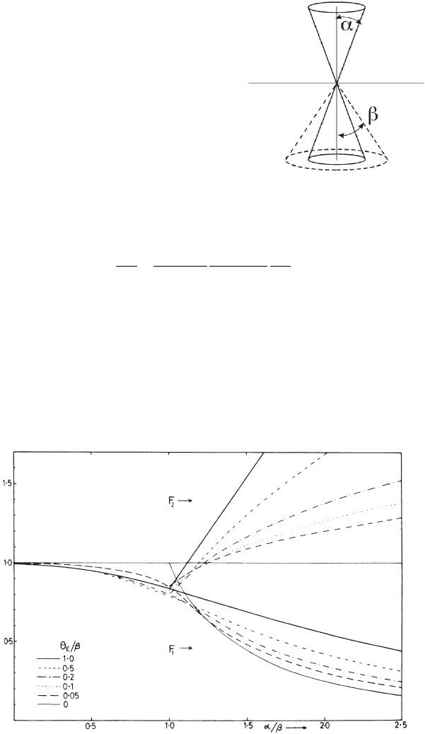
where F
1a,b
are the convergence correction factors that are dependent
on the angular scattering distribution for a particular edge via the
characteristic scattering angle θ
E
. Correction factors must therefore be
calculated for each edge of interest in the quantifi cation and can be
deduced from Figure 4–60 or from simple programs (Egerton, 1996). It
is possible to note that for edges of very similar intrinsic angular dis-
tributions (i.e., two edges close in energy will have very similar θ
E
) the
Figure 4–59. Relation between the conver-
gence and collection angles for quantifi ca-
tion of spectra accounting for the incident
beam convergence.
Figure 4–60. Correction factors F
1
useful to quantify spectra accounting for
convergence of the incident beam α. The convergence factors F
1
must be deter-
mined for each edge based on the convergence angle α relative to the collection
angle β. For each edge, the characteristic angle θ
E
relative to the collection
angle β must be used to identify the appropriate curve to be used in the quan-
tifi cation. The F
2
curves are used only for absolute quantifi cation. (From
Egerton © 1996, with permission from Springer Science+Business Media.)
346 G. Botton
ratio F
1b
/F
1a
is close to one and no effect of convergence is visible irre-
spective of the convergence angle.
For absolute quantifi cation, the correction of the expression relating
the number of atoms and the recorded intensity under the edge
[Eq. (51)] becomes necessary irrespective of the intrinsic angular
distribution
I
k
(β,∆,α) ≈ F
2
NI(β,∆)σ
k
(β,∆α) (54)
where the correction factor F
2
≈ F
1
for α ≤ β and F
2
= (α/β)
2
F
1
for α ≥ β
and can be deduced from Figure 4–60.
4.2.3 k-Factor Approach
All the expressions for relative quantifi cation in EELS are similar to
the k
AB
factor equations in EDXS microanalysis. In a similar manner, a
k-factor approach has been proposed (Malis and Titchmarsh, 1985) to
quantify EELS spectra, although the experimental and sample condi-
tions (convergence, collection, sample thickness) must be very strictly
controlled and reproducible. Once the k-factor has been determined on
the reference sample accounting for the cross-sectional ratios and pos-
sible convergence effects, the same k-factor can be used to deduce the
composition of the unknown samples as in EDXS microanalysis. Cross
section calculations using the currently available models (Hartree–
Slater or hydrogenic, see Section 3.4) and convergence corrections are
already built-in in commercial programs that control the acquisition
functions of the spectrometers. An additional calculation approach to
deduce the cross sections necessary for the quantifi cation makes use
of tabulations of the oscillator strengths obtained from optical data (see
Section 3.4.1). These methods require the acquisition of spectra in
dipole conditions (i.e., with small collection angles) and within the
energy range where the angular distribution of scattering is still domi-
nated by the Lorentzian term of Eq. (27) (i.e., near the edge threshold).
The methodology is described in Egerton (1993) and summarized in
Egerton and Leapman (1995).
4.2.4 Limitations in Analysis and Quantifi cation
Although EELS quantifi cation is not affected by absorption or fl uores-
cence, there are major drawbacks in quantitative and even qualitative
EELS analysis. The major diffi culty is the strong effect of the sample
thickness on the detection of EELS edges. For example, spectra from
specimens of very simple composition a few tens of nanometers in
thickness can reveal well-resolved edges while spectra from “thicker”
areas (as thin as 100 nm) might show no edges (Figure 4–61). This
severe limitation to the visibility of edges is due to the contribution of
multiple inelastic scattering that increases the background under the
edge. The contribution of multiple scattering, however, is not uniform
as a function of energy loss and the quantifi cation of spectra for increas-
ingly thick samples demonstrates a variation of the apparent concen-
tration with thickness. Systematic measurements of the ratio of two
elements, for example, show that samples with thickness relative to the
mean inelastic free path (Sections 3.4 and 8.2) t/λ > 0.5 are unreliable
(Figure 4–62). Even when multiple scattering effects are removed with

Chapter 4 Analytical Electron Microscopy 347
deconvolution techniques (Section 8.2), the effects persist (Egerton,
1996), suggesting that additional contributions due to the angular dis-
tributions of losses are present and cannot be neglected in correction
approaches. Calculations that include the contributions of elastic scat-
tering (Cheng and Egerton, 1993) and convolutions of the energy and
angular distributions of the scattering angle (Su et al., 1995) demon-
strate trends that are consistent with the experimental variation of the
composition with thickness. Correction programs have been devel-
oped to account for these effects (Wong and Egerton, 1995). The lack
of visibility of edges in spectra, even for pure elements, implies that
users should be particularly cautious about drawing conclusions on
the absence of elements during the analysis of point spectra and par-
ticularly with the application of energy-fi ltered imaging techniques.
In the latter condition, since spectra are usually disregarded and the
acquisition is done in an automatic procedure, erroneous results can
often be obtained if care is not taken to check the sample thickness
prior to the analysis of spectra. Relative thickness maps should
therefore be used as a routine check prior to elemental imaging to
verify the applicability of the t/λ > 0.5 condition. The procedure for
the measurement of the relative thickness is described in detail in
Section 8.2.
Figure 4–61. Variation of the
visibility of spectra with increas-
ing thickness of the sample.
Thick samples will not necessar-
ily show EELS edges even for
major constituents.
Figure 4–62. Variation in the
quantifi cation of two elements
as a function of thickness
relative to the total inelastic
mean free path.
348 G. Botton
5 Resolution in Microanalysis
5.1 EDXS Microanalysis
Two contributions determine the spatial resolution in X-ray micro-
analysis. The fi rst contribution arises from the electron beam diameter
and the second from the electron beam broadening generated when
electrons travel in the sample. The early analytical TEMs provided
beam diameters of the order of tens to a hundred nanometers and the
beam broadening within the sample was not a signifi cant issue. Mea-
surements using contamination spots demonstrated that the illumi-
nated area on the top of the sample was essentially the same as the exit
surface of the electron beam (e.g., see Stenton et al., 1981). The situation
changed radically with the development of analytical instruments
equipped with fi eld emission sources capable of achieving probe sizes
in the order of a nanometer or smaller (Section 2.1) whereby the ulti-
mate limits in spatial resolution due to the intrinsic beam broadening
could be probed.
To determine electron beam broadening, we must consider that the
trajectory of the incident beam is controlled by an elastic scattering
process that causes the deviation of the incident electrons as they travel
through the sample. Transport equations (Rez, 1984) and detailed mul-
tislice calculations (e.g., Loan et al., 1988; Mobus and Nufer, 2003;
Voyles and Muller, 2004; Dwyer and Etheridge, 2003; and Section 3.2)
have been developed to describe electron beam propagation [including
the impact of sub-Angstrom beams (Dwyer and Etheridge, 2003)] but
more extensive work has been carried out in the fi eld of AEM using
Monte Carlo simulations that consider the individual trajectories of the
incident electrons, the elastic scattering cross sections that modify the
electron trajectories, and the inelastic scattering that causes the slow-
down of the electrons (Figure 4–63). Monte Carlo approaches are easily
applicable in complex geometries of samples (for example, interfaces
and particles), although they neglect the effect of channeling of elec-
trons in crystal and therefore assume the sample is amorphous or tilted
away from a zone axis. The Monte Carlo technique is based on the
generation of random numbers that are used to calculate the scattering
angles [via equations of the elastic cross sections: Eq. (11)], the path-
length between scattering events [using the elastic mean free path of
Eq. (12)], and the energy loss between the scattering events [using the
stopping power derived from the inelastic cross sections: Eq. (19)].
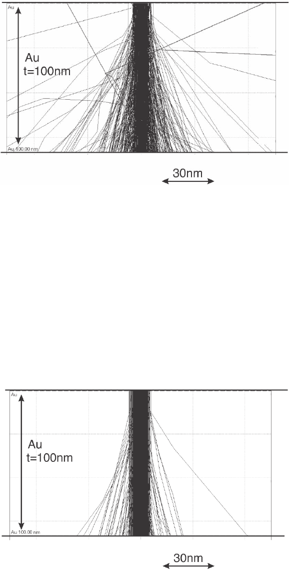
Electron trajectories simulated with the Monte Carlo method for thin
fi lms show the dependence of beam broadening on the accelerating
voltage (Figure 4–64). The effect of the average atomic number and
sample geometry can also be easily determined. With these simula-
tions, it is possible, in principle, to evaluate the exit area and the volume
containing an arbitrary fraction of electrons that will contribute to the
generation of the X-rays signal and thus the spatial resolution. For
example, the interaction volume containing 90% of scattered electrons
is typically used as a reference in the AEM literature to determine the
resolution, although more stringent criteria [with 95% of the electrons
Chapter 4 Analytical Electron Microscopy 349
(Faulkner and Norrgard, 1978) and 99% of the electrons (Reed, 1966)]
have also been proposed. Although broadening values can be retrieved
from the simulated trajectories in a few minutes of computation even
on laptop computers or through web-based programs (Hovington
et al., 1997), quick estimates of the electron beam broadening are neces-
sary to evaluate the approximate loss of spatial resolution in micro-
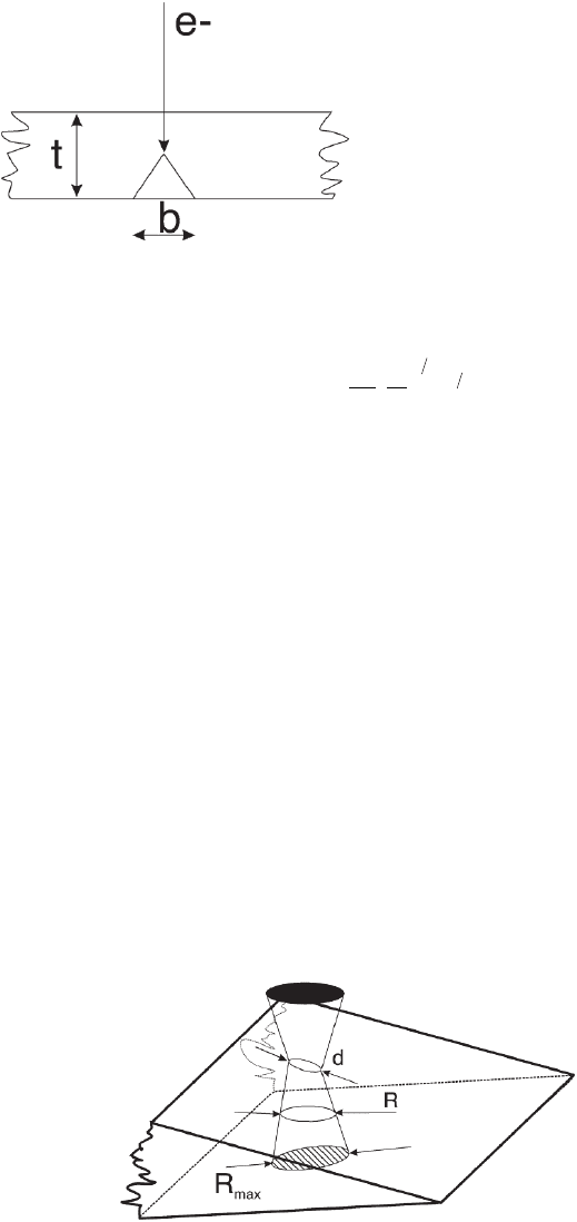
analysis. Goldstein et al. (1977) developed a simple analytical model
assuming a single scattering event in the middle of the foil thickness
Figure 4–63. Simulation of the electron trajectories in a thin sample of Au
(100 nm thickness) using the Monte Carlo method for 100-keV incident elec-
trons. Calculations of electron trajectories are carried out using the public-
domain code CASINO developed in the Gauvin group (Hovington et al., 1997)
available on the web (http://www.montecarlomodeling.mcgill.ca).
Figure 4–64. Electron trajectories in a 100-nm-thick Au Monte Carlo calcula-
tions for 300-keV incident electrons.
350 G. Botton
(Figure 4–65) to calculate the broadening of the electron beam b that
contains 90% of the scattered electrons:
b
Z
EA
t=×
(
)
721 10
5
0
12
32
.
ρ
(55)
where b is the broadening in centimeters, Z is the atomic number, A is
the atomic weight, E
0
is the accelerating voltage (electronvolts), ρ is the
density (grams/cubic centimeter), and t is the specimen thickness (cen-
timeters). This simple equation is the most common method to esti-
mate the broadening and is most suitable for foils of thickness
approximately equal to the elastic mean free path (see Section 3.2),
although refi nements were proposed to account for multiple scattering
in thicker foils (Cliff and Lorimer, 1981; Goldstein et al., 1986).
The spatial resolution can be determined based on the contribution
of both the electron beam size (d) and the electron beam broadening
(b). Early models proposed the addition of the two terms in
quadrature
R = (b
2
+ d
2
)
1/2
(56)
to yield the resolution R, but the currently adopted defi nition is based
on experimental evidence suggesting the use of a less stringent, but
still arbitrary, average sum of the two terms leading to a defi nition of
spatial resolution based on the broadening in the middle of the foil
thickness as suggested in Figure 4–66:
Figure 4–65. Simplifi ed diagram
describing the broadening of
electronic samples. The parameter
b describes the broadening at the
bottom of the foil using Eq. (55) and
the single scattering assumption.
Figure 4–66. Defi nition of parameters related to the calculation of the resolu-
tion with Eqs. (57) and (56) [where R
max
is estimated with Eq. (56) as the
maximum broadening at the bottom of the foil] and R is the broadening in the
middle of the foil. d is the diameter of the incident electron beam.
Chapter 4 Analytical Electron Microscopy 351
R
dR
=
+
max
2
(57)
where R
max
is determined from Eq. (56) as the maximum broadening
at the bottom of the foil. If 90% of the beam current distribution is
considered, the resolution becomes (Keast and Williams, 2000)
Rdbdb=++
22
3
(58)
Recent measurements with sample geometries that probe the elec-
tron beam broadening at the bottom of the foil have been published
and have led to the conclusion that although the single scattering
models predict the right beam broadening magnitude, these quantita-
tively overestimate the broadening observed experimentally (Nakata
et al., 2001). More accurate models (Keast and Williams, 2000; Doigt
and Flewitt, 1982) consider a Gaussian probe distribution of standard
deviation σ and hence a probe with an FWHM of 4.29σ propagating
the foil with a distribution accounting for beam spreading and the
incident probe distribution (Keast and Williams, 2000)
Ixyt
i
t
xy
t
b
,,
(
)
=
+
()
−+
()
+
πσ β σ β22
23
22
23
exp
(59)
where β = 500(4Z/E
0
)
2
(ρ/A) and i
b
is the incident beam current.
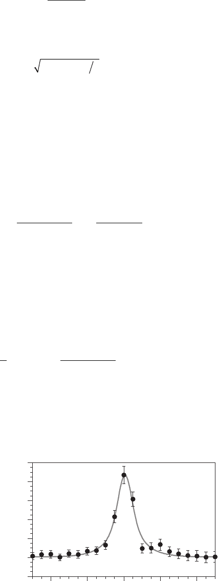
Using this more detailed electron distribution in the probe (as well
as more simple models) as it travels through the sample, Keast and
Williams (1999, 2000) determined the equilibrium segregation profi le
in grain boundaries based on composition line scans and two-
dimensional map measurements (Figure 4–67). Based on the Gaussian
intensity distribution of incident electrons the resolution, for a given
fraction of electrons Q, as a function of thickness, can be defi ned as
Q
h
R
t
dt
h
=−
−
+
()
∫
1
1
42
2
23
0
exp
σβ
(60)
where the integration is carried out over the thickness up to t = h where
R is the diameter of the cone defi ning the resolution. The distribution
is assumed circularly symmetric (since there is no dependence of the
azimuth angle in the equation) and applicable to a two-dimensional
case. By defi ning the resolution criteria based on the fraction of
-0.02
0
0.02
0.04
0.06
0.08
0.1
-4 -2 0 2 4
distance (nm)
S/Ni (counts)
Figure 4–67. Composi-
tion profi le of S segrega-
tion at an Ni grain
boundary. The profi le is
based on the modeling
of the beam profi le
using a Gaussian
probe distribution and
Eq. (59) (Keast and
Williams, 2000). (Profi le
courtesy of V. Keast,
University of Sydney.)
352 G. Botton
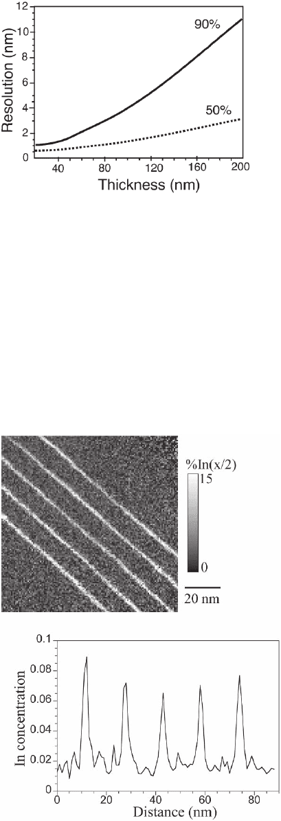
electrons within a given radius, it is possible to plot the expected reso-
lution (Figure 4–68) for a diameter containing 50% and 90% of elec-
trons. A comparison of the predictions with experimental profi les
obtained on equilibrium segregation in Cu (for FWHM and at 10%
maximum) (Keast and Williams, 2000) shows excellent agreement for
thicknesses up to the elastic mean free path.
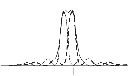
With a dedicated STEM, recent work on grain boundary segregation
and multilayer composite materials has shown that it is possible to
detect submonolayer segregation at grain boundaries (Keast et al., 1998;
Keast and Williams, 1999, 2000) and map the composition of quantum
wells in semiconductor materials (Figure 4–69). EDXS analysis with
Figure 4–68. Resolution
variation as a function of
thickness for the 50% and 90%
fraction of electrons within
the beam. (Adapted from
Keast and Williams, 2000.)
Figure 4–69. Quantita-
tive EDXS elemental
maps of In-rich quantum
wells in semiconductors
and related intensity
profi les across the
quantum wells. (Cour-
tesy of V. Keast, Univer-
sity of Sydney.)
Chapter 4 Analytical Electron Microscopy 353
aberration-corrected instruments has been recently reported by
Watanabe et al. (2005) and Watanabe and Williams (2005a).
5.2 Energy-Filtered Microscopy and EELS Microanalysis
The spatial resolution in energy loss spectroscopy measurements is
strongly dependent on the operating mode of the microscope during
EELS analysis due to the illumination conditions of the area of interest,
the effects of lens aberration, and the spectrometer coupling condi-
tions. The contribution from the microscope lens aberrations comes
essentially from the objective lens and can be calculated as follows.
With respect to an incident beam of primary energy E
0
propagating
along the optic axis, the inelastic interactions that induce an energy
loss E and scattering at an angle θ will cause a blur of an object point
(and the image on the viewing screen of the microscope) due to chro-
matic aberrations R = M
0
θ∆f where M
0
is the magnifi cation of the objec-
tive lens and ∆f = C
c
(E/E
0
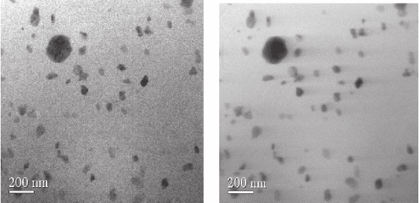
). According to the Rayleigh criterion, two
object points with blurred independent distributions can be resolved
if they are separated by a distance giving rise to an intensity drop of
20% between the maxima of the summed distributions (Figure 4–70).
The distance between these two points is defi ned as the resolution
and is obtained as (Egerton, 1996)
r
i
≈ 2θ
E
∆f ≈ C
c
(E/E
0
)
2
(61)
This resolution is expected for energy-fi ltered images acquired at an
energy loss E when the incident electron energy is kept constant, the
image is initially focused for electrons that have lost no energy (which
is also known as a zero-loss image, Section 2.4.1), and the scattering
angles are limited only by the intrinsic scattering distribution at the
energy loss E (i.e., no limiting objective aperture is used). For images
of thick samples, it should be pointed out that the inelastic losses can
constitute the most important part of the signal resulting in signifi cant
blurring of images. These contributions can be removed by energy fi l-
tering so that the inelastically scattered electrons are removed with the
Figure 4–70. Rayleigh defi nition of resolution based on the overlap of two
diffracting-limited functions placed in close proximity to each other. If the
sum of the two intensity profi les shows a dip of 20% of the maximum intensity,
the peaks are considered to be resolvable.
354 G. Botton
energy-selecting slit and the images appear sharp even for thick samples
(Figure 4–71). In typical conditions for optimum energy loss imaging
at an energy loss E, however, the incident electron energy is readjusted
so that the spectrometer is focused at the energy loss E (i.e., by raising
the high tension of the microscope so that the primary energy is now
E
0
+ E) and the electrons of the related loss are in focus. If an energy-
selecting slit ∆E wide is used, the resolution becomes (Egerton, 1996)
r
i
≈ 2θ
E
∆f ≈ (C
C
/4)(∆E/E
0
)
2
(62)
When the angular distribution of scattering is limited by an objective
aperture β smaller than the angular distribution of scattering, namely
θ
E
, other contributions become dominant (diffraction limit, spherical
aberration, delocalization of scattering) and the spatial resolution in
elemental maps is derived from a combination of all factors as described
in Section 6.2.3.
For point analyses carried out in TEM image mode, the spectrometer
entrance aperture is used to select electrons from the regions of interest
so that they enter the spectrometer for analysis. In these conditions,
the chromatic aberration will also cause contributions from areas
further away from the spectrometer aperture-delimited area due to the
signifi cant broadening of the angular distributions and high energy
losses. If no angular-selecting aperture (the objective aperture in this
case) is used, the total broadening due to angular scattering up to the
Bethe-ridge (θ
r
= θ
C
, which includes most of the scattering intensity, see
Section 3.4.1) is
r
c
≈ θ
r
∆f (63)
while the use of an angular-selecting aperture (in the objective back-
focal plane when the TEM is operated in image mode) will limit the
a) b)
Figure 4–71. (a) Energy-fi ltered image of an Al alloy with strengthening
precipitates. The image has been obtained by selecting only the electrons
that have not lost signifi cant amounts of energy (i.e., zero-loss image) and (b)
unfi ltered image (containing all the inelastically scattered electrons). Although
the total intensity is reduced, the precipitates are more clearly defi ned with
sharper edges in the zero-loss image.
