Hawkes P.W., Spence J.C.H. (Eds.) Science of Microscopy. V.1 and 2
Подождите немного. Документ загружается.

Chapter 2 Scanning Transmission Electron Microscopy 105
elemental mapping, which, given the inelastic cross sections of the
core-loss events, can be calibrated in terms of composition. Using this
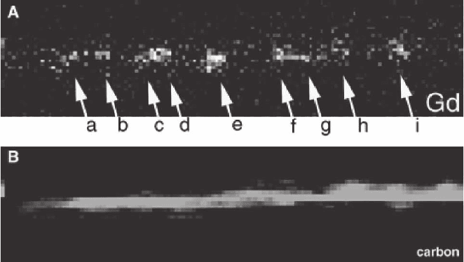
approach, individual atoms of Gd have been observed inside a carbon
nanotube structure (Suenaga et al., 2000) (Figure 2–19). A more sophis-
ticated approach is to use multivariate statistical (MSI) methods (Bonnet
et al., 1999) to analyze the compositional maps. With this approach, the
existence of phases of certain stoichiometry can be identifi ed, and
maps of the phase locations within the sample can be created. Even the
fi ne structure of core-loss edges can be used to form maps in which
only the bonding, not the composition, within the sample has changed.
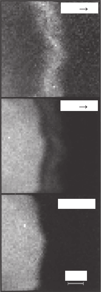
An example of this is the mapping of the sp
2
and sp
3
bonding states of
carbon at the interface of chemical vapor deposition diamond grown
on a silicon substrate (Muller et al., 1993) (Figure 2–20). The sp
2
signal
shows the presence of an amorphous carbon layer at the interface.
A similar three-dimensional data cube may also be recorded by
conventional TEM fi tted with an imaging fi lter. In this case, the image
is recorded in parallel while varying the energy loss being fi ltered for.
Both methods have advantages and disadvantages, and the choice can
depend on the desired sampling in either the energy or image dimen-
sions. The STEM does have one important advantage, however. In a
CTEM, all of the imaging optics occur after the sample, and these
optics suffer signifi cant chromatic aberration. Adjusting the system to
change the energy loss being recorded can be done by changing the
energy of the incident electrons, thus keeping the energy of the desired
inelastically scattered electrons constant within the imaging system.
However, to obtain a useful signal-to-noise ratio in energy-fi ltered
transmission electron microscopy (EFTEM), it is necessary to use a
selecting energy window that is several electronvolts in width, and
even this energy spread in the imaging system is enough to worsen
Figure 2–19. A spectrum image fi ltered for Gd (A) and C (B). Individual atoms of Gd inside a carbon
nanotube can be observed. [Reprinted from Suenaga et al. (2000), with copyright permission from
AAAS.]
106
5 nm
Diamond
C
1s p
*
C
1s
*
s
Figure 2–20. By fi ltering for spe-
cifi c peaks in the fi ne structure of
the carbon K-edge, maps of π and
σ bonded carbon can be formed.
The presence of an amorphous sp
2
bonded carbon layer at the inter-
face of a chemical vapor deposi-
tion (CVD)-grown diamond on an
Si substrate can be seen. The
diamond signal is derived by a
weighted subtraction of the π
bonding image from the σ bonding
image. [Reprinted from Muller
et al. (1993), with permission of
Nature Publishing Group.]
the spatial resolution signifi cantly. In STEM, all of the image-forming
optics are before the specimen, and the spatial resolution is not
compromised.
Inelastic scattering processes, especially single electron excitations,
have a scattering cross section that can be an order of magnitude
smaller than for elastic scattering. To obtain suffi cient signal, EELS
acquisition times may be of the order of 1 s. Collection of a spectrum
image with a large number of pixels can therefore be very slow, with
Peter D. Nellist
Chapter 2 Scanning Transmission Electron Microscopy 107
the associated problems of both sample drift, and drift of the energy
zero point due to power supplies warming up. In practice, spectrum
image acquisition software often compensates for these drifts. Sample
drift can be monitored using cross-correlations on a sharp feature in
the image. Monitoring the position of the zero-loss peak allows the
energy drift to be corrected. The advent of aberration correction will
have a major impact in this regard. Perhaps one of the most important
consequences of aberration correction is that it will increase the current
in a given sized probe by more than an order of magnitude (see Section
10.3). Fast elemental mapping through spectrum imaging will then
become a much more routine application of EELS. However, to achieve
this improvement in performance, there will have to be corresponding
improvements in the associated hardware. In general, commercially
available systems can achieve around 200 spectra per second. Some
laboratories with custom instrumentation have reported reaching 1000
spectra per second (Tencé, personal communication). Further improve-
ment will be necessary to fully make use of spectrum imaging in an
aberration corrected STEM.
7. X-Ray Analysis and Other Detected Signals in
the STEM
It is obvious that the STEM bears many resemblances to the SEM: a
focused probe is formed at a specimen and scanned in a raster while
signals are detected as a function of probe position. So far we have
discussed BF imaging, ADF imaging, and EELS. All of these methods
are unique to the STEM because they involve detection of the fast
transmitted electron through a thin sample; bulk samples are typically
used in an SEM. There are of course, a multitude of other signals that
can be detected in STEM, and many of these are also found in SEM
machines.
7.1 Energy Dispersive X-Ray Analysis
When a core electron in the sample is excited by the fast electron tra-
versing the sample, the excited system will subsequently decay with
the core hole being refi lled. This decay will release energy in the form
of an X-ray photon or an Auger electron. The energy of the particle
released will be characteristic of the core electron energy levels in the
system, and allows compositional analysis to be performed.
The analysis of the emitted X-ray photons is known as energy-
dispersive X-ray (EDX) analysis, or sometimes energy-dispersive spec-
troscopy (EDS) or X-ray EDS (XEDS). It is a ubiquitous technique for
SEM instruments and electron-probe microanalyzers. The technique
of EDX microanalysis in CTEM and STEM has been extensively covered
(Williams and Carter, 1996), and we will review here only the specifi c
features of EDX in a STEM.
The key difference between performing EDX analysis in the STEM as
opposed to the SEM is the improvement in spatial resolution (see Figure
2–21). The increased accelerating voltage and the thinner sample used

108
in STEM lead to an interaction volume that is some 10
8
times smaller
than for an SEM. Beam broadening effects will still be signifi cant for
EDX in STEM, and Eq. (6.2) provides a useful approximation in this
case. For a given fraction of the element of interest, however, the total
X-ray signal will be correspondingly smaller. For a discussion of detec-
tion limits for EDX in STEM see Watanabe and Williams (1999). A
further limitation for high-resolution STEM instruments is the geome-
try of the objective lens pole pieces between which the sample is placed.
For high resolution the pole piece gap must be small, and this limits
both the solid angle subtended by the EDX detector and the maximum
take-off angle. This imposes a further reduction on the X-ray signal
strength. A high probe current of around 1 nA is typically required for
EDX analysis, and this means that the probe size must be increased to
greater than 1 nm (see Section 10), thus losing atomic resolution sensi-
tivity. A further concern is the mounting of a large liquid nitrogen
dewar on the column for the necessary cooling of the detector. It is often
suspected that the boiling of the liquid nitrogen and the unbalancing of
the column can lead to mechanical instabilities. A positive benefi t of
EDX in STEM, however, is that windowless EDX detectors may com-
monly be used. The vacuum around the sample in STEM is typically
higher than for other electron microscopes to reduce sample contami-
nation during imaging and to reduce the gas load on the ultrahigh
vacuum of the gun. A consequence is that contamination or icing of a
windowless detector is less common.
For the reasons described above, EDX analysis capabilities are some-
times omitted from ultrahigh resolution dedicated STEM instruments,
but are common on combination CTEM/STEM instruments. A notable
exception has been the development of a 300-kV STEM instrument with
the ultimate aim of single-atom EDX detection (Lyman et al., 1994).
SEM
STEM
100 nm
1 nm
excitation volume
~ 1 mm
3
excitation volume
~ 10 nm
3
10 nm
Figure 2–21. A schematic diagram comparing the beam interaction volumes
for an SEM and a STEM. The higher accelerating voltage and thinner samples
in STEM lead to much higher spatial resolution for analysis, with an associated
loss in signal.
Peter D. Nellist
Chapter 2 Scanning Transmission Electron Microscopy 109
It is worth making a comparison between EDX and EELS for STEM
analysis. The collection effi ciency of EELS can reach 50%, compared to
around 1% for EDX, because the X-rays are emitted isotropically. EELS
is also more sensitive for light element analysis (Z < 11), and for many
transition metals and rare-earth elements that show strong spectral
features in EELS. The energy resolution in EELS is typically better than
1 eV, compared to 100–150 eV for EDX. The spectral range of EDX,
however, is higher with excitations up to 20 keV detectable, compared
with around 2 keV for EELS. Detection of a much wider range of ele-
ments is therefore possible.
7.2 Secondar y Electrons, Auger Electrons,
and Cathodoluminescence
Other methods commonly found on an SEM have also been seen on
STEM instruments. The usual imaging detector in an SEM is the sec-
ondary electron (SE) detector, and these are also found on some STEM
instruments. The fast electron incident upon the sample can excite
electrons so that they are ejected from the sample. These relatively slow
moving electrons can escape only if they are generated relatively close
to the surface of the material, and can therefore generate topographical
maps of the sample. Once again, because the interaction volume is
smaller, the use of SE in STEM can generate high-resolution topo-
graphical images of the sample surface. An intriguing experiment
involving secondary electrons has been the observation of coincidence
between secondary electron emission and primary beam energy-loss
events (Mullejans et al., 1993).
Auger electrons are ejected as an alternative to X-ray photon emission
in the decay of a core-electron excitation, and spectra can be formed and
analyzed just as for X-ray photons. The main difference, however, is that
whereas X-ray photons can escape relatively easily from a sample,
Auger electrons can escape only when they are created close to the
sample surface. It is therefore a surface technique, and is sensitive to the
state of the sample surface. Ultrahigh vacuum conditions are therefore
required, and Auger in STEM is not commonly found.
Electron-hole pairs generated in the sample by the fast electron can
decay by way of photon emission. For many semiconducting samples,
these photons will be in or near the visible spectrum and will appear
as light, known as cathodoluminescence. Although rarely used in
STEM, there has been the occasional investigation (see, for example,
Pennycook et al., 1980).
8. Electron Optics and Column Design
Having explored some of the theory and applications of the various
imaging and analytical modes in STEM, it is a good time to return to
the details of the instrument itself. The dedicated STEM instrument
provides a nice model to show the degrees of freedom in the STEM
optics, and then we go on to look at the added complexity of a hybrid
CTEM/STEM instrument.
110
8.1 The Dedicated STEM Instrument
We will start by looking at the presample or probe-forming optics of a
dedicated STEM, though it should be emphasized that most of the
comments in this section also apply to TEM/STEM instruments. In
addition to the objective lens, there are usually two condenser lenses
(Figure 2–1). The condenser lenses can be used to provide additional
demagnifi cation of the source, and thereby control the trade-off
between probe size and probe current (see Section 10.1). In principle,
only one condenser lens is required because movement of the crossover
between the condenser and objective lens (OL) either further or nearer
to the OL can be compensated by relatively small adjustments to the
OL excitation to maintain the sample focus. The inclusion of two con-
denser lenses allows the demagnifi cation to be adjusted while main-
taining a crossover at the plane of the selected area diffraction aperture.
The OL is then set such that the selected area diffraction (SAD) aper-
ture plane is optically conjugate to that of the sample.
In a conventional TEM instrument, the SAD aperture is placed after
the OL, and the OL is set to make it optically conjugate to the sample
plane. The SAD aperture then selects a region of the sample, and the
post-OL lenses are used to focus and magnify the diffraction pattern
in the back-focal plane of the OL to the viewing screen. By reciprocity,
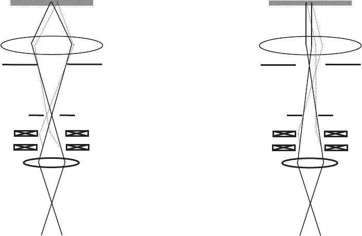
an equivalent SAD mode can be established in a dedicated STEM
(Figure 2–22). With the condenser lenses set to place a crossover at the
condenser lens
scan coils
selected area
diffraction aperture
objective
aperture
objective lens
sample
imaging mode
diffraction mode
Figure 2–22. The change from imaging to diffraction mode is shown in this schematic of part of a
STEM column. By refocusing the condenser lens on the objective lens FFP rather than the SAD aperture
plane, the objective lens generates a parallel beam at the sample rather than a focused probe. The SAD
aperture is now the beam-limiting aperture, and defi nes the illumination region on the sample.
Peter D. Nellist
Chapter 2 Scanning Transmission Electron Microscopy 111
SAD, an image can be formed with the SAD selecting a region of inter-
est in the sample. The condenser lenses are then adjusted to place a
crossover at the front focal plane of the OL, and the scan coils are set
to scan the crossover over the front focal plane. The OL then generates
a parallel pencil beam that is rocked in angle at the sample plane. In
the detector plane is therefore seen a conventional diffraction pattern
that is swept across the detector by the scan. By using a small BF detec-
tor, a scanned diffraction pattern will be formed. If a Ronchigram
camera is available in the detector plane, then the diffraction pattern
can be viewed directly and scanning is unnecessary. In practice, SAD
mode in a STEM is more commonly used for measuring the angular
range of BF and ADF detectors rather than diffraction studies of
samples. It is also often used for tilting a crystalline sample to a zone
axis if a Ronchigram camera is not available.
To avoid having to mutually align the two condenser lenses, many
users employ only one condenser at a time. Both are set to focus a
crossover at the SAD aperture plane, but the different distance between
the lenses and the SAD plane means that the overall demagnifi cation
of the source will differ. Often the two discrete probe current settings
then available are suitable for the majority of experiments. Alterna-
tively, many users, especially those with a Ronchigram camera, need
an SAD mode very infrequently. In this case, there is no requirement
for a crossover in the SAD plane, and one condenser lens can be
adjusted freely.
In more modern STEM instruments, a further gun lens is provided
in the gun acceleration area. The purpose of this lens is to focus a
crossover in the vicinity of the differential pumping aperture that is
necessary between the ultrahigh vacuum gun region and the rest of
the column. The result is that a higher total current is available for very
high current modes. For lower current, higher resolution modes, a gun
lens is not found to be necessary.
Let us now turn our attention to the objective lens and the postspeci-
men optics. The main purpose of the OL is to focus the beam to form
a small spot. Just like a conventional TEM, the OL of a STEM is designed
to minimize the spherical and chromatic aberration, while leaving a
large enough gap for sample rotation and providing a suffi cient solid
angle for X-ray detection.
An important parameter in STEM is the postsample compression.
The fi eld of the objective lens that acts on the electrons after they exit
the sample also has a focusing effect on the electrons. The result is that
the scattering angles are compressed and the virtual crossover position
moves down. Most of the VG dedicated STEM instruments have top-
entry OLs, which are consequently asymmetric in shape. The bore on
the probe forming (lower) side of the OL is smaller then on the upper
side, and therefore the fi eld is more concentrated on the lower side. The
typical postsample compression for these asymmetric lenses, typically
a factor of around 3, is comparatively low. The entrance to the EELS
spectrometer will often be up to 60 cm or more after the sample, to allow
room for defl ection coils and other detectors. A 2-mm-diameter EELS
entrance aperture then subtends a geometric entrance semiangle of
112
1.7 mrad. Including the factor of 3 compression from the OL gives a
typical collection semiangle of 5 mrad. The probe convergence angle of
an uncorrected STEM will be around 9 mrad, so the total collection effi -
ciency of the EELS system will be poor, being below 25% after account-
ing for further angular scattering from the inelastic scattering process.
After the correction of spherical aberration, the probe convergence
semiangle will rise to 20 mrad or more, and the coupling of this beam
into the EELS system will become even more ineffi cient.
A postspecimen lens would in principle allow improved coupling
into the EELS by providing further compression after the beam has left
the objective lens. However, there needs to be enough space for defl ec-
tion coils and lens windings between the lenses, so it is hard to position
a postspecimen lens closer than about 100 mm after the OL. By the time
the beam has propagated to this lens, it will be of the order of 1 mm in
diameter. This is a large diameter beam to be handled by an electron
lens, in the lower column typical widths are 50 µm or less, and large
aberrations will be introduced that will obviate the benefi t of the extra
compression. In many dedicated STEMs, therefore, postspecimen
lenses are rarely used. A more common work around solution is to
mount the sample as low in the OL as possible and to excite the OL as
hard as possible to provide the maximum compression possible, though
it is diffi cult to do this and to maintain the tilt capabilities.
A novel solution demonstrated by the Nion Co. is to use a four-
quadrupole four-octupole system to couple the postspecimen beam
to the spectrometer and provide increased compression. The four-
quadrupole system has enough degrees of freedom to provide com-
pression while also ensuring that the virtual crossover as seen by the
spectrometer is at the correct object distance. As with any postspeci-
men lens system in a top entry STEM, the beam is so wide at the lens
system that large third-order aberrations are introduced. The presence
of the octupoles allows for correction of these aberrations and addition-
ally the third-order aberrations of the spectrometer, which in turn
allows a larger physical spectrometer entrance aperture to be used.
Collection semiangles up to 20 mrad have been demonstrated with this
system (Nellist et al., 2003).
8.2 CTEM/STEM Instruments
At the time of writing, dedicated STEM columns are available from
JEOL and Hitachi. Nion Co. has a prototype aberration-corrected dedi-
cated STEM column under test, and this will soon be added to the array
of available machines. However, many researchers prefer to use a
hybrid CTEM/STEM instrument, which is supplied from all the main
manufacturers. As their name suggests, CTEM/STEM instruments
offer the capabilities of both modes in the same column.
A CTEM/STEM is essentially a CTEM column with very little
modifi cation apart from the addition of STEM detectors. When fi eld-
emission guns (FEGs) were introduced onto CTEM columns, it was
found that the beam could be focused onto the sample with spot sizes
down to 0.2 nm or better (for example, James and Browning, 1999). The
Peter D. Nellist
Chapter 2 Scanning Transmission Electron Microscopy 113
addition of a suitable scanning system and detectors thus created a
STEM. The key is that modern CTEM instruments with a side-entry
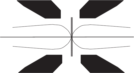
stage tend to make use of the condenser-objective lens (Figure 2–23). In
the condenser-objective lens, the fi eld is symmetric about the sample
plane, and therefore the lens is just as strong in focusing the beam to a
probe presample as it is in focusing the postsample scattered electrons
as it would do in conventional TEM mode. The condenser lenses and
gun lens play the same roles as those in the dedicated STEM. The main
difference in terminology is that what would be referred to as the objec-
tive aperture in a CTEM/STEM is referred to as the condenser aperture in
a TEM/STEM. The reason for this is that the aperture in question is
usually in or near the condenser lens closest to the OL, and this is the
condenser aperture when the column is used in CTEM mode.
An important feature of the CTEM/STEM when operating in the
STEM mode is that there are a comparatively large number of post-
specimen lenses available. The condenser-objective lens ensures that
the beam is narrow when entering these lenses, and so coupling with
high compression to an EELS spectrometer does not incur the large
aberrations discussed earlier. Further pitfalls associated with high
compression should be borne in mind, however. The chromatic aber-
ration of the coupling to the EELS will increase as the compression is
increased, leading to edges being out of focus at different energies.
Also, the scan of the probe will be magnifi ed in the dispersion plane
of the prism, so careful descan needs to be done postsample. A fi nal
feature of the extensive postsample optics is that a high magnifi cation
image of the probe can be formed in the image plane. This is not as
useful for diagnosing aberrations in the probe as one might expect
because the aberrations might well be arising from aberrations in the
TEM imaging system. Nonetheless, potential applications for such a
confocal arrangement have been discussed (see, for example, Möbus
and Nufer, 2003).
pole piece
sample
electron beam
Figure 2–23. A condenser-objective lens provides symmetrical focusing on
either side of the central plane. It can therefore be used to provide postsample
imaging, as in a CTEM, or to focus a probe at the sample, as in a STEM, or
even to provide both simultaneously if direct imaging of the STEM probe is
required.
114
9. Electron Sources
9.1 The Need for Suffi cient Brightness
Naively one might expect that the size of the electron source is not
critical to the operation of a STEM because we have condenser lenses
available in the column to increase the demagnifi cation of the source
at will, and thereby still be able to form an image of the source that is
below the diffraction limit. We will see, however, that increasing the
demagnifi cation decreases the current available in the probe, and the
performance of a STEM relies on focusing a signifi cant current into a
small spot. In fact, the crucial parameter of interest is that of brightness
(see, for example, Born and Wolf, 1980). The brightness is defi ned at
the source as
B
I
A
=
Ω
(9.1)
where I is the total current emitted, A is the area of the source over
which the electrons are emitted, and Ω is the solid angle into which
the electrons are emitted. Brightness is a useful quantity because at
any plane conjugate to the image source (which means any plane where
there is a beam crossover), brightness is conserved. This statement
holds as long as we consider only geometric optics, which means that
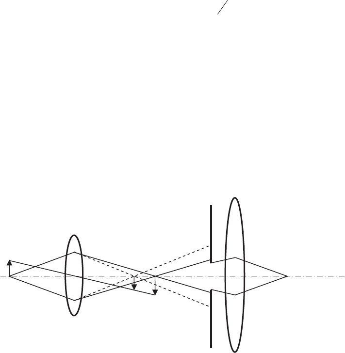
we neglect the effects of diffraction. Figure 2–24 shows schematically
how the conservation of brightness operates. As the demagnifi cation
of an electron source is increased, reducing the area A of the image,
the solid angle Ω increases in proportion. Introduction of a beam-
limiting aperture forces Ω to be constant, and therefore the total beam
current, I, decreases in proportion to the decrease in the area of the
source image.
condenser
lens
objective
aperture
objective
lens
Figure 2–24. A schematic diagram showing how beam current is lost as the source demagnifi cation
increased. Reducing the focal length of the condenser lens further demagnifi es the image of the source,
but the solid angle of the beam correspondingly increases (dashed lines). At a fi xed aperture, such as
an objective aperture, more current is lost when the beam solid angle increases.
Peter D. Nellist
