Hawkes P.W., Spence J.C.H. (Eds.) Science of Microscopy. V.1 and 2
Подождите немного. Документ загружается.

Chapter 2 Scanning Transmission Electron Microscopy 75
where φ
g
is a complex quantity expressing the amplitude and phase of
the g diffracted beam. Equation 3.4 is simply expressing the array of
discs seen in Figure 2–6.
To examine just the overlap region between the g and h diffracted
beam, let us expand (3.4) using (2.4). Since we are just interested in the
overlap region we will neglect to include the top-hat function, H(K),
which denotes the physical objective aperture, leaving
0 g 0
+ φ
h
exp[iχ(K − h) + i2π(K − h) · R
0
] (3.5)
and we fi nd the intensity by taking the modulus squared of Eq. (3.5),
0 g h g h
− χ(K − h) + 2π(h − g) · R
0
+ ∠φ
g
− ∠φ
h
] (3.6)
where ∠φ
g
denotes the phase of the g diffracted beam. The cosine
term shows that the disc overlap region contains interference features,
and that these features depend on the lens aberrations, the position
of the probe, and the phase difference between the two diffracted
beams.
If we assume that the only aberration present is defocus, then the
terms including χ in (3.6) become
χ(K − g) − χ(K − h) = πzλ (K − g)
2
− (K − h)
2
= πzλ 2K · (h − g) + |g|
2
+ |h|
2
(3.7)
Because Eq. (3.7) is linear in K, a uniform set of fringes will be observed
aligned perpendicular to the line joining the centers of the correspond-
ing discs, as seen in Figure 2–6. For interference involving the central,
or bright-fi eld, disc we can set g = 0. The spacing of fringes in the
microdiffraction pattern from interference between the BF disc and
the h diffracted beam is (zλ|h|)
−1
, which is exactly what would be
expected if the interference fringes were a shadow of the lattice planes
corresponding to the h diffracted beam projected using a point
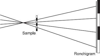
source a distance z from the sample (Figure 2–7). When the objective
aperture is removed, or if a very large aperture is used, then the inten-
sity in the detector plane is referred to as a shadow image. If the sample
is crystalline, then the shadow image consists of many crossed sets of
fringes distorted by the lens aberrations. These crystalline shadow
images are often referred to as Ronchigrams, deriving from the use of
similar images in light optics for the measurement of lens aberrations
(Ronchi, 1964). It is common in STEM for shadow images of both crys-
talline and nonperiodic samples to be referred to as Ronchigrams,
however.
The term containing R
0
in the cosine argument in Eq. (3.6) shows
that these fringes move as the probe is moved. Just as we might expect
for a shadow, we need to move the probe one lattice spacing for the
fringes all to move one fringe spacing in the Ronchigram. The idea of
the Ronchigram as a shadow image is particularly useful when con-
sidering Ronchigrams of amorphous samples (see Section 3.2). Other
aberrations, such as astigmatism or spherical aberration, will distort
Ψ(K, R ) = φ exp[iχ(K − g) + i2π(K − g) · R ]
I(K, R ) = |φ | + |φ | + 2|φ ||φ |cos[χ(K − g)
[
[ ]
]
2

76
the fringes so that they are no longer uniform. These distortions may
be a useful method of measuring lens aberrations, though the analysis
of shadow images for determining lens aberrations is more straight-
forward with nonperiodic samples (Dellby et al., 2001).
The argument of the cosine in Eq. (3.6) also contains the phase dif-
ference between the g and h diffracted beams. By measuring the posi-
tion of the fringes in all the available disc overlap regions, the phase
difference between pairs of adjacent diffracted beams can be deter-
mined. It is then straightforward to solve for the phase of all the dif-
fracted beams, thereby solving the phase problem in electron diffraction.
Knowledge of the phase of the diffracted beams allows immediate
inversion to the real-space exit-surface wavefunction. The spatial reso-
lution of such an inversion is limited only by the largest angle dif-
fracted beam that can give rise to observable fringes in the
microdiffraction pattern, which will typically be much larger than
the largest angle that can be passed through the objective lens (i.e.,
the radius of the BF disc in the microdiffraction pattern). The method
was fi rst suggested by Hoppe (1969a,b, 1982) who gave it the name
ptychography. Using this approach, Nellist et al. (1995; Nellist and
Rodenburg, 1998) were able to form an image of the atomic columns
in Si〈110〉 in a STEM that conventionally would be unable to image
them. Ptychography has not become a common method in STEM,
mainly because the phasing method described above works only for
thin samples. In thicker samples, for which dynamic diffraction theory
is applicable, the phase of the diffracted beams can depend on the
angle of the incident beam. The inherent phase of a diffracted beam
may therefore vary across its disc in a microdiffraction pattern, making
the simple phasing approach discussed above fail. Spence (1998a,b) has
discussed in principle how a crystalline microdiffraction pattern data
set can be inverted to the scattering potential for dynamically scatter-
ing samples, though as yet there has not been an experimental
demonstration.
Figure 2–7. If the probe is defocused from the sample plane, the probe cross-
over can be thought of as a point source located distant from the sample. In
the geometric optics approximation, the STEM detector plane is a shadow
image of the sample, with the shadow magnifi cation given by the ratio of the
probe-detector and probe-sample distances. If the sample is crystalline, then
the shadow image is referred to as a Ronchigram.
Peter D. Nellist
Chapter 2 Scanning Transmission Electron Microscopy 77
3.2 Ronchigrams of Noncrystalline Materials
When observing a noncrystalline sample in a Ronchigram, it is gener-
ally suffi cient to assume that most of the scattering in the sample is
at angles much smaller than the illumination convergence angles,
and that we can broadly ignore the effects of diffraction. In this case
only the BF disc is observable to any signifi cance, but it contains
an image of the sample that resembles a conventional bright-fi eld
image that would be observed in a conventional TEM at the defocus
used to record the Ronchigram (Cowley, 1979b). The magnifi cation of
the image is again given by assuming that it is a shadow projected
by a point source a distance z (the lens defocus) from the sample.
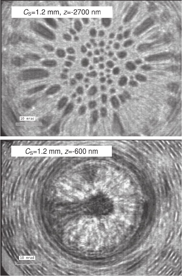
As the defocus is reduced, the magnifi cation increases (Figure 2–8)
until it passes through an infi nite magnifi cation condition when the
probe is focused exactly at the sample. For a quantitative discussion
of how Eq. (3.6) reduces to a simple shadow image in the case of pre-
dominantly low angle scattering, see Cowley (1979b) and Lupini
(2001).
Aberrations of the objective lens will cause the distance from the
sample to the crossover point of the illuminating beam to vary as a func-
tion of angle within the beam (Figure 2–3), and therefore the apparent
magnifi cation will vary within the Ronchigram. Where crossovers occur
at the sample plane, infi nite magnifi cation regions will be seen. For
example, positive spherical aberration combined with negative defocus
can give rise to rings of infi nite magnifi cation (Figure 2–8). Two infi nite
magnifi cation rings occur, one corresponding to infi nite magnifi cation
in the radial direction and one in the azimuthal direction (Cowley, 1986;
Lupini, 2001).
Measuring the local magnifi cation within a noncrystalline Ronchi-
gram can readily be done by moving the probe a known distance and
measuring the distance features move in the Ronchigram. The local
magnifi cations from different places in the Ronchigram can then be
inverted to values for aberration coeffi cients. This is the method
invented by Krivanek et al. (Dellby et al., 2001) for autotuning of a
machine, the Ronchigram of a nonperiodic sample is typically used to
align the instrument (Cowley, 1979a). The coma free axis is immedi-
ately obvious in a Ronchigram, and astigmatism and focus can be
carefully adjusted by observation of the magnifi cation of the speckle
contrast. Thicker crystalline samples also show Kikuchi lines in the
shadow image, which allows the crystal to be carefully tilted and
aligned with the microscope coma-free axis simply by observation of
the Ronchigram.
Finally it is worth noting that an electron shadow image for a weakly
scattering sample is actually an in-line hologram (Lin and Cowley,
1986) as fi rst proposed by Gabor (1948) for the correction of lens aber-
rations. The extension of resolution through the ptychographical recon-
struction described in Section (3.1) can be extended to nonperiodic
samples (Rodenburg and Bates, 1992), and has been demonstrated
experimentally (Rodenburg et al., 1993).
STEM aberration corrector. Even for a non-aberration corrected

78
a
b
Figure 2–8. Ronchigrams of Au nanoparticles on a thin C fi lm recorded at
different defocus values (a and b). Notice the change in image magnifi cation,
and the radial and azimuthal rings of infi nite magnifi cation.
4. Bright-Field Imaging and Reciprocity
In Section 3 we examined the form of the electron intensity that would
be observed in the detector plane of the instrument using an area
detector, such as a CCD. In STEM imaging we detect only a single
signal, not a two-dimensional array, and plot it as a function of the
Peter D. Nellist

Chapter 2 Scanning Transmission Electron Microscopy 79
probe position. An example of such an image is a STEM BF image, for
which we detect some or all of the BF disc in the Ronchigram. Typically
the detector will consist of a small scintillator, from which the light
generated is directed into a photomultiplier tube. Since the BF detector
will just be summing the intensity over a region of the Ronchigram,
we can use the Ronchigram formulation in Section 3 to analyze the
contrast in a BF image.
4.1 Lattice Imaging in BF STEM
In Section 3.1 we saw that if the diffracted discs in the Ronchigram
overlap then coherent interference can occur, and that the intensity in
the disc overlap regions will depend on the probe position, R
0
. If the
discs do not overlap, then there will be no interference and no depen-
dence on probe position. In this latter case, no matter where we place
a detector in the Ronchigram, there will be no change in intensity as
the probe is moved and therefore no contrast in an image.
The theory of STEM lattice imaging has been described (Spence and
Cowley, 1978). Let us fi rst consider the case of an infi nitesimal detector
right on the axis, which corresponds to the center of the Ronchigram.
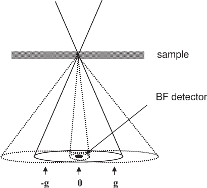
From Figure 2–9 it is clear that we will see contrast only if the diffracted
beams are less than an objective aperture radius from the optic axis.
The discs from three beams now interfere in the region detected. From
(3.5), the wavefunction at the point detected will be
Ψ(K = 0, R
0
) = 1 + φ
g
exp[iχ(−g) − i2πg · R
0
]
+ φ
−g
exp[iχ(g) + i2πg · R
0
] (4.1)
Figure 2–9. A schematic diagram showing that for a crystalline sample, a
small, axial bright-fi eld (BF) STEM detector will record changes in intensity
due to interference between three beams: the 0 unscattered beam and the +g
and -g Bragg refl ections.
80
which can also be written as the Fourier transform of the product of
the diffraction spots of the sample and the phase shift due to the lens
aberrations,
Ψ K0R K K g K g
KK
=
()
=
′
()
+
′
+
()
[
+
′
−
()
]
′
()
[]
′
∫
−
,
exp exp
0
2
δφδ φδ
χπ
gg
ii⋅⋅ RK
0
()
′
d
(4.2)
Equations (4.1) and (4.2) are identical to those for the wavefunction in
the image plane of a CTEM when forming an image of a crystalline
sample. In the simplest model of a CTEM (Spence, 1988), the sample is
illuminated with plane wave illumination. In the back focal plane of the
objective lens we could observe a diffraction pattern, and the wavefunc-
tion for this plane corresponds to the fi rst bracket in the integrand of
(4.2). The effect of the aberrations of the objective lens can then be
accommodated in the model by multiplying the wavefunction in the
back focal plane by the usual aberration phase shift term, and this can
also be seen in (4.2). The image plane wavefunction is then obtained by
taking the Fourier transform of this product. Image formation in a
STEM can be thought of as being equivalent to a CTEM with the beam
trajectories reversed in direction.
What we have shown here, for the specifi c case of BF imaging of a
crystalline sample, is the princple of reciprocity in action. When the elec-
trons are purely elastically scattered, and there is no energy loss, the
propagation of the electrons is time reversible. The implication for
STEM is that the source plane of a STEM is equivalent to the detector
plane of a CTEM and vice versa (Cowley, 1969; Zeitler and Thomson,
1970). Condenser lenses are used in a STEM to demagnify the source,
which corresponds to projector lenses being used in a CTEM for mag-
nifying the image. The objective lens of a STEM (often used with an
objective aperture) focuses the beam down to form the probe. In a
CTEM, the objective lens collects the scattered electrons and focuses
them to form a magnifi ed image. Confusion can arise with combined
CTEM/STEM instruments, in which the probe-forming optics are dis-
tinct from the image- forming optics. For example, the term objective
aperture is usually used to refer to the aperture after the objective lens
used in CTEM image formation. In STEM mode, the beam convergence
is controlled by an aperture that is usually referred to as the condenser
aperture, although by reciprocity this aperture is acting optically as an
objective aperture. The correspondence by reciprocity between CTEM
and STEM can be extended to include the effects of partial coherence.
Finite energy spread of the illumination beam in CTEM has an effect
on the image similar to that in STEM for the equivalent imaging
mode. The fi nite size of the BF detector in a STEM gives rise to limited
spatial coherence in the image (Nellist and Rodenburg, 1994), and cor-
responds to having a fi nite divergence of the illuminating beam in a
STEM. In STEM, the loss of the spatial coherence can easily be under-
stood as the averaging out of interference effects in the Ronchigram
over the area of the BF detector. At the other end of the column there
is also a correspondence between the source size in STEM and the
detector pixel size in a CTEM. Moving the position of the BF STEM
Peter D. Nellist
Chapter 2 Scanning Transmission Electron Microscopy 81
detector is equivalent to tilting the illumination in CTEM. In this way
dark-fi eld images can be recorded. A carefully chosen position for a BF
detector could also be used to detect the interference between just two
diffracted discs in the microdiffraction pattern, allowing interference
between the 0 beam and a beam scattered by up to the aperture diam-
eter to be detected. In this way higher-spatial resolution information
can be recorded, in an equivalent way to using a tilt sequence in CTEM
(Kirkland et al., 1995).
Although reciprocity ensures that there is an equivalence in the
image contrast between CTEM and STEM, it does not imply that the
effi ciency of image formation is identical. Bright-fi eld imaging in a
CTEM is effi cient with electrons because most of the scattered electrons
are collected by the objective lens and used in image formation. In
STEM, a large range of angles illuminates the sample and these are
scattered further to give an extensive Ronchigram. A BF detector
detects only a small fraction of the electrons in the Ronchigram, and
is therefore ineffi cient. Note that this comparison applies only for BF
imaging. There are other imaging modes, such as annular dark-fi eld
(Section 5), for which STEM is more effi cient.
4.2 Phase Contrast Imaging in BF STEM
Thin weakly scattering samples are often approximated as being weak
phase objects (see, for example, Cowley, 1992). Weak phase objects
simply shift the phase of the transmitted wave such that the specimen
transmittance function can be written
φ(R
0
) = 1 + iσV(R
0
) (4.3)
where σ is known as the interaction constant and has a value given by
σ = 2πmeλ/h
2
(4.4)
where the electron mass, m, and the wavelength, λ, are relativistically
corrected, and V is the projected potential of the sample. Equation (4.3)
is simply the expansion of exp[iσV(R
0
)] to fi rst order, and therefore
requires that the product σV(R
0
) is much smaller than unity. The
Fourier transform of (4.3) is
Φ(K′) = δ(K′) + iσV
˜
(K′) (4.5)
and can be substituted for the fi rst bracket in the integrand of (4.2)
Ψ K0R K K K
KR K
=
()
=
′
()
+
′
()
[
′
()
[]
′
()
′
∫
,exp
exp
.
0
0
2
δσ χ
π
iV i
id
(4.6)
Noticing that (4.6) is the Fourier transform of a product of functions,
it can be written as a convolution in R
0
.
Ψ(K = 0, R
0
) = 1 + iσV(R
0
) 䊟 FT{cos[χ(K′)] + i sin[χ(K′]} (4.7)
Taking the intensity of (4.7) gives the BF image
I(R
0
) = 1 − 2σV(R
0
) 䊟 FT{sin[χ(R
0
]} (4.8)
82
where we have neglected terms greater than fi rst order in the potential,
and made use of the fact that the sine and cosine of χ are even and
therefore their Fourier transforms are real.
Not surprisingly, we have found that imaging a weak-phase object
using an axial BF detector results in a phase contrast transfer function
(PCTF) (Spence, 1988) identical to that in CTEM, as expected from reci-
procity. Lens aberrations are acting as a phase plate to generate phase
contrast. In the absence of lens aberrations, there will be no contrast.
We can also interpret this result in terms of the Ronchigram in a STEM,
remembering that axial BF imaging requires an area of triple overlap
of discs (Figure 2–9). In the absence of lens aberrations, the interference
between the BF disc and a scattered disc will be in antiphase to that
between the BF disc and the opposite, conjugate diffracted disc, and
there will be no intensity changes as the probe is moved. Lens aberra-
tions will shift the phase of the interference fringes to give rise to image
contrast. In regions of two disc overlap, the intensity will always vary
as the probe is moved. Moving the detector to such two beam condi-
tions will then give contrast, just as two-beam tilted illumination in
CTEM will give fringes in the image. In such conditions, the diffracted
beams may be separated by up to the objective aperture diameter, and
still the fringes resolved.
4.3 Large Detector Incoherent BF STEM
Increasing the size of the BF detector reduces the degree of spatial
coherence in the image, as already discussed in Section 4.1. One expla-
nation for this is the increasing degree to which interference features in
the Ronchigram are being averaged out. Eventually the BF detector can
be large enough that the image can be described as being incoherent.
Such a large detector will be the complement of an annular dark-fi eld
detector: the BF detector corresponding to the hole in the ADF detector.
Electron absorption in samples of thicknesses usually used for high-
resolution microscopy is small compared to the transmittance, which
means that the large detector BF intensity will be
I
BF
(R
0
) = 1 − I
ADF
(R
0
) (4.9)
We will defer discussion of incoherent imaging to Section 5. It is,
however, worth noting that because I
ADF
is a small fraction of the inci-
dent intensity (typically just a few percent), the contrast in I
BF
will be
small compared to the total intensity. The image noise will scale with
the total intensity, and therefore it is likely that a large detector BF
image will have worse signal to noise than the complimentary ADF
image.
5. Annular Dark-Field Imaging
Annular dark-fi eld (ADF) imaging is by far the most ubiquitous STEM
imaging mode [see Nellist and Pennycook (2000) for a review of ADF
STEM]. It provides images that are relatively insensitive to focusing
Peter D. Nellist
Chapter 2 Scanning Transmission Electron Microscopy 83
errors, in which compositional changes are obvious in the contrast, and
atomic resolution images that are much easier to interpret in terms of
atomic structure than their high-resolution TEM (HRTEM) counter-
parts. Indeed, the ability of a STEM to perform ADF imaging is one of
the major strengths of STEM and is partly responsible for the growth
of interest in STEM over the past two decades.
The ADF detector is an annulus of scintillator material coupled to a
photomultiplier tube in a way similar to the BF detector. It therefore
measures the total electron signal scattered in angle between an inner
and an outer radius. These radii can both vary over a large range, but
typically the inner radius would be in the range of 30–100 mrad and
the outer radius 100–200 mrad. Often the center of the detector is a hole,
and electrons below the inner radius can pass through the detector for
use either to form a BF image, or more commonly to be energy ana-
lyzed to form an electron energy-loss spectrum. By combining more
than one mode in this way, the STEM makes highly effi cient use of the
transmitted electrons.
Annular dark-fi eld imaging was introduced in the fi rst STEMs built
in Crewe’s laboratory (Crewe, 1980). Initially their idea was that the
high angle elastic scattering from an atom would be proportional to
the product of the number of atoms illuminated and Z
3/2
, where Z is
the atomic number of the atoms, and this scattering would be detected
using the ADF detector. Using an energy analyzer on the lower-angle
scattering they could also separate the inelastic scattering, which was
expected to vary as the product of the number of atoms and Z
1/2
. By
forming the ratio of the two signals, it was hoped that changes in speci-
men thickness would cancel, leaving a signal purely dependent on
composition, and given the name Z contrast. Such an approach ignores
diffraction effects within the sample, which we will see later is crucial
for quantitative analysis. Nonetheless, the high-angle elastic scattering
incident on an ADF detector is highly sensitive to atomic number. As
the scattering angle increases, the scattered intensity from an atom
approaches the Z
2
dependence that would be expected for Rutherford
scattering from an unscreened Coulomb potential. In practice this limit
is not reached, and the Z exponent falls to values typically around 1.7
(see, for example, Hartel et al., 1996) due to the screening effect of the
atom core electrons. This sensitivity to atomic number results in images
in which composition changes are more strongly visible in the image
contrast than would be the case for high-resolution phase-contrast
imaging. It is for this reason that using the fi rst STEM operating at
30 kV (Crewe et al., 1970), it was possible to image single atoms of Th
on a carbon support.
Once STEM instruments became commercially available in the 1970s,
attention turned to using ADF imaging to study heterogeneous catalyst
materials (Treacy et al., 1978). Often a heterogeneous catalyst consists
of highly dispersed precious metal clusters distributed on a lighter
inorganic support such as alumina, silica, or graphite. A system con-
sisting of light and heavy atomic species such as this is an ideal subject
for study using ADF STEM. Attempts were made to quantify the
number of atoms in the metal clusters using ADF intensities. Howie
84
(1979) pointed out that if the inner radius was high enough, the thermal
diffuse scattering (TDS) of the electrons would dominate. Because TDS
is an incoherent scattering process, it was assumed that ensembles of
atoms would scatter in proportion to the number of atoms present. It
was shown, however, that diffraction effects can still have a large
impact on the intensity (Donald and Craven, 1979). Specifi cally, when
a cluster is aligned so that one of the low order crystallographic direc-
tions is aligned with the beam, a cluster is observed to be considerably
brighter in the ADF image.
An alternative approach to understanding the incoherence of ADF
imaging invokes the principle of reciprocity. Phase contrast imaging in
an HREM is an imaging mode that relies on a high degree of coherence
in order to form contrast. The specimen illumination is arranged to be
as plane wave as possible to maximize the coherence. By reciprocity, an
ADF detector in a STEM corresponds hypothetically to a large, annular,
incoherent illumination source in a CTEM. This type of source is not
really viable for a CTEM, but illumination of this sort is extremely inco-
herent, and renders the specimen effectively self-luminous as the scat-
tering from spatially separated parts of the specimen are unable to
interfere coherently. Images formed from such a sample are simpler to
interpret as they lack the complicating interference features observed
in coherent images. A light-optical analogue is to consider viewing an
object with illumination from either a laser or an incandescent light
bulb. Laser beam illumination would result in strong interference fea-
tures such as fringes and speckle. Illumination with a light bulb gives
a view much easier to interpret.
Although ADF STEM imaging is very widely used, there are still
many discrepancies between the theoretical approaches taken, which
can be very confusing when reviewing the literature. A picture of the
imaging process that bridges the gap between thinking of the incoher-
ence as arising from integration over a large detector to thinking of it as
arising from detecting predominantly incoherent TDS has yet to emerge.
Here we will present both approaches, and attempt to discuss the limi-
tations and advantages of each.
5.1 Incoherent Imaging
To highlight the difference between coherent and incoherent imaging,
we start by reexamining coherent imaging in a CTEM for a thin sample.
Consider plane wave illumination of a thin sample with a transmit-
tance function, φ(R
0
). The wavefunction in the back focal plane is given
by the Fourier transform of the transmittance function, and we can
incorporate the effect of the objective aperture and lens aberrations by
multiplying the back focal plane by the aperture function to give
Φ(K′)A(K′) (5.1)
which can be inverse Fourier transformed to the image wavefunction,
which is then a convolution between φ(R
0
) and the Fourier transform
of A(K′), which from Section 2 is P(R
0
). The image intensity is then
Peter D. Nellist
