Hawkes P.W., Spence J.C.H. (Eds.) Science of Microscopy. V.1 and 2
Подождите немного. Документ загружается.


Chapter 1 Atomic Resolution Transmission Electron Microscopy 45
optimum solution for ψ′
d
(k) can be defi ned in various ways (Saxton,
1988). In particular, a Wiener fi lter applied to a series of images (Schiske,
1973) in the presence of noise gives an optimal estimate of the recon-
structed wavefunction, expressed in the form of a weighted superposi-
tion of the individual transforms as
′
=
∑
ψ
d
() ()()kkkrc
i
i
i
(53)
in which the restoring fi lters, r
i
(k), depend on the w
i
(k) for the set of
images as
r
W C w
W W C
Ww
i
i i
i
( )
( )
*
( ) *( ) ( )
( ) ( ) ( ) ( )
() (
k
k k k k
k k k k
kk
=
− − −
− − +
=
2
ν
))() ()()
2
Cww
ii
ii
kkk=−
∑∑
(54)
From Eq. (55) above it can be seen that the effect of the restoring
fi lters on a Fourier component transmitted in only a single image is
simply to retain it after division by the corresponding transfer function
and for Fourier components present in multiple images to average the
estimates. For a Fourier component not transferred in any image the
value of the fi lter tends to zero due to the inclusion of the noise-to-
object power ratio, ν(k).
25
In the fi nal step of the overall reconstruction
process the exit-plane wavefunction itself is obtained simply by inverse
transformation.
For completeness we also provide an alternative formulation of
through focal series restoration by Spence (2002), which gives useful
insight into the behavior of the various terms and clearly illustrates the
general power of coherent detection methods.
The image amplitude under the WPOA for a single image at a
defocus, C
1n
is given as
ψσφ
φ
in p
p
iFPiW
FP iW
() (){()exp[ ( )]}
() ()exp[ (
xxkk
xk k
,C
1
=− −⋅
=⋅
1
,,
1
C
n
)]
{}
(55)
with an image intensity
IC C
CCh
nin
ini n
() ()
()
*
()
xx
xx
,,
,,
11
11
=+
=+ + +
1
1
2
ψ
ψψ
(56)
where h are the higher order, nonlinear terms.
We are now able to extract the wanted second term in Eq. (56) and
discriminate against the conjugate and the higher order terms.
To effect this we fi rst multiply the transform of the intensity by the
conjugate of the transfer function to deconvolve it and then sum over
N images recorded at different defoci.
This gives
25
This is the major advantage of the Wiener fi lter in preventing noise
amplifi cation where w
i
(k) is close to zero.
w

46
SICiWC
iW
n
nn
pp
() ( )exp[ ( )]
() () *( ) exp[
kk k
kk k
=−
=+ +− −
∑
,,
11
12δφ φ (()]
()exp[ ( )]
k
k
,
,
1
11
C
HC iW C
n
nn
nn n
n
∑∑
∑
+−
(57)
where φ(k) = 1 − iσF {φ
p
(x)} is the image we wish to restore.
Equation (57) now clearly shows the power of coherent detection.
The second term sums as N times the wanted image, whereas the
unwanted third and fourth terms sum as N
1/2
times their initial values.
The fi nal term behaves as a two-dimensional random walk. Overall
the effects of the coeffi cients of the wave aberration function appear to
have been eliminated and the only remaining resolution limiting effect
is the function P(k).
26
A further independent formulation of a linear restoration scheme
similar to that described above, known as the parabaloid method, has
also been reported (Op de Beeck et al., 1996; Thust et al., 1996a,b). This
is based on a three-dimensional Fourier transform of a through focal
series of images with the third variable conjugate to the defocus. The
exit-wave sought is localized on a parabolic shell in this three-
dimensional space and can readily be separated from the conjugate
eave and the nonlinear terms. This approach has the attraction of pro-
viding intuitive insight into how the information from a series of
images is gathered to yield the complex exit-wave, although unlike the
Wiener fi lter it is nonoptimal in its suppression of the conjugate wave
and experimental noise (Saxton, 1994).
The approaches for exit-plane wavefunction restoration described
above assume linear imaging. An alternative method has also been
An alternative imaging geometry for use in reconstruction uses a
dataset recorded for several different illumination tilts (of the same
magnitude but with different azimuths) (Kirkland et al., 1995, 1997) in
a super resolution scheme also referred to as aperture synthesis. The
basis of this approach relies on the fact that for an incident beam tilt
26
We note that the above describes a general approach of integration against
a kernel to provide a stationary phase condition for a wanted signal and as
such fi nds widespread application throughout physics.
A.I. Kirkland et al.
tions to the image intensity. In the original implementation of this latter
developed for a more general case, including the nonlinear contribu-
approach (Kirkland et al., 1985; Kirkland, 1984, 1988) the nonlinear
multiple input maximum a posteriori (MIMAP)]. The more recent
improved maximum likelihood (MAL) description (Coene et al., 1992,
calculated from the restored wave to the measured intensities of images
numerically optimized recursive solution and explicitly includes the
coupling between the exit wave and its complex conjugate.
image reconstruction is accommodated by matching the intensities
1996; Thust et al., 1996a) provides a computationally efficient and
in a focal series through minimization of a least-squares functional [the

Chapter 1 Atomic Resolution Transmission Electron Microscopy 47
of a Bragg angle Θ
b
the optic axis bisects the angle Θ = 2Θ
b
between
the incident beam and a fi rst-order Bragg beam. For this geometry the
two beams defi ne the diameter of an achromatic circle (for positive C
3
)
on which all even-order terms on the wave aberration function cancel.
However, only one sector of the diffraction pattern contributes to each
image and it is therefore necessary to record images at several (mini-
mally four) different tilt azimuths. The method is also restricted to
relatively thin samples due to the effects of parallax (Kirkland et al.,
1997). The restoring fi lters required are of a form similar to those used
for focal series reconstruction but with the transfer functions modifi ed
so as to be appropriate to tilted illumination (Saxton, 1988). The advan-
tage of this geometry is in the improved fl at information transfer, albeit
in only one direction in a single image, with the disadvantage of poorer
low-frequency transfer in the restored wave. In this way spatial fre-
quencies up to twice the conventional Scherzer limit can be recovered
from a dataset composed of four tilted illumination images recorded
with orthogonal tilt directions (Kirkland et al., 1995).
It is also worth noting that although the reconstructed wavefunction
is free of artefacts due to the lens aberrations, there still exists a sig-
nifi cant discrepancy between absolute values of the experimental and
simulated exit wavefunctions (Meyer et al., 2000a). The cause of this
discrepancy (frequently known as the Stobbs factor; Boothroyd, 1998,
2000), which is also observed in comparisons between conventional
experimental HRTEM images and simulations, remains an active
research area and recent holographic data (Lehmann and Lichte, 2002)
suggest that phonon scattering may make a substantial contribution.
4.2 Experimental Geometries
The most widely applied procedure for exit-wave reconstruction is, as
already described, to record several (or many) images at different focus
levels for which the positions of weak transfer are different. This pro-
vides almost continuous transfer in the recovered exit-wave extending
to the ultimate limits set by the axial coherence envelopes (Figure 1–17).
In practice the dataset for a focal series reconstruction is relatively
easily obtained and typically some 20 or more images separated by
close focal increments can conveniently be recorded on a CCD detector
utilizing external computer control of the microscope. The subsequent
processing involves image registration across the series (to account for
specimen drift) and the determination of the individual imaging con-
ditions (using one of the methods described earlier).
The basic methodology for recording data for use in tilt series recon-
struction is the same as that for focal series but particular attention
must be paid to the image registration (due to the distorted form of
cross-correlation functions involving tilted illumination images) and
to the initial defocus condition. In general, for this geometry optimal
transfer is achieved in the tilted illumination mode (with positive C
3
)
when C
1
= θ
2
with C
1
and θ being the defocus and illumination tilt
measured in reduced units of (
()C
3
λ
) and (λ/C
3
)
1/4
, respectively
(Hawkes, 1980) (Figure 1–17). With the development of aberration-

48
corrected instruments, this method is further enhanced by the removal
of these restrictions in the absence of C
3
. In practice a combined
defocus/tilt geometry provides optimal transfer and has the additional
benefi t of providing the required data for the determination of the
aberration coeffi cients (see earlier).
4.3 Exit-Wave Restoration of Complex Oxides
To illustrate the benefi ts of exit-wave restoration over conventional
HRTEM imaging we have chosen two examples of complex oxides.
Figure 1–18a shows a structural model of Nb
16
W
18
O
94
projected along
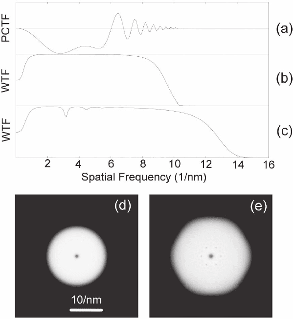
Figure 1–17. A comparison of transfer functions. (a) Conventional phase con-
trast transfer function (PCTF) for a single Scherzer defocus image. (b) Effective
wave transfer function (WTF) for a 21-member focal series with a defocus step
between images of 5 nm. (c) Effective wave transfer function for a 6 member
tilt azimuth series with a tilt of 7.7 mrad. (d) Two-dimensional representation
of the effective wave transfer function in (b). (e) Two-dimensional representa-
tion of the effective wave transfer function in (c). In all cases electron optical
parameters appropriate to a 300-kV FEGTEM were used (300 kV, C
s
= 0.57 nm,
focal spread = 4 nm, beam divergence = 0.1 mrad). (a) Scaled −1 to 1; (b and c)
scaled 0 to 1; (d and e) scaled 0 to 1 (black to white).
A.I. Kirkland et al.

Chapter 1 Atomic Resolution Transmission Electron Microscopy 49
Figure 1–18. (a) Structural model of the complex oxide Nb
16
W
18
O
94
projected along [001]. (b) Conven-
tional axial HRTEM image recorded at the Scherzer defocus of a thin crystal. (c) Reconstructed
modulus of the exit-plane wavefunction of Nb
16
W
18
O
94
with the marked area enlarged (inset), which
directly shows the cation positions (black) with improved resolution compared to the axial image. The
line indicates a stacking fault with a shift of a third of a unit cell along [010]. (d) Reconstructed phase
of the exit-plane wavefunction with the marked area enlarged (inset). The cation sites in the phase are
recovered with positive (white) contrast and additional weak between the cation atomic columns
which indicate the positions of the oxygen anions are also resolved. The reconstructed phase and
modulus are shown at the same scale. (See color plate.)

50
[001] together with an axial image recorded at the Scherzer defocus
(Figure 1–18b) and the restored exit-plane wavefunction (Figures 1-18c
and d) recovered to a resolution of ca. 0.11 nm. It is apparent that
although the basic cation lattice can be determined from the axial
image the restored modulus shows the positions of the cation columns
in projection at substantially higher resolution. Moreover, the modulus
remains directly interpretable to a greater specimen thickness than the
axial image. The restored phase shows a more complex contrast chang-
ing rapidly with specimen thickness. In addition to the strong positive
contrast (white, corresponding to a phase advance) located at the cation
sites and corresponding directly to the positions of strong negative
(black) contrast in the modulus there is additional weak contrast at
positions between the cations at the anion sublattice sites.
Our second example illustrates an experimental tilt azimuth series
restoration of an inorganic perovskite with 0.1 nm information transfer.
The basic perovskite structure is cubic with general formula ABO
3
and can be considered as an array of corner-sharing BO
6
octahedra
where B is typically a small transition metal cation (Figure 1–19). The
cuboctahedral interstices generated by this basic lattice are occupied
by larger A cations, typically alkali earth or rare earth metals. Few
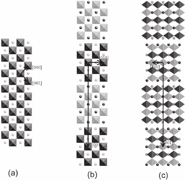
Figure 1–19. Simplifi ed structural models of A
n
B
n
O
3n+2
compounds represented by corner-sharing BO
6
octahedra and isolated A cations. Octahedra and cations drawn with light gray shading are half a
lattice plane below those with darker shading. (a) The basic perovskite ABO
3
. (b and c) The n = 5
structure A
5
B
5
O
17
with layers fi ve octahedra wide, projected in the [100] and [010] directions.
A.I. Kirkland et al.

Chapter 1 Atomic Resolution Transmission Electron Microscopy 51
perovskites are cubic and most distort to a lower symmetry to stabilize
the structure.
The layered structure described here (of general formula A
n
B
n
O
3n+2
)
is composed of slabs of perovskite sliced along the [110] plane with
the two excess oxygen anions accommodated at the interface region
between two slabs (Figure 1–19). These structures show similar distor-
tions to bulk perovskites but with additional degrees of freedom at the
interface.
The restored exit-plane wavefunction for Nd
5
Ti
5
O
17
in the [010] pro-
jection is shown in Figure 1–20. The restoration again shows improved
resolution in both modulus and phase compared to the conventional
axial image and an enhanced sensitivity to the weakly scattering
oxygen sublattice in the restored phase. Comparison with the struc-
tural model shown in Figure 1–19 shows that the Ti are bridged by O
anions and correspondingly in the restored phase, the Nd rows show
distinct cations whereas the Ti rows also show weak contrast between
the cation sites (Figure 1–20) corresponding to the anion positions. The
reconstruction also reveals local distortions present in this material by
which the Nd cations at the outside of the perovskite slabs are dis-
placed by small amounts in alternate directions (Figure 1–20).
5 Image Simulation
A key step in the process of obtaining quantitative structural informa-
tion from HRTEM images is the calculation of image simulations based
on defi ned structural models and imaging conditions for comparison
with experimental images. As already discussed, simple models for
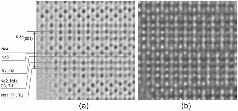
Figure 1–20. (a) Enlarged region taken from the reconstructed modulus calculated from a tilt azimuth
dataset of an Nd
4
SrTi
5
O
17
crystal edge in the [010] projection showing a small difference in positions
of the Nd(4) and Nd(5) cations at the interface between adjacent perovskite slabs. (b) Reconstructed
phase of the same region as (a) showing details of the oxygen anion sublattice between the Ti sites.
The experimental data were recorded using the tilt series geometry described in the text with a JEOL
JEM-3000F FEGTEM, 300 kV, C
3
= 0.57 mm with an injected tilt of 1.9 mrad. (See color plate.)
52
electron scattering and imaging at high resolution suffer from limita-
tions and in general, computationally tractable N-beam dynamic cal-
culations are essential to this process.
For this purpose, the multislice method is most commonly used to
compute the electron wave at the exit-plane of a specimen with known
atomic structure. This approach was fi rst suggested by Cowley and
Moodie (1957) and has found extensive use in HRTEM simulation
(Goodman and Moodie, 1974). In particular, the availability of effi cient
Fast Fourier Transform (FFT) algorithms and the general increase in
readily available computing power have made earlier constraints on its
accuracy due to limitations in the number of slices or the number of
diffracted beams immaterial.
An alternative to the multislice method is the Bloch wave approach,
fi rst introduced by Bethe (1928) and described in detail, for instance,
in Buseck et al. (1989). By analogy with the Bloch theorem in solid-state
physics, the solution of the Schrödinger equation in a periodic crystal
potential is written as a product of a plane wave and a function that
has the same periodicity as the crystal. The latter function is then
expanded into its Fourier components from which the Schrödinger
equation reduces to a matrix equation in these Fourier coeffi cients. For
simple crystals, relatively accurate solutions can be obtained using only
a few of these Bloch waves, with the minimal case requiring only two
(the two beam approximation). This approach has the advantage of
providing valuable insight into the working of dynamic electron dif-
fraction and explains phenomena such as thickness fringes. However,
for complex crystals with large unit cells, the method becomes imprac-
tical, as a large number of beams has to be used in the calculation and
the computation time for the matrix solution scales with N
3
, where N
is the number of beams included. For this reason it has only rarely been
applied to calculations of HRTEM images.
Finally we note that for the majority of cases matching of experimental
and simulated images is carried out purely visually with limited attempts
to defi ne quantitative fi gures of merit (FOM) describing the differences
between experimental and simulated images (or restored exit wavefunc-
tions). In part this is due to the large number of parameters involved in
HRTEM simulations (including atomic coordinates and those describ-
ing imaging conditions) giving rise to a large multiparameter optimiza-
tion problem with many local minima. However, a number of possible
FOMs have been reported (Mobus et al., 1998; Tang et al., 1993, 1994;
Saxton, 1997) based on comparisons in both image and Fourier space.
5.1 The Multislice Formalism
The formal basis of the multislice method (for an extensive treatment
see Kirkland, 1998) is the division of the specimen into a number of
thin slices perpendicular to the direction of the incident beam. The
effects of the specimen potential (transmission, in real space) and of
Fresnel diffraction (propagation, in Fourier space) are than treated
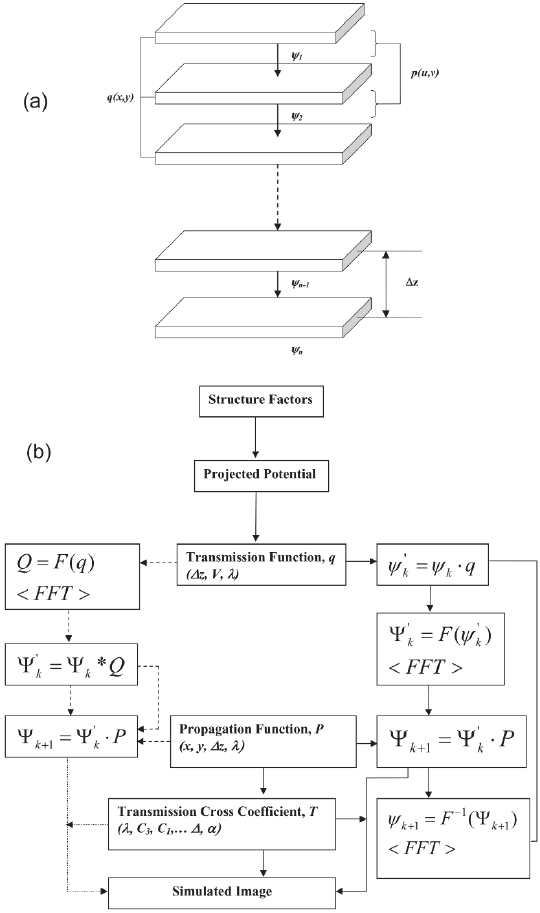
separately for each slice (Figure 1–21). It is a requirement that the indi-
vidual slices used in the simulation must be thin enough to be weak
A.I. Kirkland et al.

Chapter 1 Atomic Resolution Transmission Electron Microscopy 53
Figure 1–21. (a) Schematic diagram showing the real space transmission and
fourier space propagation steps. (b) Flow chart illustrating the steps involved
in a multislice simulation.

54
phase objects and that they obey periodic boundary conditions per-
pendicular to the incident beam direction.
The equations central to the multislice algorithm can be formally
derived starting from the Schrödinger equation for the wavefunction
ψ
f
, of an electron in the electrostatic potential V (x, y, z) of the
specimen:
−∇−
(
)
(
)
=
(
)
2
2
2m
eV x y z xyz E xyz,, ,, ,,
ff
ψψ
(58)
where m is the relativistically corrected electron mass. For high-energy
electrons, the motion is predominantly in the z direction. Hence, it
is convenient to separate the full wavefunction, ψ
f
, into a product of
the solution of the free Schrödinger equation (with V ≡ 0), which is a
plane wave propagating in the z direction, and a wavefunction ψ
that represents the effects due to the specimen and varies much more
slowly with z:
ψ
f
(x, y, z) = e
2πkz
⋅ ψ(x, y, z) (59)
where k = 1/λ is the inverse wavelength of the free electron. Substitut-
ing this into Eq. (58) and using E = h
2
k
2
/2m yields
27
−∇+
∂
∂
+
∂
∂
+
(
)
(
)
=
2
2
2
22
2
4
2
0
mz
k
z
meV x y z
xyz
xy
πψ
,,
,,
(60)
In an elastic scattering process away from the z direction, k
x
and k
y
are proportional to the scattering angle, while the change, ∆k
z
, is pro-
portional to its square. For small scattering angles therefore the term
|∂
2
ψ/∂z
2
|<<|∇
2
xy
ψ| and can be neglected in the paraxial approximation.
What remains is a fi rst-order differential equation in z
∂
(
)
∂
=+
[]
(
)
ψ
ψ
xyz
z
AB xyz
,,
,, (61)
with the operators
A
i
xy
=∇
λ
π4
2
(62)
B = iσV (x, y, z) (63)
and the interaction parameter σ defi ned here as
σ
πλ
=
2
2
me
h
(64)
This differential equation has the formal solution
ψψxyz z Az Bz dz xyz
z
zz
,, ,,+
(
)
=
′
()
+
′
()
[]
′
(
)
+
∫
∆
∆
exp (65)
When ∆z is small, this reduces to
27
It should be noted that k is defi ned here as 1/λ rather than 2π/λ and
therefore h rather than
-
h is used in the expression for E.
A.I. Kirkland et al.
