Hawkes P.W., Spence J.C.H. (Eds.) Science of Microscopy. V.1 and 2
Подождите немного. Документ загружается.


1232 S. Van Aert et al.
where g is the absolute value of the two-dimensional spatial frequency
vector g. Although this transfer function tends to zero for increasing
frequency values, it is not strictly band limited. Nevertheless, the
Gaussian approximation is suffi ciently accurate for the purpose of this
chapter. From the condition that the product of the Rayleigh resolution
limit and the cutoff frequency should approximately be equal to one,
it follows from Eq. (3) that the cutoff frequency corresponding to
Rayleigh resolution is
g
p
p
=≈
11
22
ρ
ρ
(6)
At that spatial frequency the modulus of the transfer function, which
is given by Eq. (5), is reduced to only 8%. Thus, for Gaussian point
spread functions as described by Eq. (1), the Rayleigh resolution limit
can also be defi ned as the inverse of the spatial frequency for which
the transfer function is reduced to 8% of its peak value.
The diffraction limit and its relation to Rayleigh and Sparrow resolu-
tion will now be discussed for conventional transmission electron
microscopy (TEM). Thus far, only the diffraction limited point spread
function of the imaging instrument has been taken into account.
However, for electron microscopy, this should be extended to include
the point spread function describing the effect of thermal vibrations of
the atom, the effect of the environment, and the detector (de Jong and
Dyck, 1993). Moreover, it has to be noted that the atoms are not point
scatterers. Hence, an extension from points to objects of fi nite size has
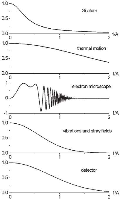
to be made. As shown in Figure 20–1, each effect contributing to the
imaging process can be represented by a transfer function, which acts
as a low pass fi lter. The transfer function of the electron microscope
consists of a damping function, which is mainly due to chromatic aber-
ration, and a phase shift, which causes the oscillations. Since there are
many ways to get rid of the oscillations, such as focal series reconstruc-
tion (Schiske, 1973; Saxton, 1978; Van Dyck and Coene, 1987; Van Dyck
et al., 1993; Coene et al., 1996; Thust et al., 1996) and correction of the
spherical aberration (Rose, 1990), the Rayleigh resolution of the electron
microscope can be assumed to be given by the so-called information
limit, which is proportional to the inverse of the highest spatial fre-
quency that is still transferred with appreciable intensity. For simplic-
ity, it will fi rst be assumed that the imaging process is linear. This
requires that the interaction between the electron and the object also
is linear, which means that there is a simple linear relation of the elec-
tron exit wave and the projected electrostatic potential. The electron exit
wave is a complex wave function in the plane at the exit face of the
object, resulting from the interaction of the electron beam with the
object. For example, the imaging process of weak phase objects, for
which the so-called weak phase object approximation holds (Buseck
et al., 1988), may be considered to be linear. If the object is a crystal,
viewed along a zone axis, the electrostatic potential of all the atoms
along the atom column is superimposed, which makes the interaction
very strong and highly nonlinear. In that particular case, due to the
focusing effect of the successive atoms, the scattering is increased to

Chapter 20 The Notion of Resolution 1233
higher angles. This effect is explained by the channeling theory (Howie,
1966; Van Dyck et al., 1989; Pennycook and Jesson, 1991; Van Dyck and
Chen, 1999). However, for amorphous objects, the atoms are stacked in
a disordered fashion, so that in projection their cores do not overlap,
except by coincidence. As a result, the interaction remains linear for
much larger object thicknesses and may be described by the weak
phase object approximation. If the imaging is linear, all transfer func-
tions have to be multiplied, or, equivalently, the point spread functions
have to be convolved. If it is assumed that all constituent point spread
functions are Gaussian, such as in Eq. (1), the resulting function is a
Gaussian as well, with a Rayleigh resolution ρ
p
determined by
ρ
2
p
= ρ
2
A
+ ρ
2
T
+ ρ
2
EM
+ ρ
2
v
+ ρ
2
D
(7)
with ρ
A
the “width” of the electrostatic potential of the atom, ρ
T
the
Rayleigh resolution limited by thermal vibrations of the atom, ρ
EM
the
Rayleigh resolution of the electron microscope, ρ
v
the Rayleigh resolu-
tion limited by the environment (vibrations and stray fi elds), and ρ
D
Figure 20–1. Transfer functions of the different subchannels of electron
microscopic imaging.
1234 S. Van Aert et al.
the Rayleigh resolution limited by the detector. Today, for the best
electron microscopes, ρ
EM
is somewhat below 1 Å (O’Keefe et al., 2001;
Kisielowski et al., 2001a; Batson et al., 2002a). In future instrumental
developments the Rayleigh and Sparrow resolution can be improved
by improving the resolutions of all different subchannels. However, a
factor that cannot be improved is ρ
A
, that is, the intrinsic “width” of
the atom itself. It is important to note that beyond a certain point, it
will be useless to further improve the Rayleigh resolution of the elec-
tron microscope since the transfer at high spatial frequencies is limited
by the scattering factor. It is already diffi cult to fi nd suitable objects
that can be used to demonstrate the true Rayleigh resolution ρ
EM
of an
electron microscope. Consider, for example, amorphous silicon. From
Figure 20–1, it follows that ρ
A
is about 1 Å. Therefore, for the best elec-
tron microscopes, it follows from Eq. (7) that for amorphous silicon
ρ
p
≈ 1 Å (8)
and from Eq. (4) that
ρ
s
≈ 0.7 Å (9)
From this example, it can be concluded that for the best electron micro-
scopes, the atoms themselves limit classical resolution criteria and
hence the diffraction limit. However, it should be noted that the discus-
sion of Rayleigh resolution and the diffraction limit is far more com-
plicated in case of nonlinear electron–object interaction, for example,
in the case of atom columns viewed along the column direction. It will
then also depend on the assumptions regarding the scattering of the
electrons on their way through the object. Furthermore, for coherent
imaging, such as in TEM, Goodman (1968) has shown that the Rayleigh
resolution will depend on the “phase distribution” associated with the
object. Depending on the relative phase associated with two atoms or
atom columns, the central dip in the composite image will be absent
or present. For particular values of the relative phase shift, the dip will
even be greater than the dip corresponding to an incoherent image of
these two atoms or atom columns. From this example, it can be con-
cluded that there is no simple generalization as to which type of
imaging, coherent or incoherent, is preferred in the sense of Rayleigh
resolution. So the assumption that incoherent imaging, for example
scanning transmission electron microscopy (STEM), will yield a
“better” resolution than coherent imaging, for example, TEM, is in
general not valid.
In the remainder of this chapter, it will be shown that by using super-
resolution algorithms, frequency components lying beyond the diffrac-
tion limit of the imaging system may be reconstructed. Then, other
defi nitions of resolution are of interest.
3.3 Superresolution
Superresolution refers to reconstructing frequency components that lie
beyond the cutoff frequency of the imaging system. At fi rst sight,
superresolution seems impossible. Knowledge of the system’s transfer
Chapter 20 The Notion of Resolution 1235
function makes it possible to reconstruct the object spectrum within
the passband of the imaging system by means of inverse fi ltering of
the image spectrum, but frequency components beyond the diffraction
limit seem irrevocably lost. Indeed, in the absence of any restriction as
to the nature of the object, there are an infi nite number of objects that
can produce the same image. Under certain conditions, however, super-
resolution is possible. The key to superresolution is prior knowledge.
For example, suppose that it is known that the object is of fi nite size,
that is, it is nonzero only in a region of fi nite extent. This single condi-
tion guarantees that the object spectrum is analytic. A well-known
property of an analytic function is that if it is known over a specifi ed
interval, it can always be reconstructed in its entirety (Castleman,
1979). This process of reconstruction is called analytic continuation. It
can be shown that this method is perfect in theory. If the images are
noise free, it leads to an exact and complete reconstruction of the object
spectrum (Harris, 1964). However, noise limits its practical use (Frieden,
1967). Nevertheless, many effective superresolution algorithms (digital
image processing methods) have been proposed in the literature [for a
review, see Frieden (1975), Hunt (1994), and Meinel (1986)]. Both empir-
ically and theoretically, it has been shown that there are certain neces-
sary conditions to be satisfi ed by a superresolution algorithm to be
successful (Hunt, 1994). First, the algorithm should explicitly utilize a
mathematical description of the image formation process that relates
object and image via the point spread function of the imaging system.
Second, the images should be suffi ciently oversampled to avoid alias-
ing after reconstruction of spatial frequencies beyond the diffraction
limit. For Nyquist sampled images (Gonzalez and Woods, 2002), the
algorithm should contain some suitable form of interpolation. Last, but
not least, the algorithm should contain prior knowledge of the object.
Examples of such prior knowledge used by superresolution algorithms
include the following:
• Finite extent of the object (as discussed above) (e.g., Harris, 1964;
Gerchberg, 1974, 1989).
• Positivity of the object (e.g., Schell, 1965; Biraud, 1969; Walsh and
Nielsen-Delaney, 1994).
• Upper and lower bounds on the object intensity (e.g., Janson et al.,
1970).
• Object statistics (e.g., Frieden, 1980; Hunt and Sementilli, 1992).
• Parametric model of the object.
Obviously, the performance of any superresolution algorithm will be
limited by noise. In the remainder of this chapter, we will assume that
the available prior knowledge of the object to be reconstructed consists
of a parametric model. Then, superresolution can be achieved by com-
puting the relatively small number of unknown parameters character-
izing the object from the available observations. In electron microscopy
these observations may be electron counting results made at the pixels
of a CCD camera. The image reconstruction problem thus becomes a
parameter estimation problem. For example, in the case of two-point
resolution, the object can be described by a two-component model
1236 S. Van Aert et al.
parametric in the locations of the point sources. Hence, if the point
spread function is known, a parametric model of the composite image
of the two-point sources can be derived. By fi tting this model to the
image optimizing some criterion of goodness of fi t, we obtain esti-
mates of the parameters of the model. For a correct model, in the
absence of noise, and apart from potential computational problems
that will be discussed in Section 4, this would result in a perfect
fi t. That is, the object parameters can be estimated with unlimited
precision so that the object can be reconstructed perfectly. This means
that there would be no limit to resolution no matter how closely the
point sources are spaced. However, in practice it is noise that limits
the accuracy and precision with which parameters can be measured
and therefore limits the resolution. This will be the subject of Sections
5 and 6.
4 Deterministic Model-Based Resolution
Classical resolution criteria disregard the possibility of using prior
knowledge to extract analytic results from observations by means of
model fi tting (den Dekker and van den Bos, 1997). In this section, prior
knowledge is taken into account in the form of a model describing the
observations. Thus far, the observations are assumed to be noise free.
Compared to Section 2, the model will be extended from two-peak
models to one or more-peak models. Instead of classical resolution, we
will speak of deterministic model-based resolution. It will be shown that
the relevant limits to deterministic model-based resolution are in any
case computational. However, if the model is inaccurate, which means
that it systematically deviates from the exact noise-free observations,
it will be shown that the relevant limits are both computational and
fundamental.
Imagine that Lord Rayleigh would image stars today. First, the image
of one star, which can be treated as a point object, will be considered.
Now, there exists a model for the object, namely that it consists of a
point. The point spread function of the telescope is also exactly known.
So, it is known how an image of one star should look like. Thus, there
is no interest in the detailed form of this image, but only in the position
of the star. The only objective of the experiment is to determine this
position. Obviously, in the absence of noise, numerically fi tting the
known one-peak model to the image with respect to the position
parameter would result in a perfect fi t. The resulting solution for this
location would be exact, and despite the blurring effect of the point
spread function, it imposes no limit to location resolution.
This line of reasoning can be extended to position measurements of
atoms or atom columns from noise-free electron microscopic observa-
tions. Suppose that these observations λ
kl
are made at the pixels (k, l)
at the position (x
k
y
l
)
T
. The model that describes these observations is
called f
kl
(τ) with τ the vector of unknown parameters, among which
are the locations of the atoms or atom columns. An example of such a
model is the following:

Chapter 20 The Notion of Resolution 1237
λτζ
η
πρ
ββ
kl kl
n
kxn lyn
f
xy
=
(
)
=+
−−
(
)
−−
(
)
22
2
22
exp
ρρ
2
1
=
∑
n
n
c
(10)
where ζ is the constant background, η
n
is the column-dependent height
of the Gaussian peak, ρ is the width of the Gaussian peak, n
c
is the total
number of atom columns, and β
xn
and β
yn
are the x- and y-coordinate
of the nth atom or atom column, respectively. The width ρ is supposed
to be identical for different atom columns. The parameter vector τ is
equal to (β
x1
. . . β
xnc
β
y1
. . . β
ync
η
1
. . . η
nc
ρ ζ)
T
and contains R = 3n
c
+ 2
elements. The unknown parameters can be measured by fi tting the
model to the observations. In a sense, we are then looking for the
optimum value of a criterion in a parameter space whose dimension is
equal to R, that is, the number of parameters to be measured. Each
possible combination of the R parameters can be represented by a point
in an R-dimensional space. The search for the global optimum of the
criterion of goodness of fi t in this space is an iterative numerical opti-
mization procedure.
The problem may be of a computational kind. The existing optimiza-
tion methods fail if the dimension of the parameter space is so high
that it is not possible to avoid ending up at a local optimum instead of
at the global optimum of the criterion of goodness of fi t, so that the
wrong structure is derived. To solve this dimensionality problem, that
is, to fi nd a pathway to the global optimum, a good starting structure
is required, that is, initial conditions should be available for the param-
eters. For example, neighboring atoms or atom columns should be
discriminated in an image. In other words, the structure has to be
resolved. This corresponds to X-ray crystallography, where it is fi rst
necessary to resolve a structure by using, for example, direct methods,
and afterward to refi ne the structure. Moreover, the computing time
needed to reach convergence of the iterative procedure increases with
the dimension of the parameter space. In the following example, the
problems related to the study of the amorphous object with atomic
resolution TEM will be discussed. More details can be found in Van
Dyck et al. (2003).
Example 1 (Amorphous Object) For an amorphous object, the number of
parameters increases with thickness. Therefore, from a certain thickness on, it
will be diffi cult to resolve the structure in projection. For example, consider
Figures 20–2 and 20–3. In Figure 20–2, the amorphous foil is thin, whereas
in Figure 20–3, it is thicker. Therefore, the number of projected atoms is larger
in Figure 20–3 than in Figure 20–2. It is clear that it will be more likely to
resolve the structure for the example given in Figure 20–2 than for that cor-
responding to Figure 20–3. To resolve the structure, it will be assumed that
the distances between neighboring projected atom positions should be larger
than or equal to the Sparrow resolution ρ
s
. The reason for choosing this crite-
rion is that the computer will then be able to distinguish the individual atoms,
since the observations are assumed to be noise free. However, it should be noted
that this criterion is not exact and, therefore, it will give only rough guidelines.
Suppose that the mean concentration of atoms per cubic ångstrom is equal to

1238 S. Van Aert et al.
V. T h e n, th e mean concentration A of projected atoms per square ångstrom is
given by
A = Vz (11)
where z is the thickness of the amorphous foil. On the other hand, if it is
assumed that each projected atom occupies a circle with a diameter equal to
the average distance d, averaged over distances between nearest-neighbor pro-
jected atoms, then
A
d
≈
(
)
1
2
2
π
(12)
From Eqs. (11) and (12), it follows that the thickness of the amorphous foil is
approximately given by
Figure 20–2. Amorphous
structure containing clearly
separable projected atoms.
Figure 20–3. Amorphous
structure containing severely
overlapping projected atoms.

Chapter 20 The Notion of Resolution 1239
z
dV
≈
4
2
π
(13)
To resolve the structure and therefore to avoid dimensionality problems, it will
be assumed that the following condition is met:
d ≥ 2ρ
s
(14)
The factor 2 is arbitrarily chosen. Requiring that d is larger than or equal to
ρ
s
would not be suffi cient. In that case, a substantial part of distances between
neighboring atoms would be smaller than ρ
s
and hence it is not possible to
resolve the structure. In principle, this can still occur if inequality (14) is ful-
fi lled, but the probability that it occurs is lower. Then, it follows from Eqs. (13)
and (14), that
z
V
≤
1
2
πρ
s
(15)
For amorphous silicon, it follows from Eq. (9) that ρ
s
is approximately equal
to 0.7 Å and, furthermore, V is approximately equal to 0.05 atoms/Å
3
. Hence,
it follows from Eq. (15) that the amorphous silicon foil should not exceed thick-
nesses of the order of 13 Å so as to avoid dimensionality problems. This thick-
ness is rather small, which means that it is unrealistic to expect that atomic
resolution TEM is able to resolve amorphous silicon samples with realistic foil
thicknesses from only one projection (Cowley, 2001). It can thus be stated that
the structure of a realistic amorphous object cannot be determined from one
image alone. However, the situation can be improved drastically by using a
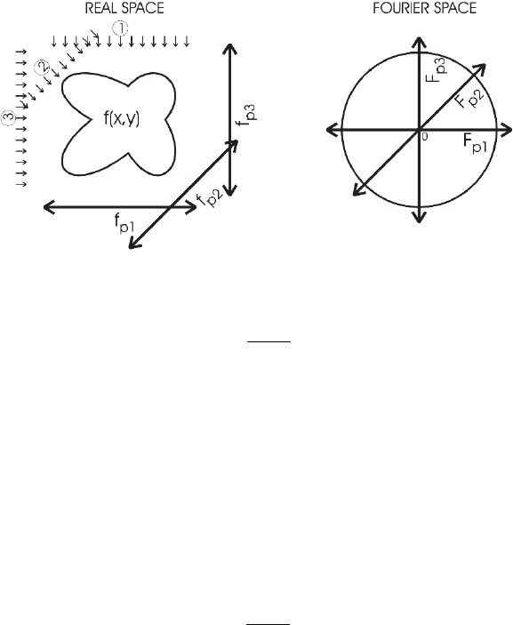
tomographic technique in which the sample is tilted and many projections from
different viewing directions are combined as shown in Figure 20–4. The
Fourier transform of a projection yields a section through the origin of the
three-dimensional Fourier space. By combining many different projections, it
is possible to reconstruct the whole Fourier space (Frank, 1992). In this way
an ideal microscope can resolve about one atom per cubic ångstrom, which is
suffi cient to resolve amorphous structures.
In the foregoing, it has been assumed that the model is accurate.
However, if the model is inaccurate, the estimated position parameters
Figure 20–4. The principle of tomography.

1240 S. Van Aert et al.
may deviate from the true positions, even if the observations would be
noise free. This is called systematic error. Obviously, systematic errors
would place fundamental limits on deterministic model-based
resolution.
In this section, it has been shown that deterministic model-based
resolution is computationally limited. Computational problems can be
overcome only if the structure can be resolved. Moreover, if the model
is inaccurate, the resolution is also fundamentally limited, since this
results in a systematic error.
5 Statistical Model-Based Resolution
In Section 4, it was assumed that the observations are noise free.
However, in any real-life experiment, the observations will “contain
errors.” Then, the resolution depends fundamentally on the signal-to-
noise ratio in the detected image. In this section, the resolution will be
considered in the framework of statistical parameter estimation theory
and will be called statistical model-based resolution. It will be shown
that the relevant limits are both computational and fundamental.
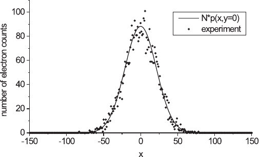
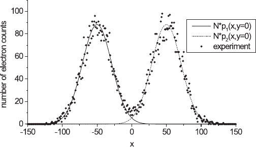
Suppose there is a CCD camera that is able to count the individual
photons forming the image of a single point object or of two point
objects. The images as measured by this camera appear as in Figure
20–5 or as in Figure 20–6 for single or two-point objects, respectively.
The noise on these images stems from the counting statistics. The posi-
tion parameters can be estimated by numerically fi tting the known
parameterized mathematical model to the images with respect to the
component positions, in the same way as expressed in Section 4.
However, if one repeated this experiment several times, one would,
due to the statistical nature of the observations, fi nd different values
Figure 20–5. Simulation experiment of the image of a point as measured by
a CCD camera (300 × 300 pixels), with pixel size ∆x = ∆y = 1. The point spread
function used is a two-dimensional normalized Gaussian function p(x, y) as
in Eq. (1) with ρ = 21. The experimental as well as the expectation values N ×
p(x, y) are shown within the section y = 0. The number of imaging particles N
is equal to 250,000.

Chapter 20 The Notion of Resolution 1241
for the position or the distance estimates for single or two-point objects,
respectively. The position and distance estimates are statistically dis-
tributed about their mean values. Then, the obvious criterion to quan-
tify statistical model-based resolution is the precision of the estimate,
which is given by the variance of this distribution, or, by its square
root, the standard deviation. In a sense, the standard deviation is the
“error bar” on the position or on the distance. Applying statistical
parameter estimation theory, the attainable precision can be adequately
quantifi ed in the form of the so-called Cramér–Rao lower bound (CRLB)
(van den Bos and den Dekker, 2001). This is a lower bound on the vari-
ance of any unbiased estimator of a parameter. It means that the vari-
ance of different estimators, such as, the least squares or the maximum
likelihood (ML) estimator, can never be lower than the theoretical
CRLB on the variance. Fortunately, the ML estimator attains the CRLB
asymptotically, that is, if the number of observations is suffi ciently
large. Note that in accordance with the available literature, the CRLB
on the variance of the position estimate of the image of a single point
object and the CRLB on the variance of the distance estimate of the
image of two point objects are measures of what is called single-source
(Falconi, 1964) and differential (two-source) resolution (Falconi, 1967),
respectively. Single-source resolution is defi ned as the instrument’s
capacity to determine the position of a point object that is observed in
a background of noise. Differential resolution is defi ned as the instru-
ment’s ability to determine the separation of two point objects.
In this section the fl uctuating behavior of the observations will be
described in Section 5.1 using parametric statistical models of observa-
tions. Next, in Section 5.2, it will be shown how an adequate expression
for the attainable statistical precision of the parameter estimates, that
is, the CRLB, can be derived from such a parametric statistical model.
Then, in Section 5.3, the ML estimator of the parameters will be derived
Figure 20–6. The results of a computer-simulated image of two neighboring
points. The experimental as well as the expectation values of the individual
peaks [N × p
1,2
(x, y)] are plotted within the section y = 0, where p
1,2
(x, y) are
two-dimensional normalized Gaussian point spread functions, as in Eq. (1).
The number of imaging particles 2N is equal to 500,000. The peaks are clearly
separable.
