Cui Dongmei. Atlas of Histology: with functional and clinical correlations. 1st ed
Подождите немного. Документ загружается.

CHAPTER 9
■
Circulatory System
175
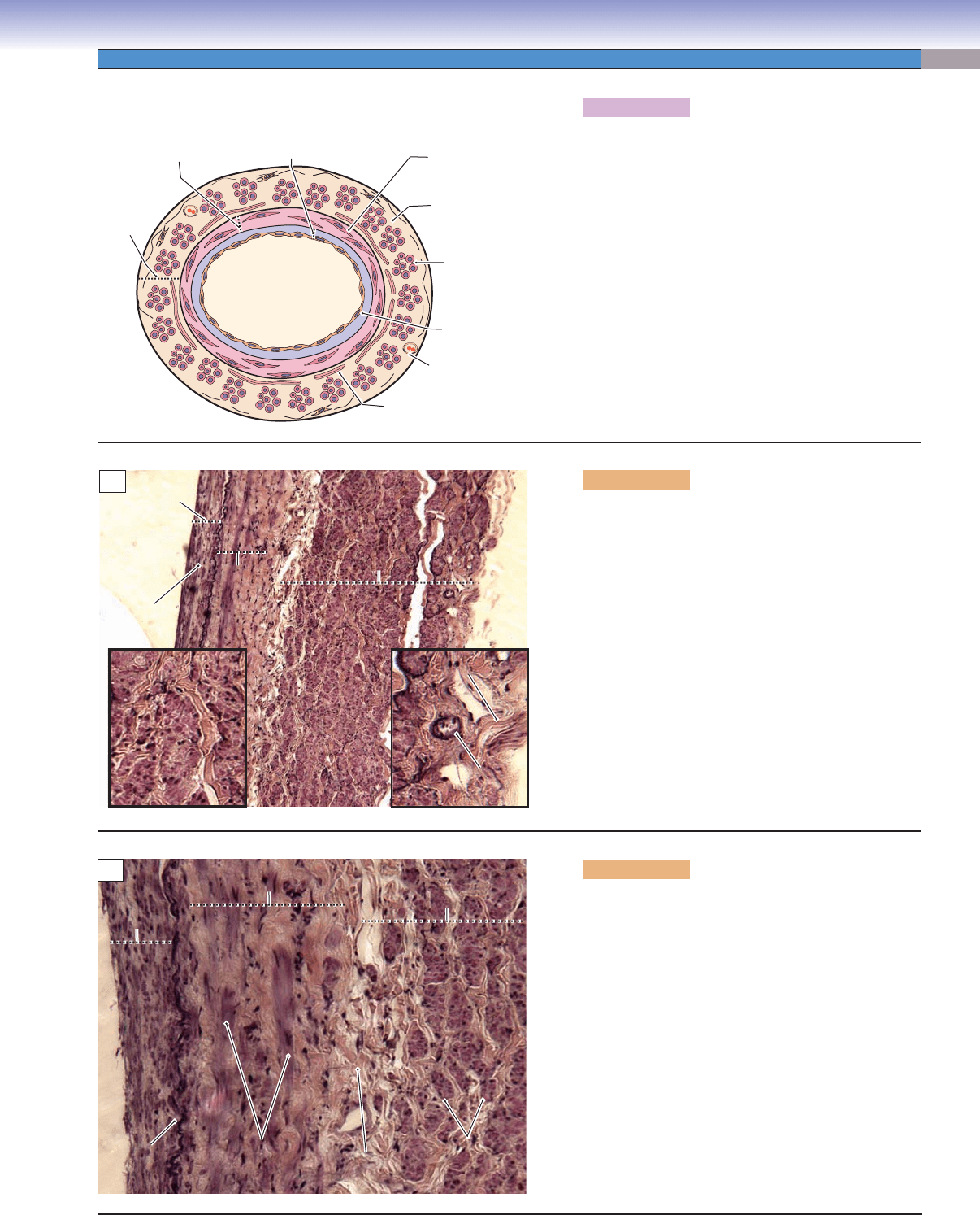
Figure 9-18A. A representation of a large vein.
The vena cava and pulmonary veins are the largest
veins in the venous system. They connect directly to
the heart. The tunica adventitia is the most prominent
and distinct layer in large veins; it contains numerous
longitudinally arranged smooth muscle bundles and
some connective tissues (mostly collagen fi bers). The
vasa vasorum are also present in the tunica adventitia;
they provide blood and nutrient supply to the wall of
the large vein. The tunica media contains a few lay-
ers of smooth muscle cells that are loosely arranged
and do not form a distinct sheath. Contraction of the
circularly arranged smooth muscle cells in the tunica
media and the longitudinally arranged smooth muscle
bundles in the tunica adventitia help to push blood
toward the heart.
D. Cui /T. Yang
Tunica
adventitia
Tunica
media
Tunica
intima
Connective tissue
Longitudinal smooth
muscle bundle
Collagen fiber
Vasa vasorum
Endothelium
Circular smooth
muscle layer
A
Subendothelial
Subendothelial
layer
layer
Connective tissue
Connective tissue
Vasa
Vasa
vasorum
vasorum
Longitudinal
Longitudinal
smooth
smooth
muscle bundles
muscle bundles
Tunica
Tunica
intima
intima
Tunica
Tunica
media
media
Tunica
Tunica
adventitia
adventitia
Connective tissue
Longitudinal
smooth
muscle bundles
Tunica
intima
Subendothelial
layer
Tunica
media
Tunica
adventitia
Vasa
vasorum
B
Figure 9-18B. Large vein, vena cava. H&E, 68;
insets 185
The tunica intima of the vena cava consists of an
endothelial layer, a thick subendothelial layer (connec-
tive tissue), and an IEL. In general, the IEL is not well
developed in veins. Although it is not commonly seen
in veins, it may still be present in some large veins.
The tunica media contains a few layers of loosely
organized smooth muscle cells; the tunica media is
much thinner compared to its tunica adventitia. There
are densely arranged smooth muscle bundles in the
tunica adventitia, which give the large vein a distinct
appearance. There is a thin layer of connective tissue
(mainly collagen fi bers) between the circular smooth
muscle and the longitudinal smooth muscle bundles in
the tunica adventitia (Fig. 9-18C). A vasa vasorum is
shown on the right, and a cross section of longitudinal
smooth muscle bundles is shown on the left.
Internal
Internal
elastic
elastic
lamina
lamina
Circular smooth
Circular smooth
muscle cells
muscle cells
Longitudinal
Longitudinal
smooth
smooth
muscle bundles
muscle bundles
Tunica
Tunica
intima
intima
Tunica
Tunica
media
media
Tunica
Tunica
adventitia
adventitia
Longitudinal
smooth
muscle bundles
Connective
Connective
tissue
tissue
Connective
tissue
Circular smooth
muscle cells
Internal
elastic
lamina
Tunica
intima
Tunica
media
Tunica
adventitia
C
Figure 9-18C. Large vein, vena cava. H&E, 186
A high power view of a large vein with smooth muscle
cells in the circular smooth muscle layer in the tunica
media. The longitudinal smooth muscle bundles are
surrounded by collagen fi bers in the tunica adventitia.
There are some connective tissues (mainly collagen
bundles) between circular smooth muscle and longi-
tudinal smooth layers. The IEL is clearly visible here
between the tunica intima and the tunica media. How-
ever, the IEL is not usually present in the veins.
CUI_Chap09.indd 175 6/17/2010 10:23:23 AM

176
UNIT 3
■
Organ Systems
Types of
Arteries and
Veins
Tunica Intima Tunica Media Tunica Adventitia Size
(Diameter)
Main Functions
Arterial System
Large/elastic
(conducting)
arteries
Endothelium;
subendothelial
layer (connective
tissue); IEL
Abundant fenestrated
elastic membranes;
interspersed smooth
muscle cells; EEL
Thinner than
its tunica media
(connective tissue);
vasa vasorum present
>10 mm Conducting blood fl ow
from the heart to the
arterial system; being
able to recoil (elastic
membranes) to buffer
pressure against arte-
rial wall and conserve
energy to force the blood
forward, even while the
ventricle is relaxing
Medium/
muscular
(distributing)
arteries
Endothelium;
subendothelial
layer (connective
tissue); prominent
complete IEL
Abundant, thick layer
of smooth muscle cells
(6–40 layers of muscle
cells); prominent EEL
in larger-sized medium
arteries
Thinner than
its tunica media
(connective tissue);
vasa vasorum not
prominent
10–0.5 mm Distributing blood
fl ow to small arteries
in various parts of
body; adjusting the
rate of blood fl ow by
vasoconstriction and
vasodilation
Small arteries Endothelium; thin
subendothelial
layer; thin IEL
3–6 layers of smooth
muscle cells; EEL
absent
Thinner than its
tunica media (loose
connective tissue);
no vasa vasorum
500–100 μm
Distributing blood
to arterioles and to
capillaries; participating
in vasoconstriction
and vasodilation to
adjust the blood fl ow
Arterioles Endothelium; no
subendothelial
layer; IEL not
prominent
1–2 layers of smooth
muscle cells; no EEL
Very thin; a sheath of
loose connective tissue
100–30 μm
Leading blood to
the capillary beds;
regulating resistance;
controlling blood fl ow
to capillaries
Venous System
Venules
( postcapillary,
collecting,
and muscular
venules)
Endothelium; no
subendothelial
layer; no valves
Very thin; smooth
muscle cells not promi-
nent
Very thin; layer of
collagen fi bers
10–100 μm
Draining exchanged
blood from capillaries
to the small veins;
promoting leukocyte
migration from blood-
stream to infl amed
tissue; primary sites for
infl ammatory response
Small veins Endothelium;
subendothelial
layer; few valves
Thin; 1–3 layers
isolated smooth muscle
cells
Thin; connective
tissue layer
0.1–1 mm Collecting blood fl ow
from venules to the
medium veins
Medium veins
Endothelium;
subendothelial
layer; increased
number of valves
Thicker than small
vein; a few layers of
smooth muscle cells
that usually do not
form a distinct sheath
Thicker than its
tunica media; variable
number and size of
longitudinal bundles of
smooth muscle mixed
with connective tissue
1–10 mm Carrying blood to large
veins leading toward
the heart; preventing
blood from backfl ow
Large veins Endothelium; thick
subendothelial
layer; large valves;
IEL may be
present
Thinner than its tunica
adventitia layer; several
layers of circularly
arranged smooth
muscle cells but do not
form a distinct sheath;
some collagen fi bers
Thickest layer in large
veins; composed of
many large bundles of
longitudinal smooth
muscle; cardiac
muscle may be present
near the atria
>10 mm Transporting blood
from the venous system
back to the heart;
forcing blood toward
the heart by
constriction of circu-
lar and longitudinal
smooth muscle
TABLE 9-1 Blood Vessels
CUI_Chap09.indd 176 6/17/2010 10:23:29 AM
CHAPTER 9
■
Circulatory System
177
The Lymphatic Vascular System
CLINICAL CORRELATION
Figure 9-19C.
Lymphangioma, Skin. H&E, 44
L
ymphangioma is a congenital malformation of the
lymphatic system that involves the skin and subcutane-
ous tissues. It occurs most often in the head and the
neck. It is characterized by multiple clusters of translu-
cent vesicles that contain lymph fl uid varying from clear
to pink to dark red. These vesicles are separated from
the normal network of lymphatic vessels but have com-
munications with the superfi cial lymph vesicles through
vertical and dilated lymph channels. Skin lesions range
from minute vesicles to cystic and cavernous spaces.
Clinically, it appears as a raised, soft, spongy, and
pinkish-white lesion. Surgical excision may be chosen if
vital structures are involved or for cosmetic correction,
but it has a high recurrence rate after surgery.
Lymphatic vessels
Lymphatic vessel
C
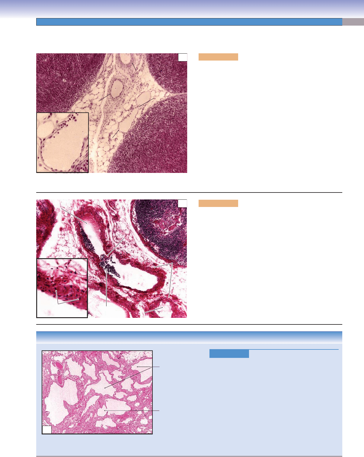
Figure 9-19A. Small lymphatic vessels, lymph node. H&E,
272; inset 767
The lymphatic vascular system is composed of lymphatic
capillaries, lymphatic vessels, and lymphatic ducts, which col-
lect and drain interstitial fl uid from the tissue into the large veins
(for subclavian veins, see Chapter 10, “Lymphoid System,”
Fig. 10-6). Lymph (fl uid in the lymphatic system) contains lym-
phocytes, immunoglobulins, plasma, foreign antigens, and other
substances. Lymphatic vessels carry lymph through the lymph
nodes along the lymphatic vessels. Lymph nodes fi lter lymph
and expose lymphocytes to antigens as part of the immune
response (see Chapter 10, “Lymphoid System”). After fi ltration,
the lymph is transported via large lymphatic vessels to lymphatic
ducts (the thoracic right lymphatic ducts) and fi nally enters the
subclavian veins and becomes part of the blood plasma. An
example of small lymphatic vessels is shown in the hilus of a
lymph node. Small lymphatic vessels have large lumens and very
thin walls, which are composed of a layer of endothelium and a
little connective tissue with a few smooth muscle cells.
Small
artery
Small
artery
Small
lymphatic
vessel
Small
lymphatic vessel
Valve
A
Smooth
Smooth
muscle
muscle
cells
cells
Lymphocytes
Lymphocytes
Valve
Valve
Lymphatic
Lymphatic
vessels
vessels
Lymphatic
vessels
Coagulated
Coagulated
plasma
plasma
Coagulated
Coagulated
plasma
plasma
Coagulated
plasma
Coagulated
plasma
Smooth
muscle cells
Lymphocytes
Valve
B
Figure 9-19B. Large lymphatic vessels, lymph node. H&E,
136; inset 422
Large lymphatic vessels have thicker walls than small
lymphatic vessels. Large lymphatic vessels are composed of
connective tissues and multiple layers of smooth muscle cells.
Contraction of smooth muscle cells helps to move the lymph
forward. Large lymphatic vessels are structurally similar to
small veins, except they have larger lumens and prominent
valves. Valves are present in all sizes of lymphatic vessels.
They prevent lymph from fl owing backward. Lymphatic ves-
sels are often distinguished by lumens that contain clusters of
lymphocytes and coagulated plasma. Lymphatic vessels can be
found in most of the tissues of the body but not in the central
nervous system, the bone marrow, or the hard tissues.
CUI_Chap09.indd 177 6/17/2010 10:23:29 AM

178
10
Introduction and Key Concepts for the Lymphoid System
Cells in the Lymphoid System
Figure 10-1A,B Lymphocytes
Figure 10-2 A Representation of Types of Lymphocytes
B Lymphocytes
Figure 10-3 A Representation of B-Lymphocyte Maturation
Synopsis 10-1 Characteristics of Types of Immunoglobulins
T Lymphocytes
Figure 10-4A A Representation of T-Lymphocyte Maturation
Figure 10-4B A Representation of Helper T-Cell and Cytotoxic T-Cell Maturation Markers
Figure 10-5A A Representation of Helper T-Lymphocyte Activation
Figure 10-5B Clinical Correlation: HIV Infection
Lymphoid Tissues and Lymphoid Organs
Figure 10-6 Overview of the Lymphoid Organs
Figure 10-7 Orientation of Detailed Lymphoid Organ Illustrations
Mucosa-Associated Lymphoid Tissue
Figure 10-8A Pharyngeal Tonsil, MALT
Figure 10-8B Palatine Tonsil, MALT
Table 10-1 Tonsils
Figure 10-9A Appendix, MALT
Figure 10-9B Clinical Correlation: Diffuse Large B-Cell Lymphoma
Figure 10-9C Clinical Correlation: Lymph Node, HIV Infection
Lymph Nodes
Figure 10-10 Overview of the Lymph Node
Synopsis 10-2 Lymphoid Organs
Lymphoid System
CUI_Chap10.indd 178 6/2/2010 4:11:23 PM

CHAPTER 10
■
Lymphoid System
179
Introduction and Key Concepts
for the Lymphoid System
The lymphoid system is composed of lymphocytes, lymphoid
organs, and lymphatic vessels. The structure of lymphatic ves-
sels is discussed in Chapter 9, “Circulatory System.” Lymphoid
organs include the bone marrow (see Chapter 8, “Blood and
Hemopoiesis”), lymph nodes, thymus, spleen, and mucosa-
associated lymphatic tissue (MALT), such as tonsils and Peyer
patches. Most lymphoid organs contain lymphatic nodules or
diffuse lymphatic tissues and play an important role in provid-
ing sites for lymphocytes to come into contact with antigens;
promote proliferation and maturation of lymphocytes; and
promote B lymphocytes to become plasma cells, which pro-
duce antibodies. Lymphoid organs can be divided into two
groups: (1) Primary lymphoid organs, also called central lym-
phoid organs, are the sites where lymphocytes differentiate and
develop the ability to recognize foreign antigens and distinguish
nonself from self. Primary lymphoid organs include the bone
marrow for B lymphocytes and the thymus for T lymphocytes.
(2) Secondary lymphoid organs, also called peripheral lym-
phoid organs, are where mature lymphocytes (both B and T
cells) encounter foreign antigens and the immune response
takes place. Secondary lymphoid organs include MALT, lymph
nodes, and the spleen.
Cells in the Lymphoid System
Lymphocytes can be classifi ed into three major types based
on their immunologic functions: B lymphocytes (B cells),
T lymphocytes (T cells), and null cells. B cells and T cells are the
two main cell types found in lymphoid organs. Lymphocytes
originate in the bone marrow and develop and mature in pri-
mary lymphoid organs. Exposure to foreign antigens initiates the
immune response in secondary lymphoid organs. It is impossible
to distinguish between the T and B cells without using immuno-
histochemical stains. However, they have a tendency to reside
in certain regions of the lymphoid organs. For example, most
B cells reside in lymphatic nodules of the secondary lymphoid
organs, whereas T cells reside in the thymus, paracortex of the
lymph nodes, and periarterial lymphatic sheath (PALS) of the
spleen. B cells participate in the humoral immune response, and
T cells are involved with the cell- mediated immune responses.
Other cells in the lymphoid organs include plasma cells and
antigen-presenting cells.
B Lymphocytes
B lymphocytes originate from precursor cells in the bone
marrow and become naive (virgin) B cells in the bone mar-
row. These B cells develop their surface antibody (Ig), which
enables them to recognize nonself antigens. If B cells rec-
ognize self-antigens during the maturation process, these
B cells will undergo apoptosis (negative selection [Fig. 10-3]).
Naive B cells migrate from the bone marrow to the second-
ary lymphoid organs through the blood circulation. If naive
B cells do not meet a specifi c foreign antigen, they will die in a
short time. If they encounter such an antigen, recognizing and
binding to the antigen will allow them to survive and become
active B cells. Activated B cells undergo cell division and dif-
ferentiate into plasma cells and memory B cells. Memory
B cells have a long life and can live for decades in circulating
blood in an inactive state. They can differentiate into plasma
cells, which produce antibodies to participate in the humoral
immune response.
T Lymphocytes
T lymphocytes also originate from precursor cells in the bone
marrow, but they do not mature in the bone marrow. Pro–T
lymphocytes enter the blood circulation and travel to their
primary lymphoid organ (thymus) to fi nish their maturation
(Fig. 10-4A). They develop into thymocytes in the cortex of the
thymus and undergo a differentiation process to become naive
(virgin) T cells. Naive T cells have surface markers on their
cytoplasmic membrane and have a short life as do naive B cells
Figure 10-11A–D Lymph Nodes
Figure 10-12A,B High Endothelial Venules (HEVs), Paracortex of a Lymph Node
Figure 10-12C Clinical Correlation: Hodgkin Lymphoma
Thymus
Figure 10-13A Thymus
Figure 10-13B Thymus, Cortex
Figure 10-13C Thymus, Medulla
Spleen
Figure 10-14A Spleen
Figure 10-14B White Pulp, Spleen
Figure 10-14C Red Pulp, Spleen
Figure 10-15A Splenic Circulation
Figure 10-15B Periarterial Lymphatic Sheath, Spleen
Figure 10-16 Red Pulp of the Spleen
Table 10-2 Lymphoid Organs
Synopsis 10-3 Pathological and Clinical Terms for the Lymphoid System
CUI_Chap10.indd 179 6/2/2010 4:11:30 PM

180
UNIT 3
■
Organ Systems
(Fig. 10-4A,B). They will die if they do not meet an antigen.
Naive T cells migrate from the thymus to secondary lymphoid
organs where they encounter foreign antigens and become active
T cells. Once T cells are activated, they can boost the action of
cytotoxic T cells and macrophages and help to expedite pro-
liferation of B lymphocytes, which increase the production of
antibodies (see Fig. 10-5A). Activated T cells undergo cell divi-
sion to become memory T cells or effector T cells.
MEMORY T CELLS have a much longer life than naive (virgin)
T cells. They can survive for a long period in an inactive state
and can differentiate into effector T cells to participate in a stron-
ger and faster secondary immune response when they encounter
the same antigen for the second time. Memory T cells include
central memory T cells and effector memory T cells. Central
memory T cells express CCR7 (chemokine receptor) surface
molecules and secrete interleukin-2 (IL-2) that stimulates B cells
to proliferate. They reside in secondary lymphoid organs, such
as the paracortex of the lymph nodes, and are capable of dif-
ferentiating into effector memory T cells. Effector memory T
cells do not express CCR7 surface molecules but secrete IL-4
(to stimulate B cells and increase immunoglobulin G [IgG] and
IgM). They often migrate to an infl ammatory site and develop
into effector T cells.
EFFECTOR T CELLS include helper T cells, cytotoxic T
cells, and regulatory (suppressor) T cells. (1) Helper T cells
have the surface marker CD4, which restricts activation
to antigens only if it is presented by another cell in associa-
tion with major histocompatibility complex (MHC) class II.
Helper T cells include Th0, Th1, and Th2 cells. Th0 cells can
differentiate into Th1 and Th2 cells; Th1 cells secrete IL-2,
interferon-g, and tumor necrosis factor and down regulate Th2
cells’ response; Th2 cells secrete IL-4, IL-5, IL-6
, and IL-10,
which help promote antibody production, stimulate prolifera-
tion of eosinophil and mast cells, and down regulate Th1 cells’
response. Helper T cells do not directly kill infected cells or
pathogens but function indirectly to promote and activate other
immune cells. (2) Cytotoxic (CD8) T cells have the CD8 surface
marker
, which restricts activation to antigen only if it is pre-
sented by another cell in association with MHC class I. They
kill target cells, such as virus-infected cells, tumor cells, and
transplanted cells (grafts). (3) Regulatory T cells are also called
suppressor T cells. They suppress the humoral and cellular
immune responses and are involved with immunological
tolerance.
Null Cells
Null cells resemble lymphocytes but do not have surface markers,
which B and T cells have. They include pluripotential hemopoi-
etic stem cells (PHSCs) and natural killer (NK) cells. PHSCs
function as stem cells and can give rise to various types of blood
cells. NK cells do not require exposure to antigens to become
activated. They function similarly to cytotoxic T cells but do
not have the surface markers CD8 or CD4. They kill invading
target cells, such as virus-infected cells and tumor cells.
Plasma Cells
Plasma cells differentiate from B cells. These activated large cells
have clock-face nuclei, abundant rough endoplasmic reticulum,
and a Golgi apparatus in the cytoplasm (see Figs. 4-2 and 4-3).
They actively produce antibodies known as immunoglobulins
(Igs), which are specifi c for each type of antigen (Fig. 10-3).
Antigen-Presenting Cells
These cells present antigens to lymphocytes. Most of them are
MHC-II class, which have surface membrane molecules MHC-II
(histocompatibility complex). These cells present antigen to
T cells (Fig.10-4B). Antigen-presenting cells include mac-
rophages, dendritic cells, Langerhans cells, and B cells. In gen-
eral, B cells are both antigen-presenting and antigen-receiving
cells. They present antigens to T cells and also receive antigens
by either binding antigen to their receptors or through antigen-
presenting cells (follicle dendritic cells). Lymphocytes are acti-
vated after receiving an antigen.
Lymphatic Tissues and
Lymphoid Organs
Mucosa-Associated Lymphatic Tissues
Diffuse lymphatic tissues or nodules are often located in the con-
nective tissue, which support the wet epithelial membranes of
the body mucosae. The lymphatic tissues found in the mucosa
of the digestive, respiratory, and genitourinary tracts are called
mucosa-associated lymphatic tissues (MALT). They can be
subdivided into gut-associated lymphatic tissue (GALT) and
bronchus-associated lymphatic tissue (BALT), according to their
locations. GALT is found in the digestive tract, such as Peyer
patches in the ileum and lymphatic nodules in the appendix and
large intestine. BALT is found in the respiratory tracts, mostly in
bronchi and bronchioles (see Chapter 11, “Respiratory System”).
Tonsils are covered by epithelium and have an incomplete cap-
sule. Most tonsils contain lymphatic nodules but some of them
have diffuse lymphatic tissues. Tonsils are located in the oral
cavity and posterior roof of the nasopharynx. Tonsils include
lingual tonsils, palatine tonsils, and a pharyngeal tonsil; they are
classifi ed as MALT (Fig. 10-8A,B and Table 10-1). MALT traps
bacteria and viruses, defends against infection, and provides sites
where lymphocytes meet antigens. Lymphatic nodules occur in
most of the secondary lymphoid organs (MALT, lymph nodes,
and spleen). Lymphatic nodules with a germinal center are called
secondary nodules. The germinal center is evidence of prolifera-
tion of lymphocytes after they encounter antigen and become
activated. Lymphatic nodules contain various stages of B cells
and most are lymphoblasts (enlarged and proliferated lympho-
cytes). The mantle zone (peripheral to the germinal center) of the
lymphatic nodule contains tightly packed small lymphocytes.
The outside of the nodules is usually surrounded by T cells. A
lymphatic nodule without a germinal center is called a primary
nodule, and it contains mostly inactivated (small) B cells.
Lymph Nodes
Lymph nodes are bean-shaped organs that are covered by a layer
of connective tissue (capsule). They are distributed throughout
the body. The regions that are associated with rich clusters of
lymph nodes include the neck (cervical nodes and pericranial
ring), axilla (axillary nodes), thorax (tracheal nodes), abdo-
men (deep nodes), groin (inguinal nodes), and femoral (fem-
oral nodes) regions. They play important roles in circulating
CUI_Chap10.indd 180 6/2/2010 4:11:30 PM

CHAPTER 10
■
Lymphoid System
181
and fi ltering lymph, defending against microbial invasion, and
providing a place for lymphocytes to meet antigens. Each lymph
node has several afferent lymphatic vessels and an efferent lym-
phatic vessel. Lymph enters a lymph node through afferent
vessels and fl ows into subcapsular sinuses and peritrabecular
sinuses and then into the medullary sinuses and exits the lymph
node through an efferent lymphatic vessel (Fig. 10-10). In gen-
eral, a lymph node can be divided into three regions: cortex,
paracortex, and medulla. (1) The cortex contains a row of lym-
phatic nodules; most of these nodules are secondary nodules.
(2) The paracortex is located between the cortex and the
medulla. Most T cells are hosted here. High endothelial venules
(HEVs) are found in this region. HEVs are postcapillary venules,
which have a cuboidal cell lining instead of the common, fl at
endothelial cell lining (Fig. 10-12A,B). They are specialized
venules, which allow lymphocytes to pass through their walls
to enter the lymphatic tissue. HEVs can also be found in other
lymphatic organs such as tonsils. (3) The medulla is composed
of medullary cords and medullary sinuses (Fig. 10-11B,D).
Medullary cords are like small islands that are surrounded by
lymphatic channels (medullary sinuses). Medullary cords con-
tain lymphocytes, plasma cells, macrophages, and dendritic
cells. Medullary sinuses are lymphatic sinuses. Bacteria and
antigens are trapped and engulfed by antigen-presenting cells
(macrophages and dendritic cells) in the medulla.
Thymus
The thymus is the primary lymphoid organ for maturation of
T cells (Fig. 10-13A–C). It is located in the superior mediastinum.
The thymus continues to grow until puberty and then gradually
atrophies. In elderly individuals, a large portion of the thymus
tissue is replaced by adipose tissue. The thymus is covered by a
thin layer of connective tissue (capsule) and has two lobes. Each
lobe is composed of many lobules, and the lobules can be divided
into a cortex and medulla. Unlike other lymphatic organs, the
thymus does not have lymphatic nodules. Its stroma is com-
posed of a framework of epithelial reticular cells derived from
the endoderm. (1) The cortex contains thymocytes (developing
T cells), macrophages, dendritic cells, and epithelial reticular
cells. T-cell maturation occurs in the cortex. (2) The medulla
contains virgin T cells, which have developed and migrated from
thymocytes in the cortex. The medulla also contains a large
number of epithelial reticular cells. Hassall corpuscles (thymic
corpuscles), which are formed by concentrically arranged type
VI epithelial reticular cells, are found in the medulla. There are
several types of epithelial reticular cells in the thymus: Types I to
type III epithelial reticular cells are located in the cortex, type IV
in the junction of the cortex and medulla, and types V and VI
epithelial reticular cells in the medulla (Fig. 10-13B,C).
Spleen
The spleen is a large and highly vascularized lymphoid organ,
located in the superior left quadrant of the abdomen. It is cov-
ered by a thick layer of dense connective tissue (capsule). The
spleen does not have a cortex and medulla; it is organized into
two regions: white pulp and red pulp (Fig. 10-14A–C). (1) The
white pulp is an immune component in the spleen, composed
of nodules, central arteries, and a periarterial lymphatic sheath
(PALS). Lymphatic nodules are often secondary nodules, which
have germinal centers and are often called splenic nodules.
Central arteries pass through the white pulp and give rise to
sinuses in the marginal zone (peripheral region of the nodule).
The central artery also gives rise to the penicillar arterioles in the
red pulp (Fig. 10-15A). The PALS is a sheath of concentrated
T cells surrounding a central artery. (2) The red pulp is the main
region; it fi lters antigens and particulate materials, engulfs aged
erythrocytes, and serves as reservoir for erythrocytes and plate-
lets. It is composed of splenic sinuses and splenic cords (Billroth
cords). Splenic sinuses are venous sinuses (discontinuous capil-
laries) that have large lumens and large gaps between endothe-
lial cells, which permit large proteins and cells to pass through
the walls of sinuses. Splenic cords are formed by a framework
of reticular tissue, which contains lymphocytes, plasma cells,
macrophages, and other blood cells.
CUI_Chap10.indd 181 6/2/2010 4:11:30 PM
182
UNIT 3
■
Organ Systems
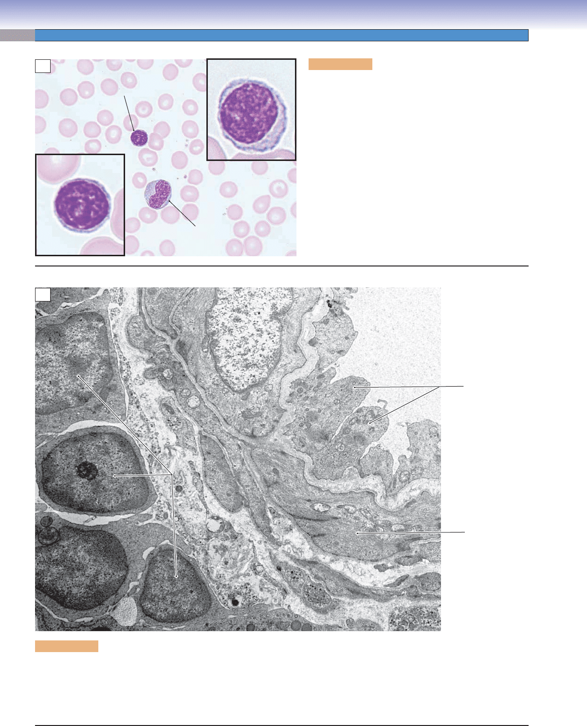
Figure 10-1A. Lymphocytes. H&E, 702; inset, 2,176
Lymphocytes can be found in blood and lymph circulation
as well as in lymphoid organs. There are many types and
subtypes of lymphocytes, which can be classifi ed into three
major types based on their immunologic functions: B lym-
phocytes (B cells), T lymphocytes (T cells), and null cells.
Their sizes vary they are morphologically similar to each
other. B and T cells cannot be distinguished from each
other in routine H&E stain. Shown here is an example
of lymphocytes in a blood smear. Lymphocytes have rela-
tively large and round nuclei, and they have a small rim of
cytoplasm surrounding the nucleus. The two insets in the
same magnifi cation show the size variation of the lympho-
cytes. A monocyte is also seen in this specimen. Monocytes
differentiate into tissue macrophages or special forms of
macrophages, such as Kupffer cells in the liver, microglia in
the nervous tissue, and osteoclasts in the bone tissue.
Lymphocyte
Larger lymphocyte
Smaller lymphocyte
Monocyte
A
Endothelium
Smooth
muscle
Lumen of the
Lumen of the
central arteriole
central arteriole
Lumen of the
central arteriole
Lymphocytes
Lymphocytes
of PALS
of PALS
Lymphocytes
of PALS
B
Figure 10-1B. Lymphocytes in spleen. TEM, 8,800.
The lymphocytes in the left part of this fi eld are part of the PALS that surrounds a central arteriole. Part of the wall of the arteriole
occupies the central part of the fi eld, and part of its lumen is seen to the right. The lymphocytes here appear to be inactive, judging
from the scant amount of cytoplasm and the small nuclei containing little euchromatin. However, one of the lymphocytes does dis-
play a small nucleolus, suggesting at least a basal level of protein synthesis. Although T and B lymphocytes are not distinguishable
morphologically, the location of these lymphocytes in the PALS indicates that they are T lymphocytes.
CUI_Chap10.indd 182 6/2/2010 4:11:30 PM
CHAPTER 10
■
Lymphoid System
183
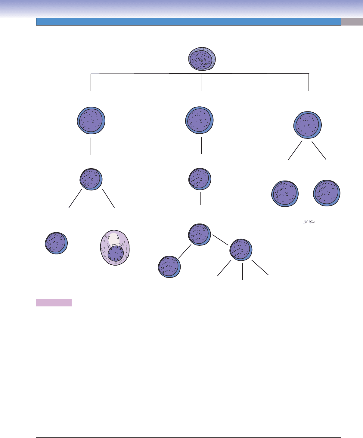
Figure 10-2. A representation of types of lymphocytes.
B lymphocytes (B cells), T lymphocytes (T cells), and null cells are three major cell types in the immune system. Each of these
cells originates from precursor cells in the bone marrow. B and T lymphocytes are the main cell types located in lymphoid organs.
(1) B cells mature and become naive (virgin) B cells (immunocompetent cells that have not been previously exposed to foreign
antigen) in the bone marrow; they migrate to secondary lymph organs and may meet with antigens. B cells that become activated
by exposure to antigens differentiate into memory B cells and effector B cells (plasma cells). (2) T cells differentiate from pro–T
lymphocytes, which have migrated from the bone marrow into the thymus through the circulatory system. Thymocytes (developing
lymphocytes) differentiate to naive (virgin) T cells in the thymus and then migrate to secondary lymphoid organs where they may be
activated by exposure to foreign antigens. Activated T cells can differentiate into both memory T cells and effector T cells. Effector T
cells include helper T cells, cytotoxic T cells, and regulatory (suppressor) T cells. B and T cells share some common features. Each B
and T cell is programmed to respond to a particular antigenic determinant. Each naive B cell or T cell is relatively short lived unless
it becomes activated by contact with the antigen it recognizes. Both types give rise to both memory cells and effector cells if they
interact with an antigen (“antigen dependent”). Both B and T cells reside in specifi c regions in secondary lymphoid organs. However,
there are some important differences between B and T cells. B-cell antigen recognition is mediated by Ig molecules in their surface
membranes, whereas T-cell antigen recognition is mediated by the T-cell receptor (TCR), and activation requires presentation of
the antigen in association with an MHC molecule on the surface of another cell. Finally, activated B cells function by differentiating
into antibody-secreting plasma cells (humoral immune response), whereas activated T cells can differentiate into several functional
forms: helper T cells, cytotoxic T cells, or suppressor T cells (cell-mediated immune responses). (3) Null cells are described in detail
in the introduction.
Null cells
(bone marrow,
circulation)
Pro–T lymphocytes
(bone marrow)
Pro–B lymphocytes
(bone marrow, fetal liver)
Hematopoietic stem cells
(bone marrow)
Virgin (inactive) B lymphocytes
(circulation to lymphoid organs)
T lymphoblasts
(circulation to thymus)
Pluripotential
hemopoietic
stem cells (PHSCs)
Natural killer
(NK) cells
Plasma cells
(lymphoid organs to
connective tissue)
Virgin
T cells
(inactive)
Effector
T cells
Memory B cells
(lymphoid organs to
circulation)
Memory
T cells
Helper T cells
(Th0, Th1, Th2)
Cytotoxic
T cells
Regulatory
(suppressor)
T cells
CUI_Chap10.indd 183 6/2/2010 4:11:35 PM
184
UNIT 3
■
Organ Systems
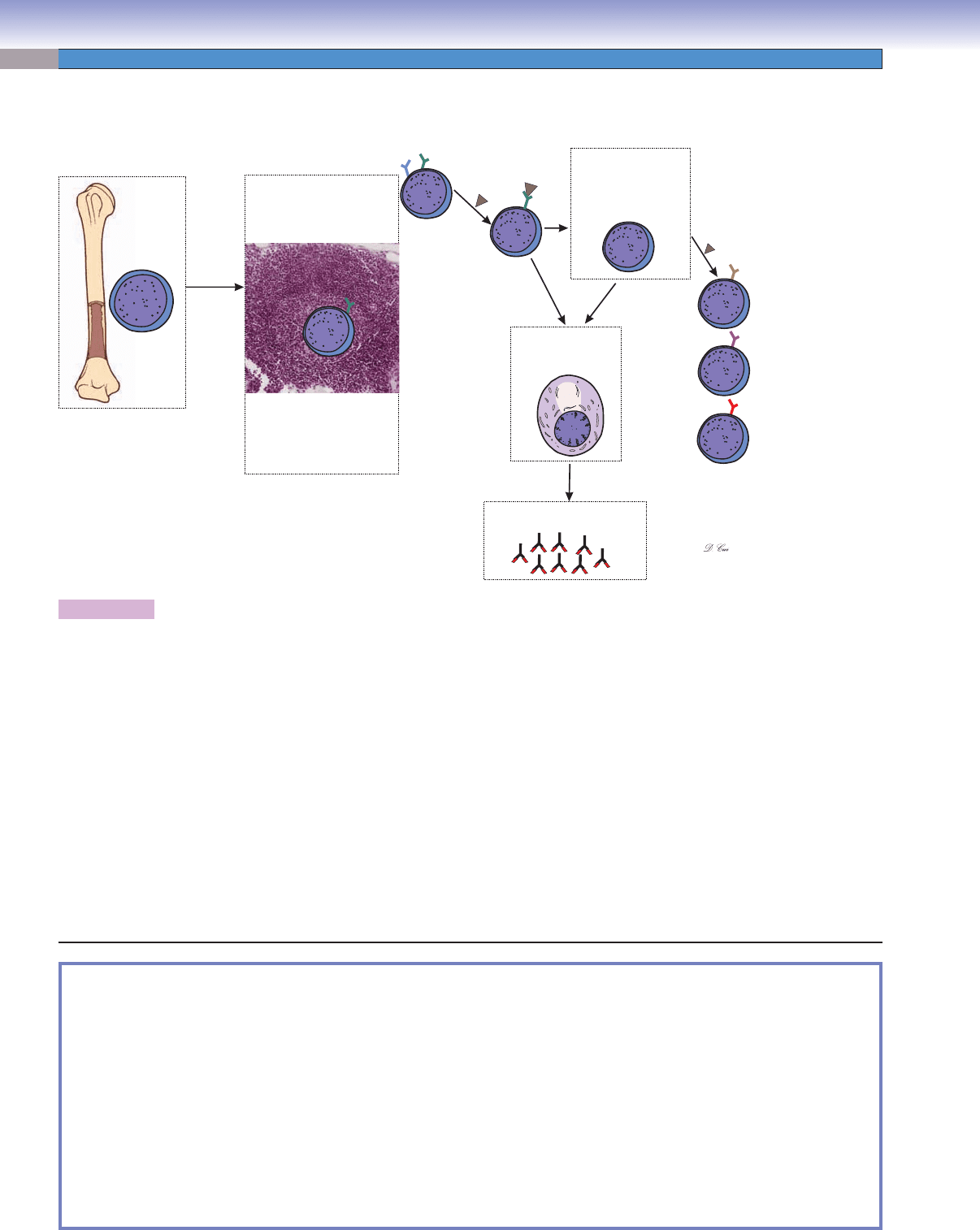
Figure 10-3. A representation of B-lymphocyte maturation. H&E, 83
B lymphocytes (B cells) originate and mature in the bone marrow. Because naive (virgin) B lymphocytes differentiate from
precursor cells (pro–B lymphocytes), they become randomly programmed to recognize a specifi c antigenic determinant. Dur-
ing the B-cell maturation process, they are subjected to negative selection, through which those B cells that happen to recognize
self-antigens are induced to undergo apoptosis. Naive B lymphocytes are immunocompetent cells with specifi c antibodies (Igs)
inserted into their plasma membrane as receptors. Each B lymphocyte has the ability to recognize and respond to a particular
antigen. After newly matured B lymphocytes leave the bone marrow, they use the vasculature and their own motility to recirculate
through the peripheral lymphoid organs (lymph nodes, spleen, MALT, etc.). This continual wandering increases the likelihood that
a lymphocyte will encounter its antigen if the antigen has gained entry into the body. Naive B cells die in a few days or weeks if
they do not meet their antigen, but those that encounter their specifi c antigen under favorable conditions will become activated. B
cells that are activated by an encounter with antigens undergo cell division and differentiation. Some descendants of an activated
B cell become memory B cells; others differentiate into effector B cells, that is, plasma cells, which are able to produce and secrete
antibodies. Antibodies secreted by plasma cells become widely distributed throughout the body so that foreign antigens are unlikely
to evade binding by antibodies and the defense mechanisms that are triggered by antibody binding. Memory B cells have a much
longer life than naive B cells; they enter the blood circulation in an inactive state and may live and recirculate for decades. If there is a
subsequent encounter with the same antigen, memory B cells rapidly divide and differentiate into plasma cells that secrete antibodies
in great quantity, thereby producing a much quicker and more powerful secondary immune response.
Pro–B lymphocytes
Pro–B lymphocytes
in bone marrow
in bone marrow
Pro–B lymphocytes
in bone marrow
Circulation
Activated
B cells
Plasma cell in the
lymphoid organs and
connective tissue
Antibodies secreted into blood,
lymph, or connective tissue
Memory B cells in the
circulation
and
lymph organs
M
B lymphocytes in
lymph nodes, spleen,
and other lymphoid organs
Binding antigens to antibody;
inactive B lymphocytes
become activated B cells
IgE
IgM
IgD
IgG
First
encounter
with an
antigen
Second
encounter
with same
antigen
IgA
SYNOPSIS 10-1 Characteristics of Types of Immunoglobulins
There are fi ve types of Igs classifi ed by their heavy chains:
IgG
■ : This is the most abundant type of Ig in blood serum and the only one that is able to cross the placenta. It is a major
Ig during the secondary immune response and has high antigen-binding specifi city.
IgA
■ : This is the major type of Ig in external secretions (milk, saliva, tears, sweat, and mucus) of epithelial cells, including
gland epithelial cells. Its main function is to protect mucosal (epithelial) surfaces. It includes subclasses IgA1 and IgA2.
IgM
■ : This is the principal Ig in the primary immune response; it is most effective in activating a complement but with lower
antigen-binding specifi city. It activates macrophages and serves as an antigen receptor on the B cell surfaces.
IgE
■ : This is found only in small amounts in blood serum; it binds to Fc receptors of the mast cells and basophils and plays
an important role in allergic reactions (see mast cell, Fig. 4-4B).
IgD
■ : This has a low concentration in blood serum; it serves along with IgM as an antigen receptor on the membranes of
mature B cells.
B Lymphocytes
CUI_Chap10.indd 184 6/2/2010 4:11:36 PM