Cui Dongmei. Atlas of Histology: with functional and clinical correlations. 1st ed
Подождите немного. Документ загружается.

155
UNIT 3
■
Organ Systems
9
Circulatory System
Introduction and Key Concepts for the Circulatory System
The Cardiovascular System
Figure 9-1 Overview of the Cardiovascular System
Figure 9-2 The Heart and Its Impulse Conductive Function
Synopsis 9-1 Structure and Functions of the Heart
Figure 9-3A Layers of the Heart Wall: Endocardium, Ventricle
Figure 9-3B Layers of the Heart Wall: Myocardium, Ventricle
Figure 9-3C Layers of the Heart Wall: Epicardium, Ventricle
Figure 9-4A Purkinje Fibers and Intercalated Disks
Figure 9-4B Cardiac Valves
Figure 9-4C Clinical Correlation: Myocardial Infarction
Figure 9-5 A Representation of the General Structure of Blood Vessel Layers (Tunicae) and a
Comparison of the Medium Artery and the Medium Vein
Figure 9-6 A Representation of Types of Blood Vessels: Arteries, Veins, and Capillaries
The Arterial System
Figure 9-7A–C Large Arteries (Elastic Arteries)
Figure 9-8A–C Medium Arteries (Muscular Arteries)
Figure 9-9A Medium Artery
Figure 9-9B Small Artery
Figure 9-10A–C Small Arteries and Arterioles
Figure 9-11 Arteriole
Synopsis 9-2 Functions of Endothelium in Blood Vessels
Figure 9-12A Clinical Correlation: Coronary Artery Atherosclerosis
Figure 9-12B Clinical Correlation: Polyarteritis Nodosa (Vasculitis)
Synopsis 9-3 Pathological and Clinical Terms for the Circulatory System
CUI_Chap09.indd 155 6/17/2010 10:22:07 AM

156
UNIT 3
■
Organ Systems
The Capillary System
Figure 9-13A,B Continuous Capillaries
Figure 9-14A,B Fenestrated Capillaries
Figure 9-15A,B Discontinuous (Sinusoidal) Capillaries
The Venous System
Figure 9-16A–C Venules and Small Veins
Figure 9-17A–C Medium Veins
Figure 9-18A–C Large Veins
Table 9-1 Blood Vessels
The Lymphatic Vascular System
Figure 9-19A,B Lymphatic Vessels
Figure 9-19C Clinical Correlation: Lymphangioma
Introduction and Key Concepts
for the Circulatory System
The circulatory system includes the cardiovascular system
and the lymphatic vascular system. The cardiovascular system
includes the heart and the arterial, capillary, and venous sys-
tems. Blood is transported from the heart through the arterial
system to the capillaries, where exchange of gases, nutrients,
and other substances takes place. Blood is carried back to the
heart by the venous system. Blood fl ows through two routes:
(1) The systemic circulation system transports oxygenated
blood from the heart to the capillaries in the tissues and
organs of the body and then collects and carries the blood
back to the heart (Fig. 9-1). (2) The pulmonary circulation
system transports deoxygenated blood from the heart to the
capillaries of the lungs. After gas exchange, blood is carried
back to the heart (see Fig. 9-1). The lymphatic vascular sys-
tem consists of lymphatic capillaries, lymphatic vessels, and
lymphatic ducts. This system collects lymph (excess tissue
fl uid) from the tissues of all organs (except the nervous sys-
tem, bone marrow, and hard tissues) by lymphatic capillar-
ies and then transports it through lymphatic vessels to the
lymphatic ducts, which eventually empty the lymph into the
venous system. The collected lymph passes through lymph
organs, where it is fi ltered, and lymphocytes are exposed to
antigens. Lymphopoiesis and the immune response occur here
(Fig. 9-19A,B).
The Cardiovascular System
The Heart
The heart contains four chambers: the left and right atria and
the left and right ventricles. The atria receive blood fl ow dis-
charged from the venous system, whereas the ventricles pump
blood into the arterial system (Fig. 9-1). The wall of the heart
is composed of three layers: endocardium (innermost layer),
myocardium (middle layer), and epicardium (outermost layer).
(1) Endocardium consists of endothelium, subendothelial
connective tissue, and subendocardium (Purkinje fi bers, small
coronary blood vessels, and nerve fi bers). (2) Myocardium, the
thickest layer of the heart, contains an abundance of cardiac
muscle cells (Fig. 9-3A,B). Cardiac muscle contracts producing
heart beats, which are generated and regulated by the heart
conductive system including the sinoatrial (SA) node, the
atrioventricular (AV) node, the AV bundle, and Purkinje fi bers
(Fig. 9-2). (3) Epicardium is covered by mesothelium and con-
tains fi brous connective tissue, nerves, coronary vessels, and
adipose tissue (Fig. 9-3C).
Types of Blood Vessels
THE ARTERIAL SYSTEM is composed of large (conducting)
arteries, medium (distributing) arteries, small arteries, and arte-
rioles. The arterial system conducts blood (under higher pres-
sure than veins) from the ventricles to the capillary networks.
The walls of arteries can be generally divided into three layers:
tunica intima, tunica media, and tunica adventitia (Figs. 9-5
and 9-6).
Large arteries are also called elastic arteries because of the
large quantity of elastic material in their walls (Fig. 9-7A–C).
They have a thick tunica media with numerous elastic mem-
branes. The internal and external elastic laminae are hard to
distinguish from the nearby elastic membranes. Large arter-
ies conduct blood from the ventricles into the medium arter-
ies. Rich elastic materials in large arteries enable the vessels to
recoil to accommodate pressure changes and maintain a con-
tinuous fl ow of blood during ventricular diastole (relaxation).
Medium arteries are also called muscular arteries because
of their thick tunica media, which contains circularly arranged
multiple layers of smooth muscle cells in a distinct sheath
(Figs. 9-8 to 9-9A). Internal and external elastic laminae are
easy to distinguish from nearby tissues.
Small arteries and arterioles are smaller diameter vessels.
The walls of small arteries contain two to six layers of smooth
muscle cells (Figs. 9-9B and 9-10A,B). Arterioles are the small-
est components of the arterial system, with only one or two
layers of smooth muscle cells (Figs. 9-10A,C, and 9-11). They
control the blood fl ow into the capillaries.
CUI_Chap09.indd 156 6/17/2010 10:22:18 AM

CHAPTER 9
■
Circulatory System
157
THE CAPILLARY SYSTEM contains continuous capillaries,
which have a continuous basal lamina and complete endothe-
lial cells. These structures allow only a very limited amount of
materials to pass through the capillary walls (Fig. 9-13A,B).
Fenestrated capillaries have a continuous basal lamina
and fenestrated endothelial cells (perforated by small pores).
A greater range of substances can, therefore, pass through the
capillary walls (Fig. 9-14A,B).
Discontinuous (sinusoidal) capillaries have a discontinuous
(or missing) basal lamina and incomplete endothelial cells
(perforated by large pores), which allow proteins and other
materials, even cells, to pass through the capillary walls freely
(Fig. 9-15A,B).
THE VENOUS SYSTEM is composed of venules and small,
medium, and large veins. Veins collect blood (under lower pres-
sure than arteries) from capillaries and transport it to the heart.
Veins have larger, fl at lumens and thinner walls than their com-
panion arteries (Figs. 9-5 and 9-6).
Venules and small veins have very thin walls and few valves.
Venules drain exchanged blood from the capillaries to the small
veins. Venules are the primary sites of many infl ammatory
reactions (Fig. 9-16A–C).
Medium veins have various diameters, and their structure
varies based on their size and location. Segments of medium
veins in some locations have a thick tunica adventitia with a
few longitudinal smooth muscle bundles (Fig. 9-17A–C). Valves
are prominent in medium veins, with an abundance in the
extremities to prevent blood from fl owing backwards.
Large veins have a thick tunica adventitia with numerous
longitudinal smooth muscle bundles, which help to force blood
to fl ow toward the heart (Fig. 9-18A–C). There are some large
valves in the large veins.
The Lymphatic Vascular
System
Lymphatic Vessels
The lymphatic vessels have a structure similar to small veins
(Fig. 9-19A,B). They have a large lumen, and valves are plenti-
ful in all sizes of lymphatic vessels.
CUI_Chap09.indd 157 6/17/2010 10:22:18 AM
158
UNIT 3
■
Organ Systems
T. Yang
Large veins (vena cava)
Large (pulmonary) veins
Large arteries (pulmonary artery)
Large arteries
Aorta
Arterioles
Arterioles
Small arteries
Small arteries
RA
LA
RV
LV
: right atrium
: left atrium
: right ventricle
: left ventricle
M
edium
arteries
Medium
arteries
Pulmonary circulation
Systemic circulation
Medium veins
Medium veins
Small veins
Small veins
Venules
Venules
Deoxygenated blood in pulmonary circulation system
Oxygenated blood in pulmonary circulation system
Oxygenated blood in systemic circulation system
Deoxygenated blood in systemic circulation system
Systemic circulation system
Organs and tissues of the body
Lungs
RA
LA
LV
RV
Pulmonary
capillaries
Systemic
capillaries
Pulmonary circulation system
Left
ventricle
Right
ventricle
Small arteries
Small arteries
Arterioles
Arterioles
Venules
Venules
Small
veins
Small
veins
Medium
veins
Medium
veins
Large veins
(vena cava, etc.)
Large veins
(pulmonary veins)
Right
atrium
Left
atrium
Systemic capillaries
(exchange of gases
and other substances)
Pulmonary capillaries
(gas exchange)
Large/elastic arteries
(aorta, etc.)
Large/elastic arteries
(pulmonary arteries)
Medium systemic
arteries (muscular arteries)
Medium pulmonary
arteries (muscular arteries)
Pulmonary circulation system
Systemic circulation system
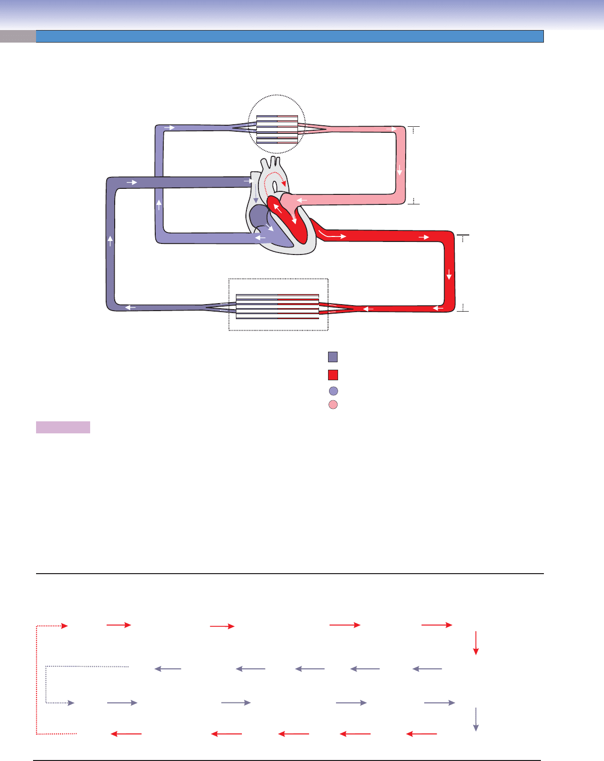
Figure 9-1. Overview of the cardiovascular system.
The cardiovascular system consists of the heart, arterial system, venous system, and capillary system. The heart is composed of two
atria and two ventricles. The right atrium receives blood from the body, and the left atrium receives blood from the lungs; the left
ventricle pumps blood to the body, and the right ventricle pumps blood to the lungs. The arterial system conducts blood from the
heart to capillaries in the body and the lungs. This system includes large arteries (elastic arteries), medium arteries (muscular arter-
ies), small arteries, and arterioles. The venous system carries blood from the capillary system in the body and lungs to the heart.
It includes venules, small veins, medium veins, and large veins. The capillary system is located between arterioles and venules and
often forms capillary beds where exchange of gases and various substances and movement of blood cells (diapedesis) take place. It
includes continuous, fenestrated, and sinusoidal (discontinuous) capillaries. Blood fl ows through two routes: (1) the systemic circu-
lation system, which supplies oxygenated blood from the heart to the organs and tissues of the body and then carries deoxygenated
blood back to the heart and (2) the pulmonary circulation system, which sends deoxygenated blood from the heart to the lungs for
gas exchange and then returns oxygenated blood to the heart.
Systemic and Pulmonary Circulation Systems
The Cardiovascular System
CUI_Chap09.indd 158 6/17/2010 10:22:18 AM
CHAPTER 9
■
Circulatory System
159
SYNOPSIS 9-1 Structure and Functions of the Heart
The heart is composed of two ■ atria (singular: atrium) and two ventricles.
The wall of the heart consists of
■ endocardium (inner layer), myocardium (cardiac muscle layer), and epicardium (outer
layer).
Endocardium
■ is composed of endothelium, subendothelium (a thin layer of connective tissue), and subendocardium.
Purkinje fi bers are located in the subendocardium.
Myocardium
■ is composed of cardiac muscles, the thickest layer of the heart; cardiac muscles are branched and connected
to each other end to end by intercalated disks (junction complexes). Cardiac muscle fi bers require high oxygen supply.
Epicardium
■ is composed of mesothelium and a thicker layer of connective tissue, which contains coronary vessels, nerves,
and adipose tissue.
The
■ conductive system of the heart consists of groups of specially modifi ed cardiac muscle fi bers, the SA node, AV node,
AV bundle, and Purkinje fi bers.
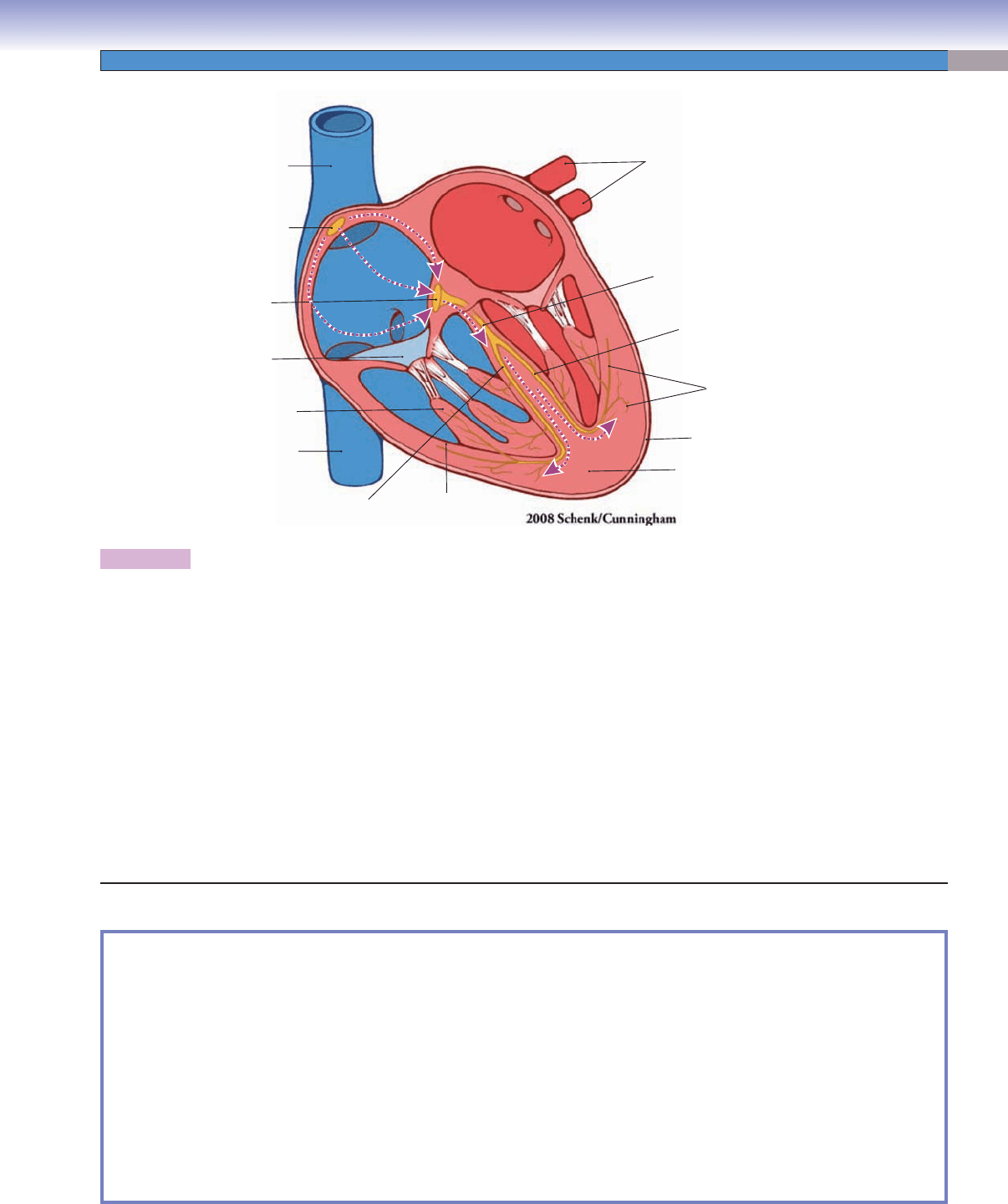
Figure 9-2. The heart and its impulse conductive function.
The contractions of the right and left atria and ventricles must be coordinated precisely in order for blood to be pumped effi ciently to
the lungs for oxygenation and then to the rest of the body. A network of specially modifi ed cardiac muscle fi bers generates and con-
ducts electrical impulses that provide the necessary coordination. This network includes the SA node, the AV node, the AV bundle,
and Purkinje fi bers. (1) The SA node is located in the right atrial wall near the opening of the superior vena cava. The SA node is
important for initiating the heartbeat impulse and controlling its frequency; it is also called the “pacemaker” of the heart. (2) The AV
node is also located in the right atrial wall, medial to the right AV valve, and along the lower part of the interatrial septum. (3) The
AV bundle arises from the AV node and divides into two branches (right and left) to run along the sides of the interventricular sep-
tum. (4) Purkinje fi bers are the terminal branches of the right and left branches of the AV bundle. Purkinje fi bers run along the infe-
rior and lateral wall of the ventricle and extend to the papillary muscles. These fi bers are modifi ed large cardiac muscle cells, which
contain numerous gap junctions and well-developed intercalated disks. The SA node generates the heartbeat signal, which quickly
spreads to adjacent cardiac muscle cells in the myocardium of both atria to cause the atria to contract. Impulses are also picked up
by the AV node and travel along the AV bundle. These impulses pass through the right and left bundle branches to Purkinje fi bers
in the ventricles, where they induce contraction of the cardiac muscles of the ventricles. There is a time delay, which allows the atria
to contract fi rst to empty blood into the ventricles, before the ventricles contract. This time delay ensures that blood fl ows smoothly
from the atria to the ventricles and then to the conductive arteries.
Superior
vena cava
Inferior
vena cava
Sinoatrial (SA)
node
Atrioventricular
(AV) node
Atrioventricular
(AV) bundle
Left bundle branch
Purkinje fibers
Left pulmonary veins
Right bundle
branch
Papillary
muscle
Cardiac
valves
RV:
LV:
right ventricle
left ventricle
LV
Left
atrium
RV
Right
atrium
Myocardium
Epicardium
Endocardium
CUI_Chap09.indd 159 6/17/2010 10:22:19 AM
160
UNIT 3
■
Organ Systems
A
Myocardium
Myocardium
Myocardium
Endocardium
Endocardium
Endocardium
Myocardium
Myocardium
Myocardium
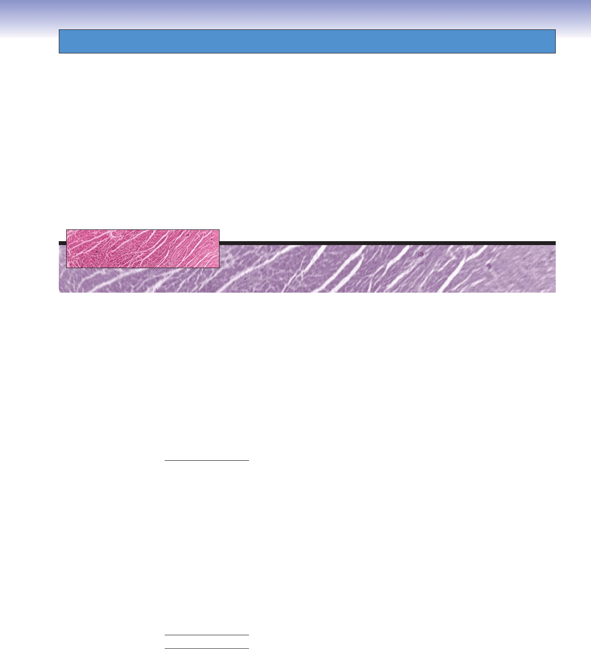
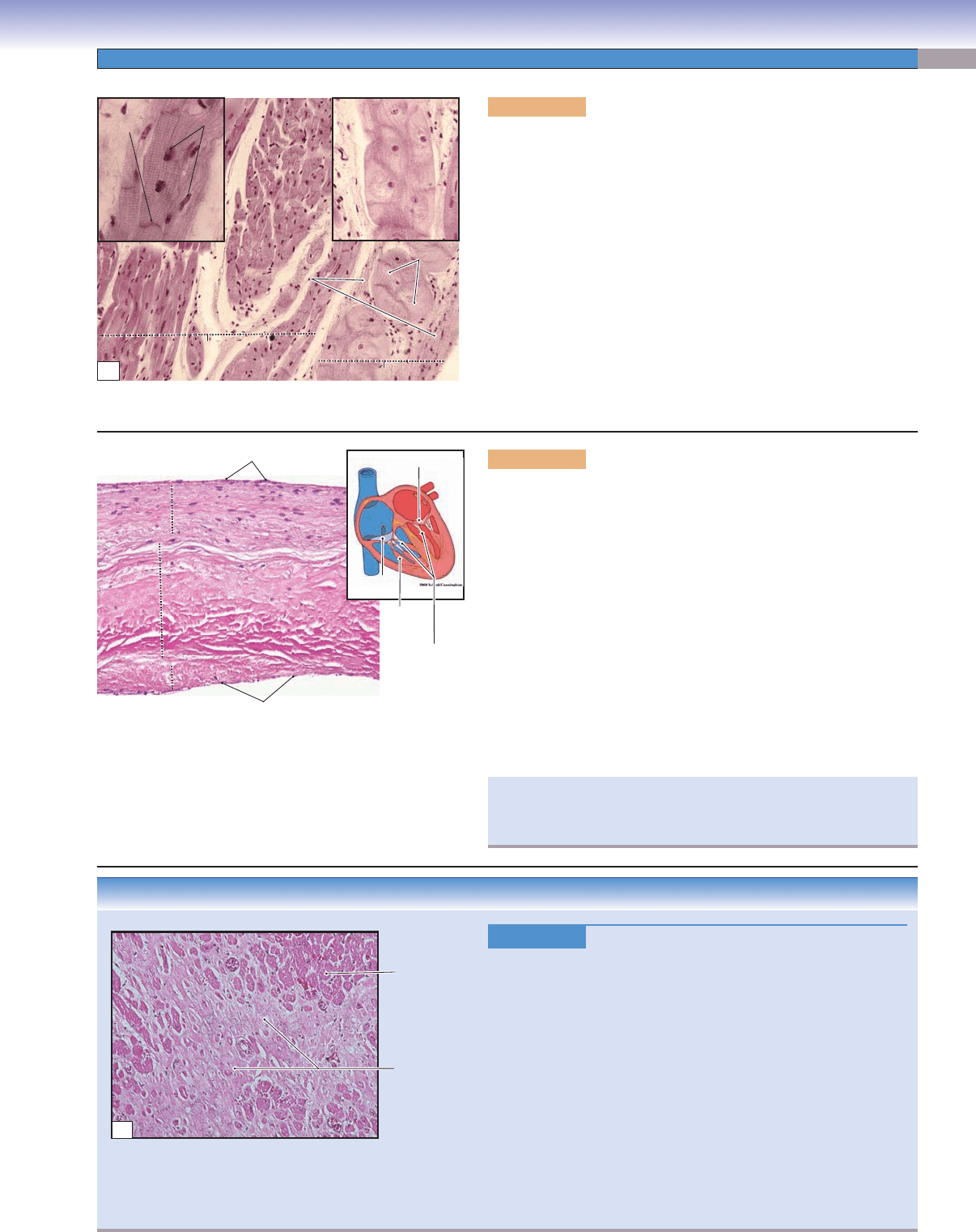
Figure 9-3A. Layers of the heart wall: endocardium, ventri-
cle. H&E, 68
The wall of the heart is much thicker than the wall of the large
vessels and is composed of three basic layers, as are the blood
vessels: endocardium (equivalent to the tunica intima), myocar-
dium (equivalent to the tunica media), and epicardium (equiva-
lent to the tunica adventitia). The endocardium is the innermost
layer of the heart wall, which lines the lumen of the heart. This
layer consists of endothelium (simple squamous epithelium),
subendothelial connective tissue, and subendocardium. The
subendocardium is in contact with the cardiac muscle and con-
tains small coronary blood vessels, nerves, and Purkinje fi bers
in certain areas (Fig. 9-4A). Some adipose cells are also pres-
ent within the connective tissue. The endocardium provides a
smooth lining for the four chambers of the heart and provides a
covering for the AV valves.
B
Nuclei of the cardiac muscle cells
Nuclei of the cardiac muscle cells
Nuclei of the cardiac muscle cells
Branched
Branched
cardiac muscle
cardiac muscle
Branched
cardiac muscle
Intercalated disk
Intercalated disk
Intercalated disk
Figure 9-3B. Layers of the heart wall: myocardium, ventri-
cle. H&E, 272; insets 786
Myocardium is the thickest layer of the heart wall and makes up
the bulk of the heart. It consists of cardiac muscle cells that are
arranged in branching columns. The ends of the cardiac mus-
cle cells are connected to each other by intercalated disks. The
inset shows cardiac muscles with their characteristic striations
and an intercalated disk (Fig. 9-4A). These muscles contract
to pump blood out of the ventricles of the heart and distribute
blood to the tissues and organs of the body. Myocardium of the
left ventricle wall is the thickest because of the fact that it must
pump the blood a great distance and overcome the high pressure
and resistance of the systemic circulation. In general, the atria
have thinner walls than the ventricles. Myocardium of the right
atrium is the thinnest because of the relatively low pressure and
resistance of the blood circulation.
C
Epicardium
Epicardium
(visceral layer)
(visceral layer)
Myocardium
Myocardium
Epicardium
(visceral layer)
Myocardium
Lumens of the
blood vessels
Figure 9-3C. Layers of the heart wall: epicardium, ventricle.
H&E, 68
Epicardium surrounds the heart. It is a layer of connective tissue
that contains nerves, blood vessels, and adipocytes. The inner
surface of the epicardium is connected with cardiac muscle, and
the outer surface is covered by mesothelium (see Fig. 3-2) that
faces the pericardial cavity. Mesothelium secretes a fl uid known
as pericardial fl uid, which provides lubrication and reduces fric-
tion between the epicardium (visceral pericardium) and the pari-
etal pericardium during the movements caused by heart contrac-
tion. Epicardium covers and protects the heart and allows small
blood vessels and nerves to pass through to provide nutrients
and nerve innervation.
Pericardial effusion refers to excess fl uid in the pericardial
cavity due to infl ammation of the pericardium (pericarditis);
hemopericardium is a condition in which blood is trapped in
the pericardial cavity. In either case, compression of the thin-
walled atria and vena cava can result in cardiac tamponade
and failure of circulation.
CUI_Chap09.indd 160 6/17/2010 10:22:20 AM
CHAPTER 9
■
Circulatory System
161
Figure 9-4C.
Myocardial Infarction. H&E, 68
Myocardial infarction (MI), also known as a heart attack, occurs
when the blood supply to part of the heart is completely or partially
blocked because of atherosclerosis or the rupture of an atheroscle-
rotic plaque and the formation of a blood clot. Symptoms range from
characteristic chest discomfort (angina) and shortness of breath to
sudden death. Coronary artery disease (CAD) is the most common
underlying cause of this medical emergency. Atherosclerotic plaques,
which consist of lipids, fi broblasts, collagen, and white blood cells
(especially macrophages), are found in the wall of an affected cor-
onary artery. The plaques cause luminal narrowing or occlusion
(Fig. 9-12A). The histologic appearance of MI depends on the age
of the infarct. Early changes include edema and hypereosinophilia,
followed by infi ltration by neutrophils, coagulation necrosis, phago-
cytosis of dead cells by macrophages, formation of granulation tis-
sue, and, ultimately, the formation of a dense collagenous scar.
Necrotic
cardiac muscle
Remaining
normal cardiac
muscle cells
C
Spongiosa
Spongiosa
Spongiosa
Endothelium
Papillary
muscle
Mitral valves
Tricuspid
valve
Chordae
tendineae
Endothelium
Fibrosa
Fibrosa
Fibrosa
Ventricularis
Ventricularis
Ventricularis
B
Figure 9-4B. Cardiac valves, aortic valve. H&E, 134
There are four valves in the heart: two AV valves (mitral and tricuspid
valves) in the chambers and two semilunar valves (aortic and pul-
monary valves) in the arteries leaving the heart. Heart valves consist
of connective tissues and both surfaces are covered by endothelium.
They are composed of three layers: (1) Spongiosa consists of loosely
arranged collagen and elastic fi bers and the surface is covered by the
endothelium. This layer is continuous with the atrial or blood vessel
side. (2) Fibrosa is the core of the heart valve, which contains dense
irregular connective tissue. (3) Ventricularis is a dense connective tis-
sue layer with many elastic and collagen fi bers. The surface of the
ventricularis is covered by endothelium. The heart valves open and
close to allow the blood to fl ow through the openings and to prevent
the backfl ow of blood. Shown is an example of the aortic valve. The
aortic valve has three cusps and lies between the left ventricle and the
aorta. The tricuspid and mitral valves are anchored to the ventricle
wall by chordae tendineae, which are attached to papillary muscles.
Common heart valve diseases include calcifi c aortic valve disease,
valvitis, and rheumatic heart disease. These diseases can lead to aortic
stenosis, aortic regurgitation, embolism, and mitral valve stenosis.
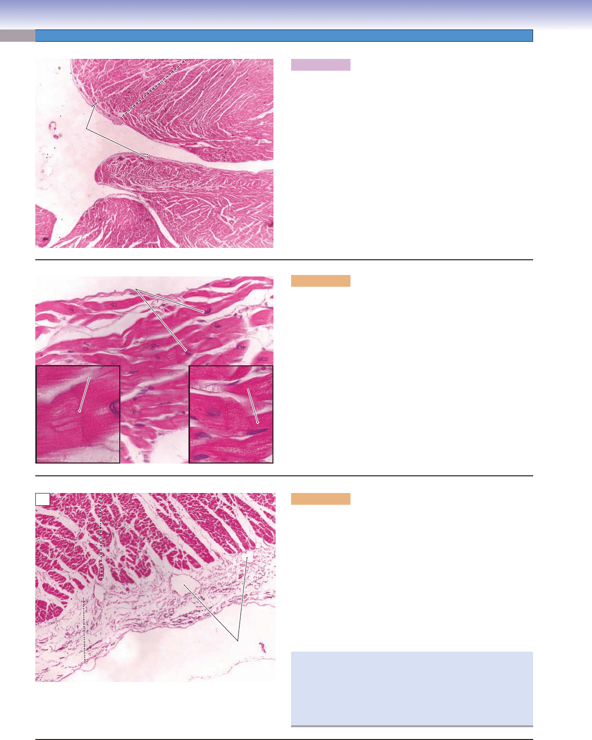
Figure 9-4A. Purkinje fi bers and intercalated disks, ventricle.
H&E, 136; inset (right) 198, inset (left) 229
Purkinje fi bers (impulse-conducting fi bers) are large, modifi ed car-
diac muscle cells, which are part of the heart conducting system. They
are terminal branches of the AV bundle branches (Fig. 9-2), located in
subendocardial connective tissue. Purkinje fi bers often appear large in
size and cluster as groups. Each cell has only one or two nuclei and pale-
staining cytoplasm because it has fewer myofi brils than regular cardiac
muscle cells. Purkinje fi bers work together with other impulse- conducting
structures to regulate the heartbeats by conveying impulses to neigh-
boring cardiac muscle cells (Fig. 9-2). Intercalated disks are specialized
junctional complexes that contain fascia adherens, desmosomes (macula
adherens), and gap junctions, which provide connection and communi-
cation between the cardiac muscle cells. Intercalated disks bind cardiac
muscle cells together in an entire unit to prevent muscle cells being pulled
apart during contraction. They also provide ion exchange through gap
junctions, allowing electrical impulses to pass from one cell to another.
Nuclei of
Nuclei of
muscle cells
muscle cells
Intercalated
Intercalated
disk
disk
Purkinje
Purkinje
fibers
fibers
Purkinje fibers
Purkinje fibers
Purkinje fibers
Purkinje fibers
Subendocardial
Subendocardial
connective
connective
tissue
tissue
Cardiac muscle
Cardiac muscle
(cross section)
(cross section)
Purkinje
fibers
Purkinje fibersPurkinje fibers
Subendocardial
connective
tissue
Cardiac muscle
(cross section)
Cardiac muscle
Cardiac muscle
(longitudinal section)
(longitudinal section)
Cardiac muscle
(longitudinal section)
Intercalated
disk
Nuclei of
muscle cells
Myocardium
Myocardium
Myocardium
Endocardium
Endocardium
Endocardium
A
CLINICAL CORRELATIONS
CUI_Chap09.indd 161 6/17/2010 10:22:28 AM
162
UNIT 3
■
Organ Systems
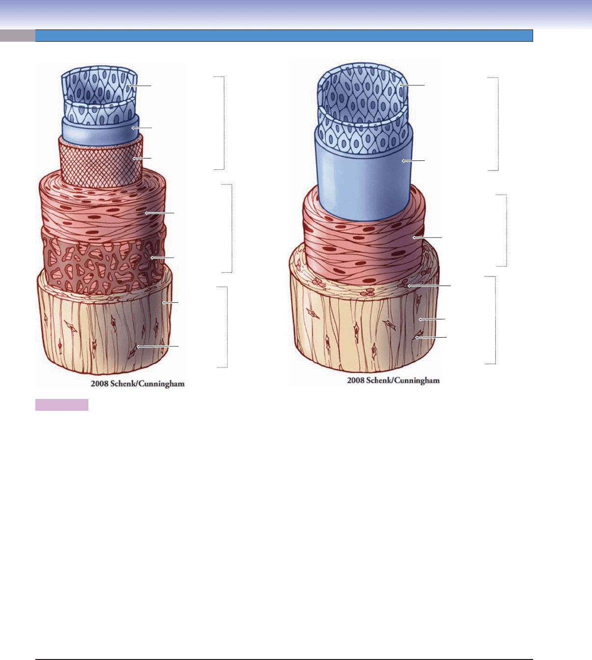
Figure 9-5. A representation of the general structure of blood vessel layers (tunicae) and a comparison of the medium artery and
the medium vein.
The walls of the blood vessels can be divided into three layers (tunicae). The innermost layer in contact with the blood is called the
tunica intima. This layer contains endothelium and subendothelial connective tissue and may contain an internal elastic lamina (IEL)
in some vessels, particularly arteries. The endothelium is a layer of simple squamous cells, which forms the smooth surface of the
lumen. Subendothelial connective tissue is a thin layer of connective tissue beneath the endothelium. The IEL, if present, is a sheet
of elastic material that divides the tunica intima from the tunica media. The middle layer is called the tunica media. It primarily
contains circularly arranged smooth muscle cells (except in elastic arteries). Contraction and relaxation of these smooth muscle cells
will change the vessel diameter and affect blood pressure. The outermost layer, the tunica adventitia, is a layer of connective tissue
dominated by collagenous and elastic fi bers. In large and some medium veins, the tunica adventitia may also contain longitudinal
smooth muscle bundles. Tunica adventitia surrounds and covers the vessels for protection. Occasionally, small blood vessels called
vasa vasorum are found in the tunica adventitia of large vessels. The vasa vasorum provide oxygen and nutrients for the large vessel
walls when the distance from the lumen is great and it is diffi cult to get nutrients from diffusion. Some differences between a medium
artery and a medium vein are listed here: (1) the artery has a smaller and more rounded lumen, whereas the vein has a larger and
oval or irregular-shaped lumen; (2) the artery has a thicker wall than does the vein; (3) in the artery, the tunica media is much thicker
than the tunica adventitia, but in the vein, the tunica adventitia is much thicker than the tunica media; (4) circularly oriented smooth
muscle cells form uniform sheets in the tunica media of the artery; however, smooth muscle cells are fewer and do not form a distinct
sheet in the vein; (5) There are a few longitudinal smooth muscle bundles in the tunica adventitia of medium veins in some locations,
whereas these smooth muscle bundles are more abundant in large veins. This pattern does not occur in arteries. Longitudinal smooth
muscle bundles in the tunica adventitia of the vein contract to help push blood back to the heart; and (6) valves are present in many
veins, especially in the medium veins of the extremities. Their function is to prevent gravitational backfl ow of the blood and to help
blood return to the heart. Arteries do not have valves (except the aortic and pulmonary valves).
Fibroblast
Fibroblast
Tunica
adventitia
Tunica
adventitia
Tunica
media
Tunica
media
Tunica
intima
Tunica
intima
External elastic
lamina ( )EEL
Internal elastic
lamina ( )IEL
Connective
tissue
Connective
tissue
smooth muscle
with connective
tissue
Circular
smooth muscle
Circular
Longitudinal
smooth
muscle
Endothelial cells
Endothelial cells
Subendothelial
layer
Subendothelial
layer
CUI_Chap09.indd 162 6/17/2010 10:22:35 AM
CHAPTER 9
■
Circulatory System
163
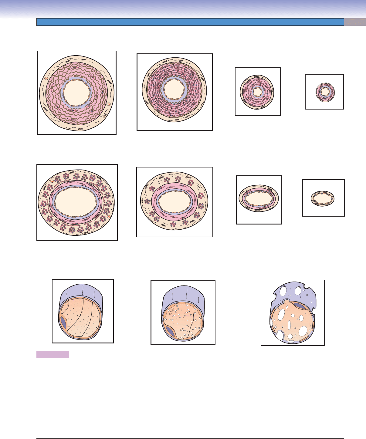
Figure 9-6. A representation of types of blood vessels: arteries, veins, and capillaries.
Blood vessels make up the arterial, the venous, and the capillary systems. In general, arteries have smaller, rounder lumens than
veins; their tunica media are thicker than the tunica adventitia, and the IEL are prominent. Veins have larger, fl atter lumens than
arteries; longitudinal smooth muscle bundles may be present in the tunica adventitia, which is the most dominant layer in large
and some medium veins. The tunica adventitia is much thicker in the veins than the tunica media. For details, also see Table 9-1.
Exchange of gases, nutrients, and materials occurs in the capillaries. Three types of capillaries are illustrated here. Continuous cap-
illaries have complete endothelial cells and continuous basal laminae. Fenestrated capillaries have continuous basal laminae with
fenestrated endothelial cells (perforated by small pores); discontinuous (sinusoidal) capillaries have incomplete endothelial cells
perforated by large pores, and part of the cytoplasm may be missing. The basal lamina is discontinuous. Discontinuous capillaries
have gaps between endothelial cells, and their lumen sizes are much larger than the other two types. Blood vessel sizes are not drawn
to scale.
D. Cui /T. Yang
D. Cui /T. Yang
T. Yang
Large artery
(elastic artery)
Large vein
Medium artery
(muscular artery)
Medium vein
Small artery
Small vein
Arteriole
Venule
Continuous capillary
Fenestrated capillary
Discontinuous (sinusoidal)
capillary
CUI_Chap09.indd 163 6/17/2010 10:22:38 AM
164
UNIT 3
■
Organ Systems
The Arterial System
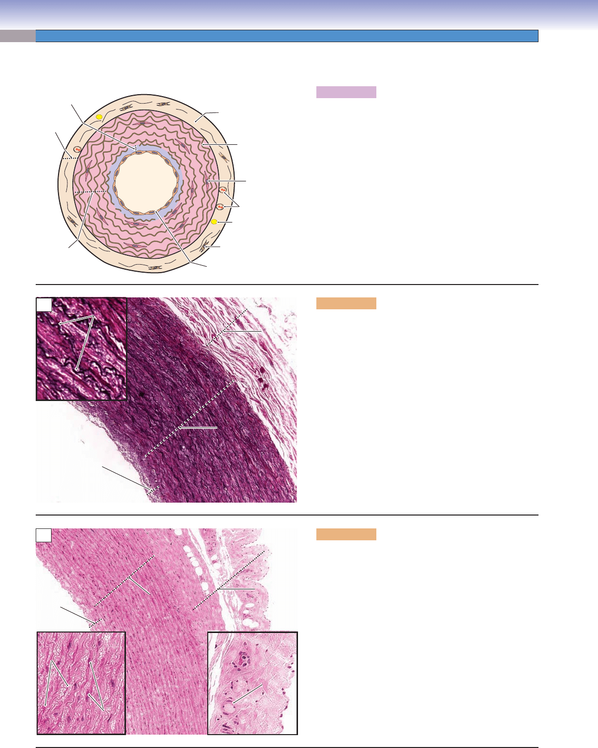
Figure 9-7A. A representation of a large (elastic) artery.
Large arteries (elastic arteries) are also referred to as
conducting arteries, because of their function in conduct-
ing blood fl ow out of the heart. There are abundant elastic
laminae (thick bundles of elastic fi bers) in the tunica media
of large arteries, which enable the walls to recoil to resist
pressure as well as to maintain arterial pressure during
diastole when the ventricles are relaxed and pressure is not
generated by the heart. Small vessels located in the tunica
adventitia are called vasa vasorum; they supply the tissue
of the artery wall with oxygen and nutrients when they are
far from the lumen and diffusion of nutrients is diffi cult.
The aorta, pulmonary arteries, and their main branches
are good examples of elastic arteries.
D. Cui /T. Yang
Vasa vasorum
Nerve
Fibroblast
Elastic lamina
Connective tissue
Smooth
muscle cell
Endothelial cell
Tunica
adventitia
Tunica
media
Tunica
intima
A
Elastic
Elastic
laminae
laminae
Tunica
Tunica
adventitia
adventitia
Tunica
Tunica
media
media
Tunica
Tunica
intima
intima
Elastic
laminae
Tunica
adventitia
Tunica
media
Tunica
intima
B
Figure 9-7B. Large (elastic) artery, carotid artery. Elastic
stain, 68; inset 242
This is an example of a large artery (elastic artery) from a
portion of the carotid artery. The carotid artery generally
has the characteristics of elastic arteries. The tunica media
of the vessel wall has multiple layers of elastic lamina that
are well organized in concentric fashion. These elastic
laminae are sheets of fenestrated elastic material produced
by smooth muscle cells in the tunica media. These cells are
interspersed between the elastic laminae (see Fig. 4-9B).
The elastic laminae in this specimen are revealed as par-
allel wavy black sheets by a special elastic stain; smooth
muscle cells are not visible here with this type of stain. The
tunica adventitia of an elastic artery consists of loosely
arranged connective tissue fi bers. These fi bers are mainly
collagen fi bers, with a small number of elastic fi bers that
are produced by fi broblasts.
Tunica
Tunica
adventitia
adventitia
Tunica
Tunica
media
media
Vasa
vasorum
Smooth
Smooth
muscle cells
muscle cells
Elastic
Elastic
laminae
laminae
Tunica
adventitia
Tunica
media
Tunica
intima
C
Smooth
muscle cells
Elastic
laminae
Figure 9-7C. Large (elastic) artery, aorta. H&E, 68;
insets 198
An example of a large artery (aorta) is shown. The elastic
laminae are pink and are more diffi cult to distinguish with
routine H&E stain than they are with elastic stains. The
dark nuclei in the tunica media belong to smooth muscle
cells (left inset), which produce the elastic membrane.
There are some vasa vasorum deep in the tunica adven-
titia (right inset), which provide oxygen and nutrients for
the tunica media and tunica adventitia of the large artery
wall. Nerves belonging to the autonomic nervous system
may occasionally be found in the tunica adventitia (see
Fig. 9-7A).
CUI_Chap09.indd 164 6/17/2010 10:22:40 AM