Cui Dongmei. Atlas of Histology: with functional and clinical correlations. 1st ed
Подождите немного. Документ загружается.

CHAPTER 7
■
Nervous Tissue
125
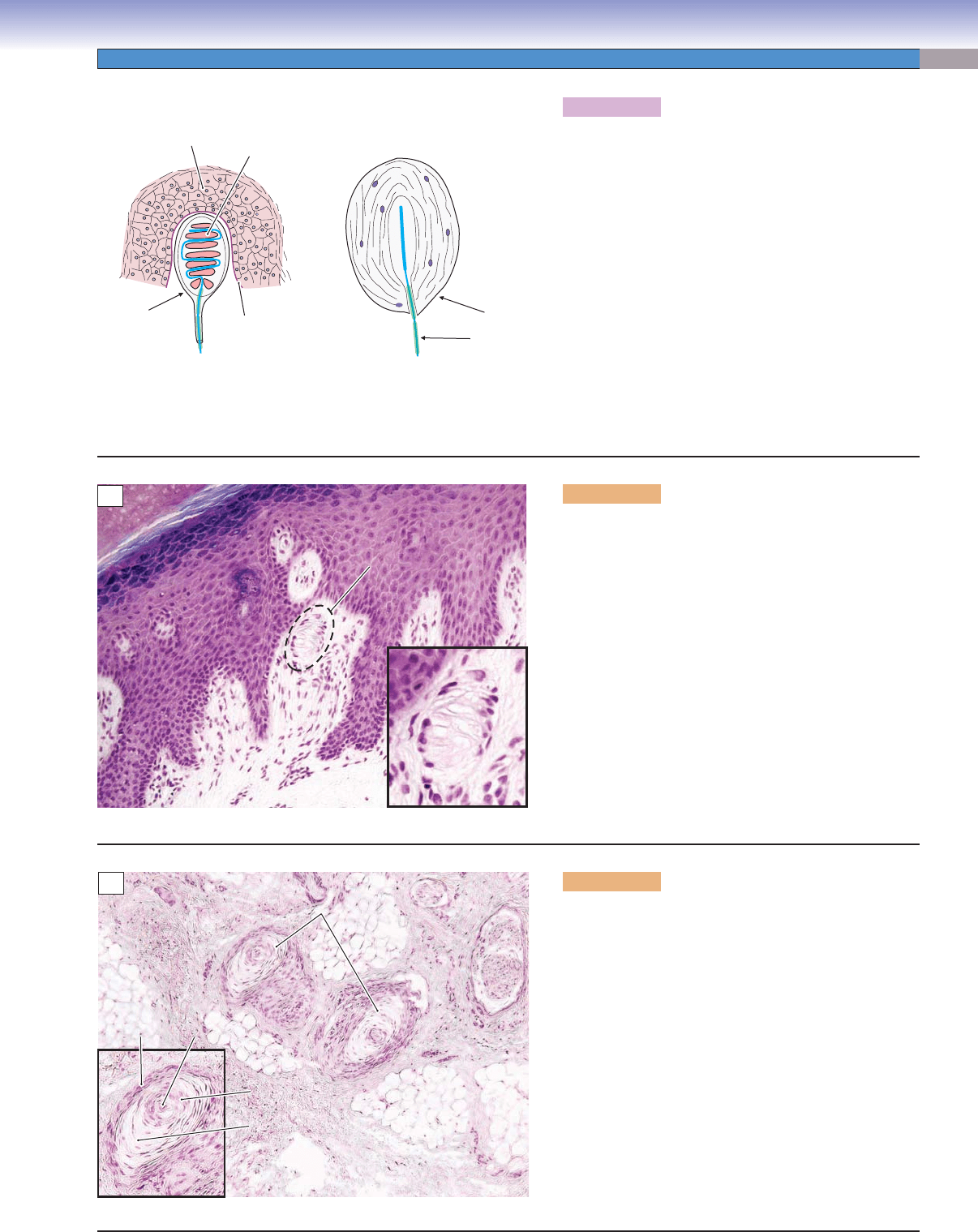
Figure 7-8A. Peripheral sensory receptors.
Axons of the neuron cell bodies in the posterior root
ganglia carry information from sensory receptors in the
skin, muscles, joints, viscera, blood vessels, mesentaries,
and other connective tissues. Sensory receptors associ-
ated with these axons may consist of encapsulated axon
endings, specialized endings around hair follicles, endings
in conjunction with specialized cells (e.g., Merkel cells),
muscle spindles, and specialized regions of axonal mem-
branes termed free nerve endings (see Figs. 6-7A,B and
13-1). Meissner corpuscles and Pacinian corpuscles are
two types of encapsulated endings and are easier to see
using light microscopy than other receptors in the skin.
In these receptors, the axonal membrane is specialized to
change its permeability in response to mechanical pres-
sure (the regions of specialization are indicated by the
thicker blue lines). The connective tissue capsules help to
focus mechanical force on the axonal membrane.
J
. Lynch &T. Yang
Meissner corpuscle
Basement
membrane
Pacinian corpuscle
Epidermis
Capsule
Capsule
Myelin
Schwann
cell
A
Epidermis
Epidermis
Meissner
Meissner
corpusle
corpusle
Epidermis
Meissner
corpusle
Dermis
B
Figure 7-8B. Meissner corpuscle, thick skin of palm.
H&E, 136; inset 360
Meissner corpuscles are encapsulated, quickly adapting
mechanoreceptors that are sensitive to light touch and
low-frequency vibration. They are important for the
sensation of discriminative touch. Meissner corpuscles
are located in the dermal ridges, at the interface of the
dermis and epidermis. Only the nuclei of the capsu-
lar connective tissue cells are visible after H&E stains.
Using special stains, the corpuscle looks like a stack of
pancakes (Schwann cells, Fig. 7-8A) with one or more
axons intertwining among them. The main axons associ-
ated with Meissner corpuscles are large (6–12 μm) and
heavily myelinated, hence their rapid conduction veloci-
ties. Meissner corpuscles are found in all parts of the skin
of the hand and foot, in the lips, and in a number of
other locations but are most concentrated in thick, hair-
less (glabrous) skin.
Pacinian corpuscles
Capsule
Axon
Core
Growth zone
Adipose tissue
Connective tissue
C
Figure 7-8C. Pacinian corpuscle, thick skin of palm.
H&E, 68; inset 105
Pacinian corpuscles are large, encapsulated structures that
detect very light touch and vibration. They are located
primarily in the hypodermis of the palms of the hands
and fi ngers and the soles of the feet but are also found in
other areas of skin as well as in the periostea and mes-
entery. They are much larger than Meissner corpuscles,
sometimes reaching 2 mm in length (note different mag-
nifi cation). Pacinian corpuscles consist of a specialized
zone of axonal membrane that is exquisitely sensitive
to pressure (Fig. 7-8A) surrounded by a layered cellular
structure that consists of a central core immediately sur-
rounding the axon, an intermediate growth zone, and an
outer capsule. Around 60 to 100 layers are formed by
very thin cells that overlap at their edges, giving the Pacin-
ian corpuscle the appearance of an onion when sectioned.
This sample is from the hypodermis of the skin.
CUI_Chap07.indd 125 6/2/2010 6:32:35 PM
126
UNIT 2
■
Basic Tissues
Central Nervous System
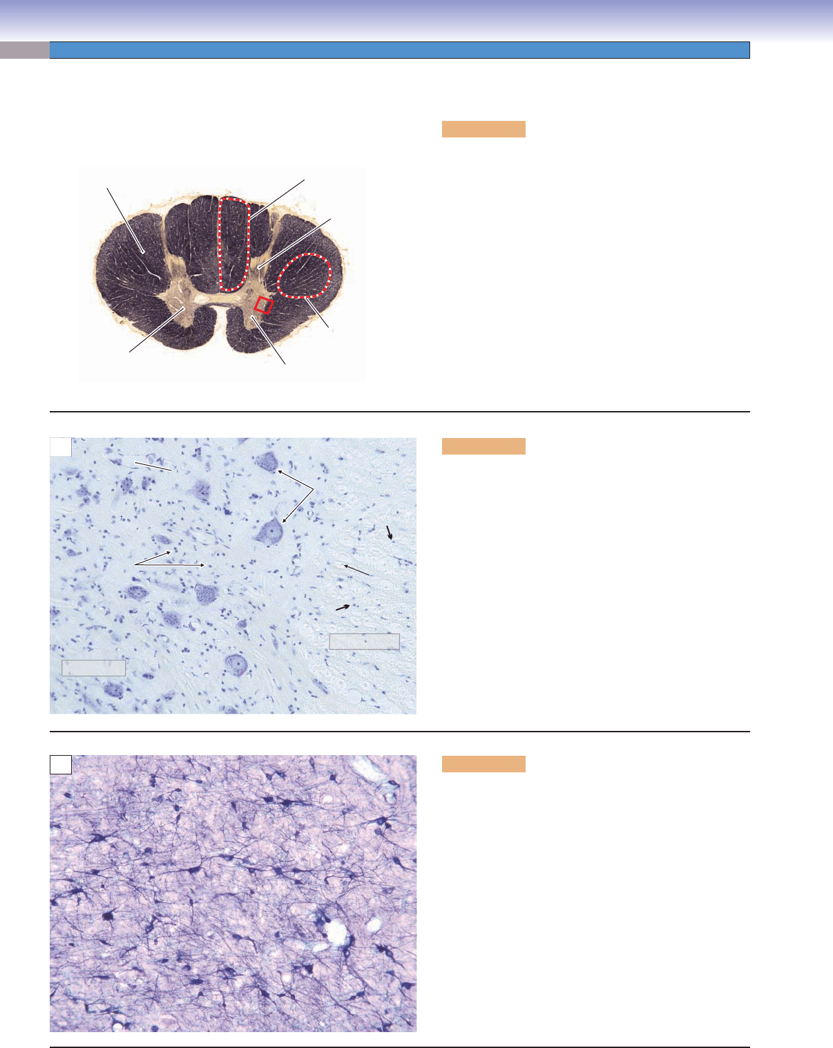
Figure 7-9A. Spinal cord. Myelin stain, 7
In this section through an upper thoracic level of the spinal
cord (Fig. 7-4), the white matter (fi ber pathways) has been
stained dark brown with a myelin stain. The gray matter
is a region of densely packed neuron cell bodies, and this
stain has consequently left the gray matter in the center of
the cord relatively unaffected. The anterior horn of the gray
matter contains motor neurons that innervate muscle fi bers;
the posterior horn contains interneurons in both sensory
and motor pathways. The white matter consists of nerve
fi bers carrying sensory information from receptors in skin
and muscles up to the brain (e.g., the gracile fasciculus) or
nerve fi bers carrying motor information down from the
brain to interneurons and motor neurons in the gray matter
of the spinal cord (e.g., the corticospinal tract). The red rect-
angle indicates the position of the tissue in Figure 7-9B that
has been stained with a Nissl stain for neuron cell bodies.
White matter
(axons)
Gray matter
(neuron cell bodies)
Posterior
Gracile
fasciculus
Posterior
horn
Anterior
horn
Corticospinal
tract
Anterior
A
Gray matter
Glia cell nuclei
Capillary
Axon
White matter
Neuron cell bodies
B
Figure 7-9B. Spinal cord. Nissl stain, 136
Nissl stains, such as thionin or cresyl violet, react with
nucleic acids (RNA, DNA) and, therefore, stain the rough
endoplasmic reticulum, nuclei, and nucleoli of neurons.
This renders the neuron cell bodies visible, along with the
nuclei of glial cells and nuclei of epithelial cells in blood
vessels. The large cell bodies in this section belong to motor
neurons in the anterior horn of the spinal cord (red rect-
angle in Fig. 7-9A). Also visible are the nuclei of glial cells
(astrocytes and oligodendrocytes) in the gray matter on
the left side of the picture and in the white matter on the
right side of the picture (small arrows). It is important
to keep in mind when looking at myelin-stained sections
such as in Figure 7-9A that the light-colored areas, where
nothing seems to be stained, are in fact fi lled with neuron
cell bodies similar to the ones illustrated here.
C
Figure 7-9C. Neurons in the reticular formation of
the brainstem. NADPH histochemical stain, 68
Neurons use a wide variety of neurotransmitters. In
addition to stains that react with structural components
of a nerve cell, it is possible to use histochemical reac-
tions to visualize the presence of particular neurotrans-
mitters. This photomicrograph illustrates an example of
such a reaction, in which only those neurons that generate
the neurotransmitter nitric oxide (NO) are visualized. In
this case, an enzyme necessary for the synthesis of NO,
NADPH diaphorase, was labeled using a blue chroma-
gen (colored substance). The neurons are colored in their
entirety because NO is a novel neurotransmitter that is not
bound in vesicles as are most neurotransmitters, but rather
is synthesized everywhere in the cell and leaks through the
cell membrane when it is released. It often functions to
modulate the action of other neurotransmitters.
CUI_Chap07.indd 126 6/2/2010 6:32:40 PM
CHAPTER 7
■
Nervous Tissue
127
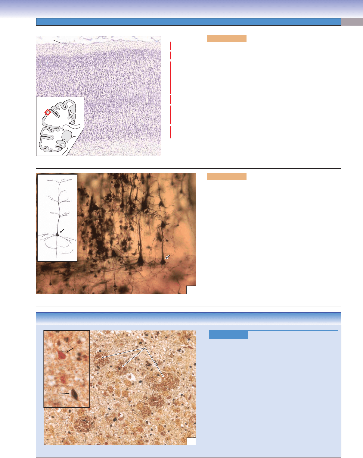
Figure 7-10A. Cerebral cortex. Nissl stain, 24
The cerebral cortex is a layer of densely packed neurons
about 2 mm thick that forms the surface of the hemi-
spheres of the brain. The cortex is organized into layers
(indicated by red lines and Roman numerals) on the basis
of the size, shape, and packing density of the neurons
in different regions of the cortex. For example, layers II
and IV in this photomicrograph consist of small, tightly
packed neurons (mainly granule cells), whereas layers III
and V consist of larger neurons (mostly pyramidal cells).
This example is taken from the association cortex of the
parietal lobe (red box in inset). Layer I consists predomi-
nantly of horizontally running dendrites and axons; the
nuclei in this region belong to glial cells. The white matter
consists of axons entering and leaving this small region of
cortex and connecting it with other cortical regions, with
subcortical structures such as the thalamus and with the
spinal cord.
I
II
Pia
White matter (myelinated axons)
III
IV
V
VI
A
CLINICAL CORRELATION
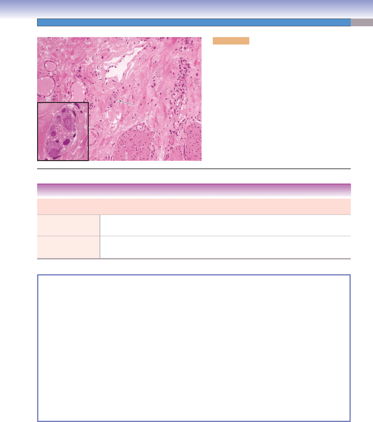
Figure 7-10C.
Alzheimer Disease. Bielschowsky
stain, 272; inset 550
Alzheimer disease is the most common form of
dementia in the elderly (60%–80% of cases). Patients
show progressive memory loss, personality changes,
and cognitive impairments. Pathologically, Alzheimer
disease is characterized by extracellular deposition
of Ab protein (plaques), intracellular neurofi brillary
tangles (inset: N, normal neuron; T, neuron cell body
fi lled with dark-staining tangles), and loss of neurons
and synapses in the cerebral cortex and some subcor-
tical regions. Gross anatomy shows atrophy of the
affected regions. The most prominent theory is that
mutation of genes such as those located on chromo-
some 21 lead to overproduction of Aβ precursor.
There is no cure for Alzheimer disease. Treatments
include pharmaceutical and psychosocial therapies
and supportive caregiving.
Amyloid plaques
Amyloid plaques
Amyloid plaques
N
T
C
S
S
AD
BD
S
S
A
B
Figure 7-10B. Cerebral cortex, pyramidal cells. Golgi
preparation, 136
The most numerous neurons in the cerebral cortex are
pyramidal cells, named for their triangular cell bodies
(singular, soma; plural somata; S in the inset and photo-
micrograph). Pyramidal cells are also characterized by a
long apical dendrite (AD) that extends into layer I and
has many lateral branches; several basal dendrites (BD)
that extend laterally from the base of the soma; and a long
axon (A) that leaves the cortex and extends, in the white
matter, either to some other region of the cortex or to sub-
cortical structures, such as the basal nuclei, brainstem, or
spinal cord. Pyramidal cells are found in layers II through
VI of the cortex but are most obvious in layers III and V.
Somata range in size from 10 to over 50 μm, with the larg-
est located in layer V of primary motor cortex. The sam-
ple shown here is taken from layer III. It includes several
medium pyramidal cells and many small pyramidal cells.
CUI_Chap07.indd 127 6/2/2010 6:32:45 PM
128
UNIT 2
■
Basic Tissues
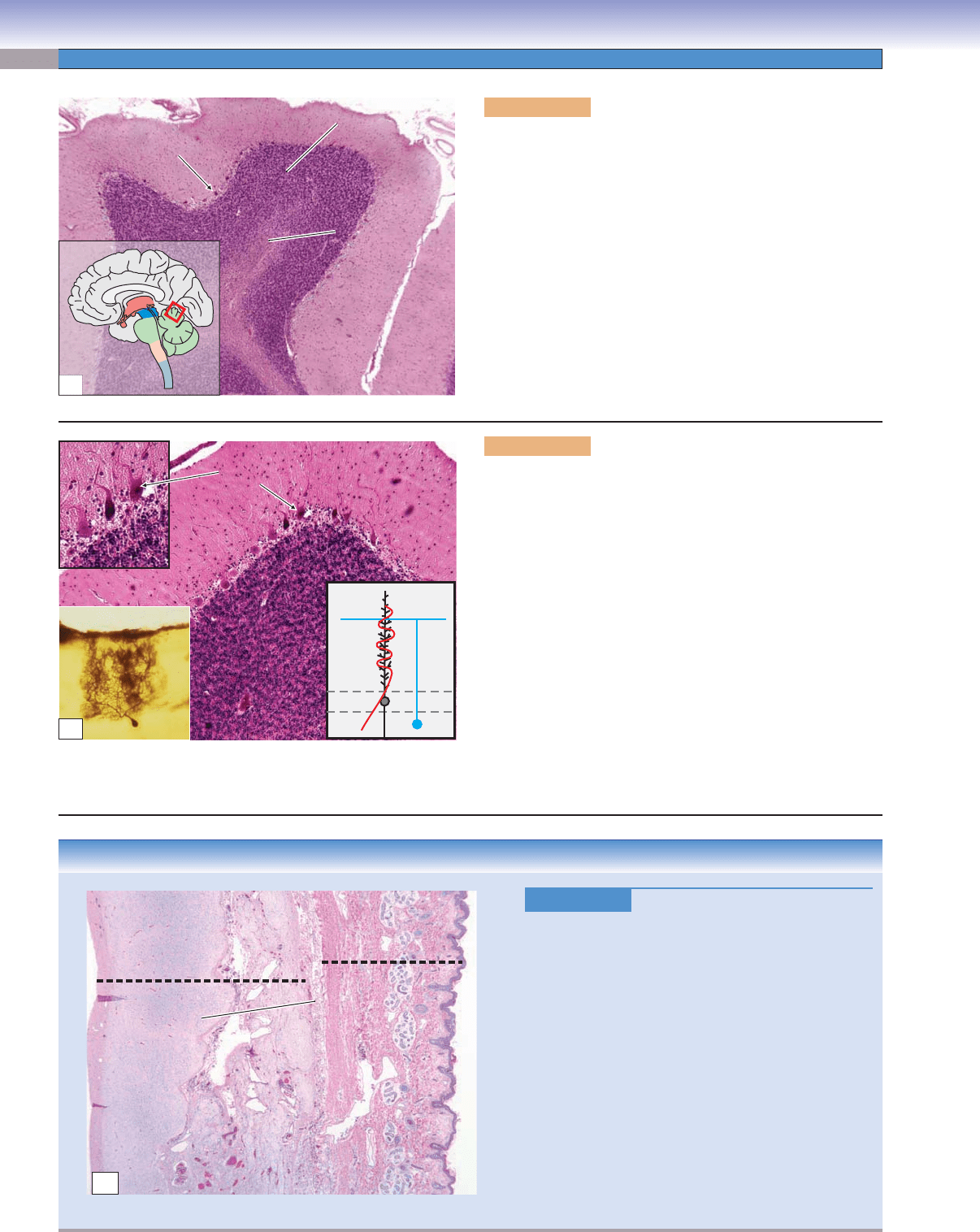
Figure 7-11A. Cerebellar folium. Nissl stain, 34
The cerebellum (green structure in inset) is a large, complex struc-
ture that lies beneath the posterior portion of the cerebral hemi-
spheres. Its name means “little brain.” It is critical for smooth,
coordinated movements and participates, to a lesser extent, in many
other functions. The structural organization of the cerebellum is
similar to that of the cerebrum in that it consists of white matter,
a cortex, and subcortical nuclei. However, the organization of the
cortex is very different from that of the cerebral cortex. There are
only three layers, the granule cell layer, the Purkinje cell layer, and
the molecular layer. The granule cell layer contains an enormous
number of very small, tightly packed cells and their dendrites. The
Purkinje cell layer, at the interface of the granule cell and molecular
layers, is only one cell deep. The molecular layer contains primarily
axons and dendrites, with only very few neuron cell bodies. The
red rectangle indicates the position of the tissue in this fi gure.
Purkinje cell
Purkinje cell
Molecular layer
Molecular layer
Granule cell
Granule cell
layer
layer
White matter
White matter
White matter
Molecular layer
Purkinje cell
Granule cell
layer
A
CLINICAL CORRELATION
Figure 7-11C.
Encephalocele. H&E, 17
In some embryos, the neuroectoderm does not sepa-
rate from the surface ectoderm during early stages of
development. A defect in the calvarium may occur as
the bone of the skull is formed, through which CSF
,
meninges, and brain tissue may protrude. This case
illustrates immature brain tissue and its meningeal
covering, which have herniated through a bony defect
in the occipital bone. These neuroectodermal tissues
are found directly beneath the dense subcutaneous
connective tissue of overlying skin. Encephaloceles
are most frequently found in the occipital region. In
their several variations, they may contain (1) only CSF
and meninges, (2) CSF, meninges, and brain substance
(occipital lobe or cerebellum), or (3) CSF, meninges,
brain, and part of the ventricular system. The more
elaborate the encephalocele, the more debilitating and
diffi cult it is to treat.
Skin
Skin
Skin
Abnormal
nervous tissue
Meninges
C
M
3
P
G
Purkinje cell
Purkinje cell
bodies (P)
bodies (P)
Granule cell
Granule cell
layer (G)
layer (G)
Molecular layer (M)
Molecular layer (M)
1
1
Molecular layer (M)
Granule cell
layer (G)
1
2
Purkinje cell
bodies (P)
B
Figure 7-11B. Cerebellar cortex. Nissl stain, 68, inset 1,
124; inset 2, Golgi, 74
Purkinje cells and granule cells are the most obvious neurons in
the cerebellar cortex. Purkinje cells, among the largest neurons
in the CNS, lie between the granule cell layer and the molecular
layer (inset 1). Purkinje cells have widely branching dendritic
trees that extend through the entire depth of the molecular layer
(inset 2). The dendritic tree of a Purkinje cell is shaped like
a paper fan. The wide part of the “fan” is seen when cutting
across the long axis of a folium (inset 2). The edge of the fan is
seen when cutting parallel to the long axis of the folium (inset 3).
Granule cells (blue, inset 3) send their axons into the molecular
layer in which they divide and run parallel to the long axis of the
folium, making synaptic contact with hundreds or thousands of
Purkinje cell dendrites. Another major element in the basic cer-
ebellar cortex circuitry is the climbing fi ber (red, inset 3). These
axons originate in the inferior olivary nucleus. Each climbing
fi ber encircles the dendrites of a single Purkinje cell. Purkinje cell
axons provide the sole output pathway of the cerebellar cortex.
CUI_Chap07.indd 128 6/2/2010 6:32:51 PM
CHAPTER 7
■
Nervous Tissue
129
CLINICAL CORRELATION
Figure 7-12C.
Meningitis. H&E, 34; inset 147
Meningitis is an infl
ammatory disease of the meninges
that is largely sequestered in the SAS. The infl amma-
tion is usually caused by infection by viruses, bacteria,
or fungal agents. Signs and symptoms include fever,
headache, irritability, photophobia, neck stiffness
(meningismus), vomiting, altered mental status, and
cutaneous hemorrhages (purpura). Complications may
lead to deafness, epilepsy, and hydrocephalus. Patho-
logically, the meningitis affects the pia- arachnoid and
the CSF in the SAS and may extend into the cerebral
ventricles. Characteristic fi ndings include perivascular
cuffs of acute and chronic infl ammatory cells (inset,
from red box), which distend the arachnoid space.
Lumbar puncture (spinal tap) for CSF is an impor-
tant diagnostic tool. Treatment is usually supportive
for viral meningitis and includes antibiotics or other
agents that can cross the blood-brain barrier for bac-
terial or fungal meningitis.
Arachnoid space
Arachnoid space
filled with
filled with
inflammatory cells
inflammatory cells
and proteinaceous fluid
and proteinaceous fluid
Pia mater
Pia mater
Spinal cord
Spinal cord
white matter
white matter
Spinal cord
white matter
Pia mater
Arachnoid
membrane
Arachnoid space
filled with
inflammatory cells
and proteinaceous fluid
C
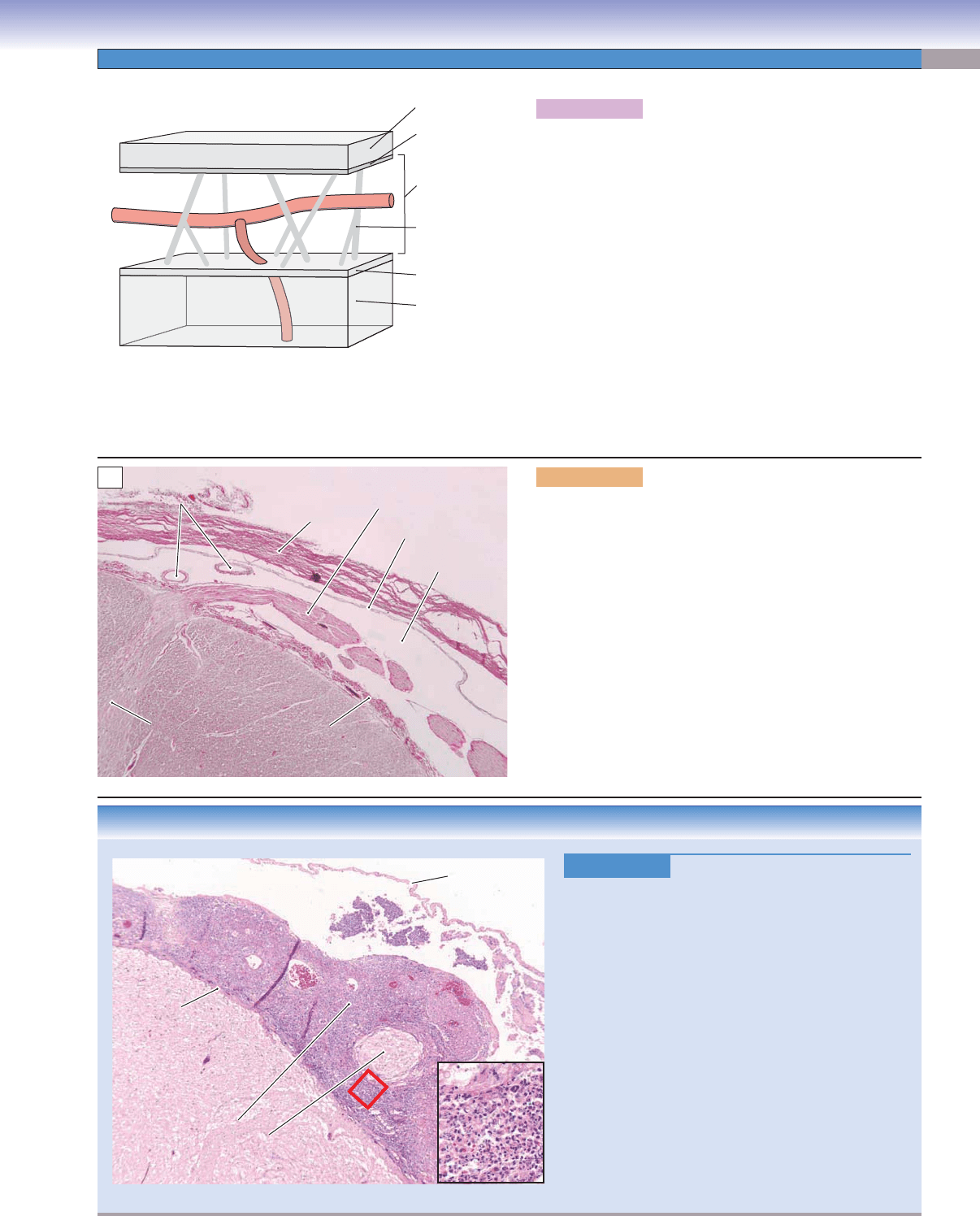
Figure 7-12A. Dura mater, arachnoid, and pia mater.
The leathery outer meningeal layer, the dura mater (or dura),
consists of elongated fi broblasts and large amounts of extracel-
lular collagen. It is tenuously attached to the arachnoid barrier
layer; there is no “subdural space” in the normal state. The dura
is tenaciously attached to the skull at the base of the brain and
at the sutures and is less tightly adherent to the skull in other
regions. The arachnoid barrier layer consist of two to three
layers of cells that are attached to each other by many con-
tinuous tight junctions, hence its “barrier” nature to CSF. The
sub arachnoid space (SAS) is located between the arachnoid
and the pia, contains blood vessels and CSF, and is traversed by
arachnoid trabeculae. The pia mater generally consists of one
to two layers of fl attened fi broblasts that are adherent to the
surface of the brain and spinal cord. Blood vessels located in the
SAS are frequently covered by thin layers of pia. The interface
between the pia and the nervous tissue is characterized by a glial
limiting membrane (glia limitans), as shown in Figure 7-13A.
J. Lynch
Dura mater
Arachnoid
barrier cells
Pia mater
Nervous tissue
(brain or spinal
cord)
Subarachnoid
space
Blood
vessel
Arachnoid
trabeculum
A
Posterior horn
Posterior horn
Spinal cord
Spinal cord
white matter
white matter
Pia
Pia
Blood vessels
Blood vessels
Dura
Epidural space
Spinal cord
white matter
Posterior horn
Blood vessels
Pia
Arachnoid
Subarachnoid
space
Posterior root
of spinal nerve
B
Figure 7-12B. Spinal meninges in the region of the
posterior roots. H&E, 34
At spinal levels, the dura mater forms a tubular sac enclosing
the spinal cord. However, in contrast to the cranial dura, which
adheres to the skull, the spinal dura is separated from the ver-
tebral bodies by an epidural space. Both the spinal and the cra-
nial dura consist of many elongated fi broblasts and abundant
extracellular collagen. The arachnoid barrier cell layer is basi-
cally the same at cranial and spinal levels. In the normal state,
the barrier cell layer is attached to the dura, although only
weakly. There is no naturally occurring subdural space; what
appears to be a space here is a tissue preparation artifact. The
pia mater on the cord is continuous with the epineurium on
the posterior root, the latter representing a part of a peripheral
nerve. The SAS is located between the pia and the arachnoid
and at this point contains the posterior roots.
CUI_Chap07.indd 129 6/2/2010 6:32:56 PM
130
UNIT 2
■
Basic Tissues
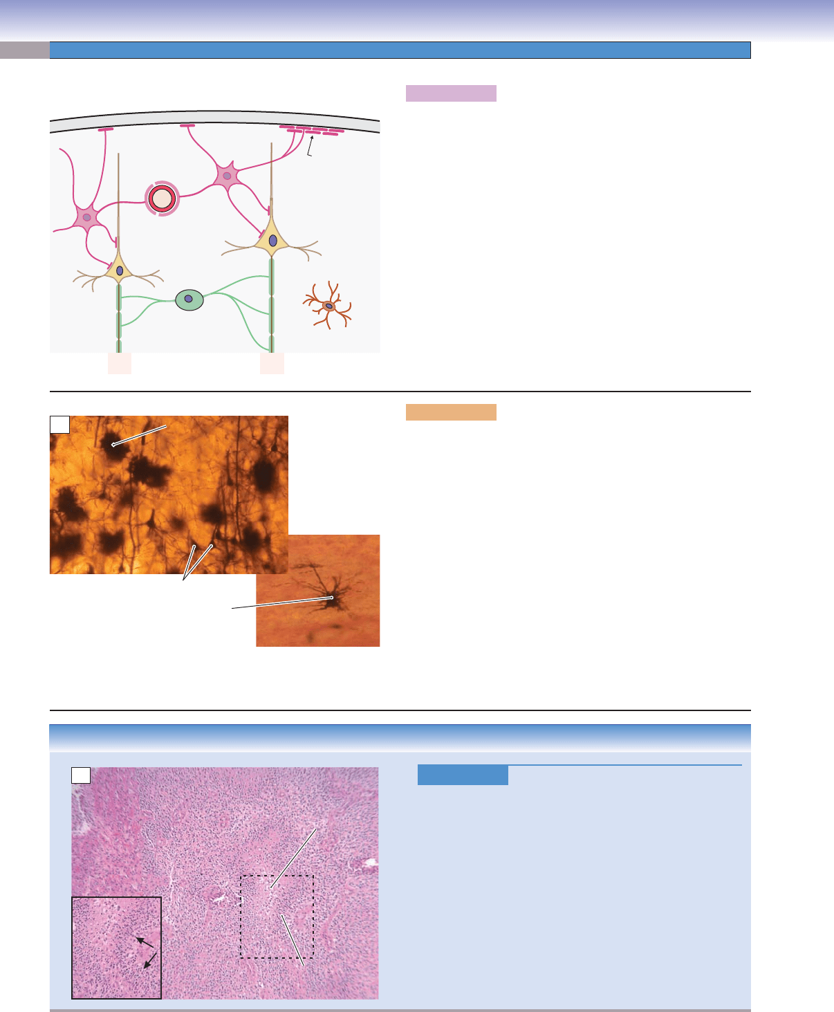
Figure 7-13A. Types of glial cells.
Glial cells (also called neuroglia or glia) are nonneuronal cells that
aid in transferring nutrients from capillaries to neurons, main-
tain the blood-brain barrier, regulate the intercellular environ-
ment, provide myelin insulation for axons, provide mechanical
support to neurons, act as phagocytes to remove pathogens and
dead neurons, play a role in presenting antigens in the immune
system, and perform numerous other functions. Recent evidence
suggests that glial cells even participate in some aspects of synap-
tic transmission. It is thought that there are as many as 10 times
the number of glial cells as neurons in the nervous system. The
major types of CNS glial cells are astrocytes (described below),
oligodendrocytes (which produce myelin), and microglia (which
act as phagocytes and elements of the immune system). Other
glialike cells include radial glia cells and ependymal cells in the
CNS and Schwann cells and satellite cells in the PNS.
J. Lynch
Pia
Astrocyte
Glia limitans
Capillary
Oligodendrocyte
Microglia
Pyramidal
cell
A
Protoplasmic astrocyte
Protoplasmic astrocyte
Protoplasmic astrocyte
Pyramidal cells
Cerebral cortex, layer III
Fibrous astrocyte
in cerebellar white matter
B
Figure 7-13B. Astrocytes. Golgi preparations, upper left,
136; lower right, 204
Astrocytes are found throughout the CNS. Astrocyte processes
called end-feet form contacts with capillaries that help produce
the blood-brain barrier and form contacts on neurons that play a
role in supplying nutrients to these cells. Astrocytes regulate the
ionic composition and pH of the extracellular environment and
secrete various neuroactive substances. Astrocyte end-feet form
the glia limitans, a coating of the inner surface of the pia mater
that surrounds the brain and spinal cord (Fig. 7-13A). Finally,
astrocytes play an important role in neurotransmitter metabo-
lism and in the modulation of synaptic transmission. There are
two types of astrocytes. Protoplasmic astrocytes, in gray matter,
have short, thick processes that are densely clustered and highly
branched, giving them a cloudlike appearance. Fibrous astro-
cytes, in white matter, have long, thin processes with relatively
few branchings. The two types of astrocytes have similar func-
tions but differ in some special properties.
CLINICAL CORRELATION
Figure 7-13C.
Glioblastoma. H&E, 68; inset 84
Glioblastoma (a form of astrocytoma) is a highly malignant
tumor that arises in the brain from neoplastic astrocytes. Sev-
eral features of this tumor help the pathologist arrive at the
diagnosis. In the center of this micrograph, the tumor cells
are necrotic. At the edges of the zone of necrosis, nonnecrotic
tumor cells align themselves in a striking parallel array, like
the pickets of a fence. This confi guration is called palisading
(arrows, inset). Beyond the area of palisading, living tumor
cells commonly surround complex abnormal vascular struc-
tures (glomeruloid vascular structures) that resemble the glom-
eruli of the kidney in their tortuous arrangement of capillar-
ies. Increased mitosis among the viable tumor cells also aids
in the diagnostic process. Increasing the life span of patients
with glioblastoma is currently an area of intensive research.
Live
Live
tumor cells
tumor cells
Zone of
Zone of
palisading
palisading
Necrotic
Necrotic
tissue
tissue
Necrotic
tissue
Necrotic
tissue
Live
tumor cells
Zone of
palisading
Abnormal
Abnormal
capillaries
capillaries
Abnormal
capillaries
C
CUI_Chap07.indd 130 6/2/2010 6:32:59 PM
CHAPTER 7
■
Nervous Tissue
131
J. Lynch
1
2
3
4
5
6
7
8
9
10
11
12
Preganglionic cholinergic fibers
Postganglionic cholinergic fibers
Postganglionic adrenergic fibers
Eye
Lacrimal gland
Submandibular gland
Sublingual gland
Parotid gland
Oral mucosa
NOTE: In addition to the other
all chain ganglia include
connections diagramed above,
postganglionic fibers that go to
the skin (e.g., sweat glands)
and to blood vessels in the
these three ganglia.
body wall, as indicated in
Ciliary ganglion
Pterygopalatine ganglion
Submandibular ganglion
Otic ganglion
III
VII
X
IX
Larynx,
trachea,
bronchi
Heart
Liver
Pancreas
Intestines
Myenteric plexus,
submucosal plexus
Pelvic
splanchnic
nerves
Stomach
Kidney
Bladder
Celiac
ganglion
Sympathetic
chain ganglia
Inferior
mesenteric
ganglion
Adrenal
Superior
mesenteric
ganglion
Cervical
ganglia
C
T
L
S
Genitalia
Sympathetic Division Parasympathetic Division
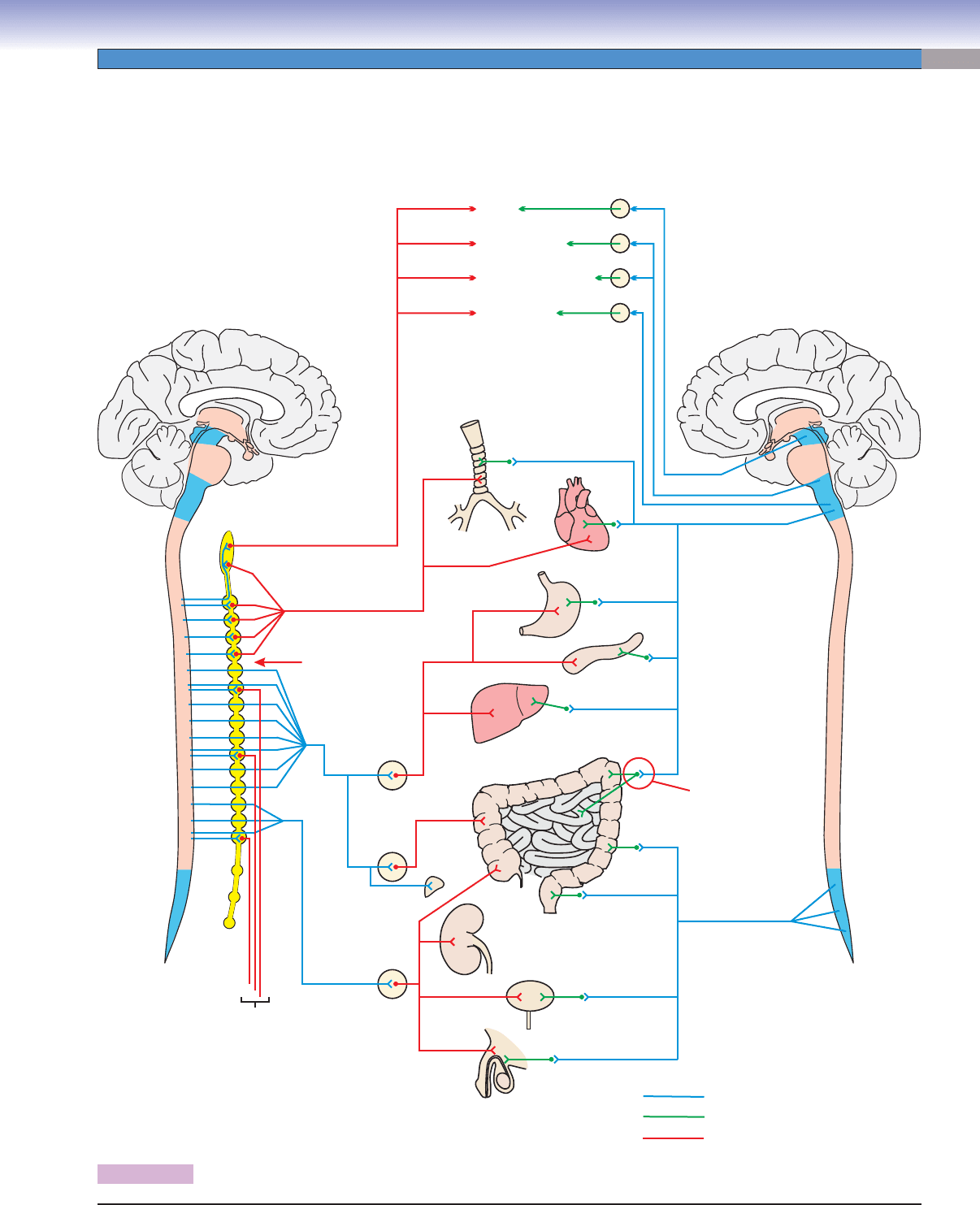
Figure 7-14. Overview of the autonomic nervous system (continues on page 131).
Autonomic Nervous System
CUI_Chap07.indd 131 6/2/2010 6:33:01 PM
132
UNIT 2
■
Basic Tissues
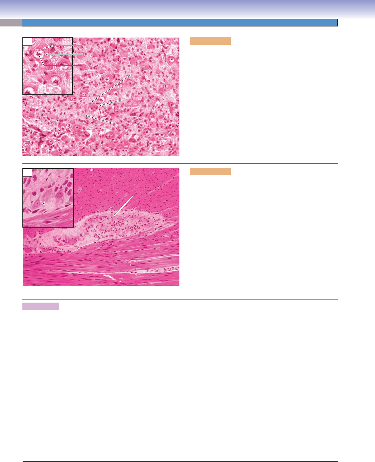
Figure 7-15A. Sympathetic ganglion. H&E, 272; inset
520
This section from a sympathetic chain ganglion shows small
to medium-sized visceromotor neuron cell bodies that give rise
to postganglionic axons. These neurons receive synapses from
preganglionic sympathetic axons that originate in the lateral horn
of the thoracic and upper lumbar spinal cord. The preganglionic
axons are myelinated; the postganglionic axons are unmyeli-
nated. These motor neurons are multipolar (in contrast to the
unipolar sensory neurons in the posterior root ganglia), although
the dendrites are not visible in this H&E stain. The sizes of the
cell bodies are more uniform than in the sensory ganglia, and
the cell bodies and axons are distributed more evenly across the
ganglia rather than being grouped into clumps as in the sensory
ganglia. The satellite cells are not distributed so evenly around
the neuron cell bodies as they are in the sensory ganglia.
Nucleolus
Nucleolus
Nucleus
Nucleus
Cytoplasm
Cytoplasm
Axons
Axons
Satellite
Satellite
cell nucleus
cell nucleus
Sympathetic
Sympathetic
motor neuron
motor neuron
Sympathetic
motor neuron
Axons
Satellite
cell nucleus
Cytoplasm
Nucleus
Nucleolus
A
Longitudinal smooth
Longitudinal smooth
muscle layer
muscle layer
Myenteric plexus
Myenteric plexus
Circular smooth
Circular smooth
muscle layer
muscle layer
Circular smooth
muscle layer
Myenteric plexus
P
P
S
S
Longitudinal smooth
muscle layer
P
S
B
Figure 7-15B. Myenteric plexus (Auerbach plexus).
H&E, 136; inset 300
The enteric division lacks the discrete, encapsulated ganglia that
characterize the sympathetic division. Its visceromotor neurons
are distributed in a network of plexuses that are distributed
within the walls of the gastrointestinal tract. Most neurons in
the enteric division are found in the myenteric and submucosal
plexuses. The myenteric plexuses lie between the circular smooth
muscle layer and the longitudinal smooth muscle layer of the
intestine (see Chapter 15 “Digestive Tract”; see also Chapter 6,
“Muscle,” Fig. 6-10B). These plexuses are clusters of parasym-
pathetic postganglionic motor neurons; sensory neurons, which
receive input from chemoreceptors and mechanoreceptors in
the intestinal wall; and local circuit neurons (interneurons).
Interneurons can process neural signals within a plexus and can
also mediate the coordination of multiple plexuses. (Inset: P,
multipolar postganglionic motor neuron; S, satellite cell.)
Figure 7-14. Overview of the autonomic nervous system (Continued).
The ANS is composed of three divisions: sympathetic, parasympathetic, and enteric. The sympathetic and parasympathetic divi-
sions function under direct CNS control; the enteric division functions somewhat more independently. The sympathetic division
includes preganglionic neurons with cell bodies in the lateral horn of the thoracic and upper lumbar spinal cord (Fig. 7-4). Some of
these neurons synapse on postganglionic neurons in the sympathetic chain ganglia (Figs. 7-4 and 7-15A); others continue past these
ganglia and synapse in prevertebral sympathetic ganglia (e.g., celiac ganglion, mesenteric ganglia) near the organs to be innervated.
Postganglionic neurons send axons to internal organs, glands, and blood vessels. Effects of sympathetic activity include increas-
ing cardiac output, blood pressure, and bronchial diameter; decreasing gut peristalsis; and, in general, preparing the individual for
strenuous activity, sometimes called the “fi ght-or-fl ight” reaction. The parasympathetic division has a markedly different organiza-
tion. Preganglionic fi bers originate in brainstem nuclei associated with cranial nerves III, VII, IX, and X and in the sacral parasym-
pathetic nucleus (which occupies a position in the sacral spinal cord similar to that of the lateral horn in the thoracic cord). The
parasympathetic preganglionic fi bers that supply the head region synapse in discrete ganglia (Fig. 7-14) and the postganglionic fi bers
end in glands and smooth muscle. By contrast, parasympathetic preganglionic fi bers that travel in cranial nerve X (vagus nerve)
and the pelvic splanchnic nerves send signals to the viscera and blood vessels within the body cavity. These fi bers do not synapse in
discrete ganglia but rather in small plexuses of postganglionic cell bodies that lie in or adjacent to the walls of their target organs
(Figs. 7-15B and 7-16A). The general effect of parasympathetic activity is the opposite of sympathetic activity and tends to return
the internal organs and cardiovascular system to a baseline level of function. The enteric division consists of a vast number of neu-
rons arranged in a network of plexuses in the walls of the gut. Some of these plexuses are shared with the parasympathetic division.
The activity of the enteric division is modulated in a general way by the sympathetic and parasympathetic divisions, but it is able to
act independently and refl exively to move boluses of food substances through the gastrointestinal tract by peristaltic action and to
control absorption, local blood fl ow, and secretion in response to the chemical composition of the bolus.
CUI_Chap07.indd 132 6/2/2010 6:33:02 PM
CHAPTER 7
■
Nervous Tissue
133
Figure 7-16. Submucosal plexus (of Meissner). H&E,
136; inset 453
The submucosal plexuses (of Meissner) are located in the
submucosal layer of the intestine (see Chapter 15, “Diges-
tive Tract”). These plexuses are similar to the myen-
teric plexuses in that they are unencapsulated clusters
of parasympathetic postganglionic motor neurons; sen-
sory neurons, which receive input from chemoreceptors
and mechanoreceptors in the intestinal wall; and local
circuit neurons (interneurons). The postganglionic motor
neurons may innervate smooth muscle to either increase
or decrease muscle activity and may also innervate secre-
tory cells in the walls of the intestine.
Submucosal
plexus
Submucosal
connective tissue
SYNOPSIS 7-1 Pathological and Clinical Terms for the Nervous System
Ataxia ■ : Inability to coordinate the muscles properly in the execution of a voluntary movement.
Gliosis
■ : The multiplication of astrocytes as a response to injury in the brain, as exemplifi ed by the feltwork of astrocytic
cell bodies and processes in a demyelinated plaque of MS.
Glomeruloid vascular structures
■ : Complex arrays of capillaries that resemble the glomeruli of a kidney, are another
hallmark of the glioblastoma.
Neurofi brillary tangle
■ : The helical arrangement of abnormally phosphorylated neurofi laments found in many hippocampal
and cortical neurons of an Alzheimer patient.
Neuropathy
■ : A disease involving the cranial or spinal nerves.
Onion bulb
■ : After repeated cycles of demyelination and remyelination, thin layers of Schwann cell cytoplasm form
concentric circles around a central axon. The appearance of the structure resembles a cross section of an onion bulb and
its nested leaves.
Palisading
■ : The alignment of viable tumor cells at the edge of a necrotic focus in glioblastoma, a diagnostic hallmark of
this grade 4 (most malignant) astrocytoma, a family of glial tumors derived from the astrocyte.
Plaque
■ : This word indicates a lesion, in several pathological contexts. (1) An atherosclerotic plaque is the hard, calcifi ed
buildup of fatty material in large arteries, such as the coronary arteries or aorta. (2) A neuritic plaque is the extracellular
knot of phosphorylated axons and dendrites, often with a central deposit of amyloid protein, found in great numbers
in brains of patients with Alzheimer disease. (3) In MS, a demyelinated plaque is an irregular zone of axons, often in a
periventricular location, that have lost their sheaths of myelin.
Type of Ganglion Cell Body
Arrangement
Cell Body
Characteristics
Satellite Cells Synapses in
Ganglion
Posterior root
(sensory)
Arranged into groups
within ganglion, cell
bodies variable in size
Round cell body,
central nucleus
Complete capsule of
satellite cells
No synapses
Autonomic
( visceromotor)
Evenly distributed
within ganglion,
uniform in size
Multipolar cell body,
eccentric nucleus
Incomplete capsule of
satellite cells
Numerous synaptic
contacts
TABLE 7-1 Comparison of Posterior Root and Autonomic Ganglia
CUI_Chap07.indd 133 6/2/2010 6:33:05 PM
134
8
Blood and
Hemopoiesis
Peripheral Blood Cells
Introduction and Key Concepts for Peripheral Blood Cells
Figure 8-1 Overview of Peripheral Blood Cell Types
Erythrocytes and Platelets
Figure 8-2A Erythrocytes (Red Blood Cells)
Figure 8-2B Platelets (Thrombocytes)
Figure 8-2C Clinical Correlation: Sickle Cell Anemia
Figure 8-3A Erythrocytes, Small Artery
Figure 8-3B Erythrocytes and a Platelet in a Small Blood Vessel
Leukocytes: Agranulocytes
Figure 8-4A Lymphocytes
Figure 8-4B Monocytes
Figure 8-4C Clinical Correlation: Chronic Lymphocytic Leukemia
Leukocytes: Granulocytes
Figure 8-5A,B Neutrophils
Figure 8-6 Neutrophil Phagocytosis
Figure 8-7A Eosinophil
Figure 8-7B Basophil
Synopsis 8-1 Life Spans, Counts, and Sizes of Blood Cells
Figure 8-8 Eosinophil in Connective Tissue
Table 8-1 Leukocytes
Hemopoiesis
Introduction and Key Concepts for Hemopoiesis
Figure 8-9A A Representation of Erythropoiesis
Figure 8-9B Thrombopoiesis
CUI_Chap08.indd 134 6/17/2010 10:19:31 AM