Cui Dongmei. Atlas of Histology: with functional and clinical correlations. 1st ed
Подождите немного. Документ загружается.

CHAPTER 5
■
Cartilage and Bone
95
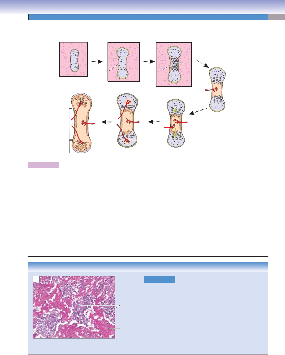
Figure 5-13A. A representation of the development of a long bone.
Most long bones are formed by endochondral ossifi cation, a process of bone formation involving hyaline cartilage serving as a cartilage
model, cartilage proliferation and calcifi cation, and gradual replacement by bone. Long bone formation includes the following steps: (1)
Cartilage model: A small piece of hyaline cartilage is formed by mesenchymal tissue, and the outer part of this tissue condenses to form
a perichondrium. (2) Developing cartilage model: Cartilage assumes the shape for the future bone. (3) Formation of bone collar: As the
cartilage proliferates, the perichondrium in the middle shaft region transforms into periosteum. Osteoprogenitor cells in the periosteum
differentiate into osteoblasts, which start to form the bone collar (periosteal bone) by intramembranous ossifi cation. (4) Formation of
primary ossifi cation centers: The cartilage plate (epiphyseal plate) continues to proliferate and then calcify (see Fig. 5-12A,B). The bone
collar (contains bone matrix, osteoblasts, and osteoclasts) triggers blood vessels to invade and create a primary marrow cavity. (5) For-
mation of bony trabeculae: Osteoprogenitor cells in the periosteum migrate with blood vessels into the region of the calcifi ed cartilage.
These cells become osteoblasts and begin to deposit osteoid (prebone) on the surface of the calcifi ed cartilage matrix. At the same time,
osteoclasts remove dead chondrocytes and extra calcifi ed cartilage matrix, thereby producing bony trabeculae. (6) Formation of second-
ary ossifi cation centers: A similar bone ossifi cation takes place at the distal ends of long bones (epiphyses) called secondary ossifi cation
centers. (7) Continuation of primary and secondary ossifi cation: Repetition of the endochondral ossifi cation process results in more
bone being produced and more cartilage being absorbed in both primary and secondary ossifi cation centers. Finally, the cartilage in the
epiphyseal plates disappears, and the primary ossifi cation center meets the secondary ossifi cation center at about age 20 in humans.
D. Cui
D. Cui
Blood vessel
Bony
trabeculae
Secondary
ossification
center
Perichondrium
Perichondrium
Perichondrium
Periosteum
Periosteum
Periosteum
Bone
collar
Diaphysis
Epiphysis
Primary
ossification
center
1. Cartilage model
2. Developing
cartilage model
3. Formation
of bone collar
4. Formation of
primary ossification
center
7. Continuous primary and
secondary ossification
6. Formation of the
secondary ossification center
5. Formation of
bony trabeculae
A
CLINICAL CORRELATION
Figure 5-13B.
Osteosarcoma.
Osteosarcoma, also known as osteogenic sarcoma, is the most
common primary malignant neoplasm of bone and occurs most
commonly in the second decade of life. Conventional osteosarcoma
tends to affect the long bones, including the distal femur, proxi-
mal tibia, and proximal humerus, and is most often a disease of
the metaphysis (Fig. 5-8). Clinically, patients may experience pain,
decreased range of motion, edema, and localized warmth. Histolog-
ically, the tumor cells tend to be pleomorphic with a variety of sizes
and shapes. Central to the diagnosis of osteosarcoma is the presence
of osteoid (prebone) produced by the malignant cells (tumor cells).
Osteoid is a dense, pink, amorphous material. Conventional osteo-
sarcoma is an aggressive tumor and preferentially metastasizes to
the lungs. Treatment involves surgery and chemotherapy.
B
Osteoid
Tumor cells
CUI_Chap05.indd 95 6/2/2010 6:30:31 PM
96
UNIT 2
■
Basic Tissues
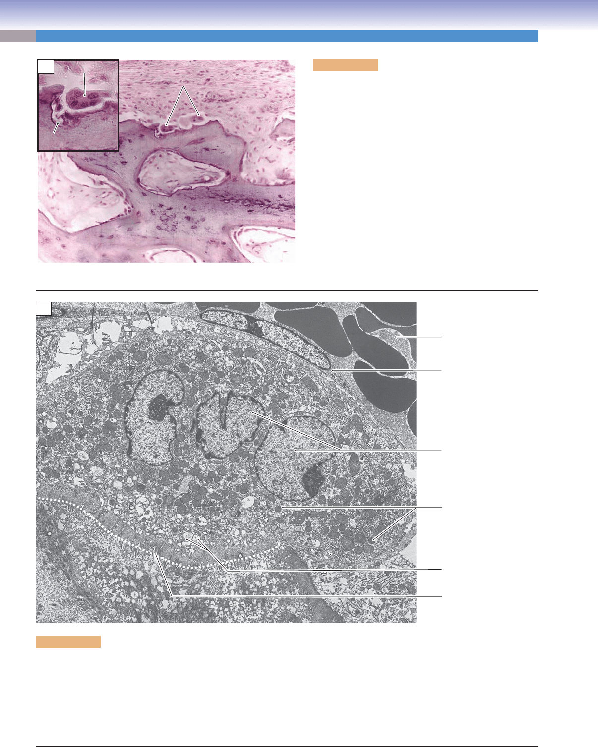
Figure 5-14A. Bone remodeling, nasal. H&E, 136;
inset 363
Bone remodeling is necessary during bone formation in
order to mold the bone into a proper shape to carry out its
function. Remodeling usually occurs on the surface of the
bone where osteoblasts and osteoclasts play different roles.
In order to achieve a certain shape, bone matrix is continu-
ally being deposited by osteoblasts in one region, and, at
the same time, bone matrix is being absorbed by osteoclasts
in another area. Osteoclasts are large, multinucleated cells,
which originate from monocytes and act as phagocytes. They
often sit in the Howship lacunae (eroded grooves produced
by ongoing reabsorption) on the bone surface. Osteoclasts
are under the infl uence of the hormone calcitonin, which is
synthesized by the thyroid gland, and parathyroid hormone
produced by the parathyroid gland. Calcitonin directly
inhibits osteoclast activity and reduces bone reabsorption.
Parathyroid hormone indirectly increases osteoclast activity
and increases bone reabsorption.
Bone matrix
Bone matrix
Bone matrix
Howship
Howship
lacuna
lacuna
Howship
lacuna
Osteoclasts
Osteoclasts
Osteoclasts
Osteoclast
Osteoclast
Osteoclast
A
Figure 5-14B. Osteoclast. EM, 14,000
Osteoclasts are large, multinucleated cells derived from cells seen in circulating blood as monocytes, which are derived, in turn, from
progenitor cells in the bone marrow. Key features in identifying an osteoclast are multiple nuclei, abundance of mitochondria in the
cytoplasm, and intimate attachment to the surface of bone matrix. The mitochondria provide the energy for pumping protons into
the space adjacent to the bone matrix. The cytoplasm near the matrix contains lysosomes, the acid hydrolases of which are secreted
into the space adjacent to the bone matrix. This area of the cytoplasm also contains numerous electron lucent vacuoles that prob-
ably refl ect endocytosis of degraded matrix components. In an active osteoclast, the plasmalemma in the central part of the interface
between the cell and the matrix is highly folded into a ruffl ed border, a structure that is not discernible in this electron micrograph.
B
Euchromatin
Lysosomes
Vacuoles
Howship lacuna
Endothelial cell
Lumen of venule
Bone matrix
Bone matrix
Bone matrix
CUI_Chap05.indd 96 6/2/2010 6:30:32 PM

CHAPTER 5
■
Cartilage and Bone
97
Types of Bone Gross Appearance
(Shape)
Characteristics Main Locations Main Functions
Classifi cation Based on Gross Appearance
Compact bone Uniform; no trabeculae and
spicules
Higher density; lamellae
arranged in circular pattern
Outer portion of
the bone (cortical
bone)
Protection and support
Cancellous
(spongy) bone
Irregular shape; trabeculae
and spicules present;
surrounded by the bone
marrow cavities
Lower density; lamellae
arranged in parallel pattern
Inner core of the
bone (medullary
bone)
Support; blood cell
production
Classifi cation Based on Shape
Long bone Longer than it is wide Consists of diaphysis (long
shaft) and two epiphyses at
the ends
Limbs and fi ngers Support and movement
Short bone Short, cube shaped A thin layer of compact
bone outside and thick
cancellous bone inside
Wrist and ankle
bones
Movement
Flat bone Flat, thin Two parallel layers of
compact bone separated by
a layer of cancellous bone
Many bones of the
skull, ribs, scapulae
Support; protection
of brain and other
soft tissues; blood cell
production
Irregular bone Irregular shape Consists of thin layer of
compact bone outside and
cancellous bone inside
Vertebrae and
bones of the pelvis
Support; protection
of the spinal cord and
pelvic viscera; blood
cell production
Classifi cation Based on Microscopic Observation
Primary bone
(immature
bone)
Irregular arrangement Lamellae without organized
pattern; not heavily
mineralized
Developing fetus Bone development
Secondary bone
(mature bone)
Regular arrangement Well-organized lamellar
pattern; heavily mineralized
Adults Protection and support
TABLE 5-2 Bone
SYNOPSIS 5-4 Pathological and Clinical Terms for Cartilage and Bone
Eburnation ■ : In osteoarthritis, the loss of the articular cartilage results in the exposure of the subchondral bone, which
becomes worn and polished (Fig. 5-4B).
Fibrillation
■ : Early degenerative change in the process of osteoarthritis by which the articular cartilage becomes worn and
produces a papillary appearance; fragments of degenerated cartilage may be released into the joint space (Fig. 5-4B).
Neoplasm
■ : Abnormal tissue arising from a single aberrant cell; neoplasms may be benign or malignant. Malignant
neoplasms are capable of destructive growth and metastasis (Fig. 5-13B).
Achondroplasia
■ : An autosomal-dominant genetic disorder that causes dwarfi sm. The fi broblast growth factor receptor
gene 3 (FGFR3) is affected, resulting in abnormal cartilage formation and short stature.
Osteoporosis
■ : A bone disease characterized by reduced bone mineral density, thinned bone cortex, and trabeculae. It causes
an increased risk of fracture, especially in postmenopausal women.
Osteomalacia
■ : A bone condition caused by impaired mineralization. It causes rickets in children and bone softening in
adults. Vitamin D defi ciency and insuffi cient Ca
++
ions are the most common causes of the condition.
Paget disease
■ : A chronic disorder characterized by excessive breakdown and formation of bone tissue that typically results
in enlarged and deformed bones. The blood alkaline phosphatase level in patients is usually above normal.
Parosteal osteosarcoma
■ : A malignant bone tumor, usually occurring on the surface of the metaphysis of a long bone
(Fig. 5-8).
CUI_Chap05.indd 97 6/2/2010 6:30:36 PM
98
Introduction and Key Concepts for Muscle
Figure 6-1 Overview of Muscle Types
Skeletal Muscle
Figure 6-2 Organization of Skeletal Muscle
Figure 6-3A Longitudinal Section of Striated Muscle
Figure 6-3B Transverse Section of Skeletal Muscle (Tongue)
Figure 6-3C Clinical Correlation: Muscular Dystrophy
Figure 6-4A Skeletal Muscle, Striations
Figure 6-4B Skeletal Muscle—Sarcomeres, Myofi laments
Figure 6-4C Muscle Contraction
Figure 6-5 Muscle Contraction: Transverse Tubule System and
Sarcoplasmic Reticulum
Figure 6-6A Motor Endplates on Skeletal Muscle
Figure 6-6B Neuromuscular Junction
Figure 6-6C Clinical Correlation: Myasthenia Gravis
Figure 6-7A,B Muscle Spindles
Cardiac Muscle
Figure 6-8A Organization of Cardiac Muscle—A Branching Network of Interconnected
Muscle Cells
Figure 6-8B Cardiac Muscle, Longitudinal Section
Figure 6-8C Cardiac Muscle, Transverse Section
Figure 6-9 Characteristics of Cardiac Muscle
Muscle
6
CUI_Chap06.indd 98 6/2/2010 4:08:26 PM

CHAPTER 6
■
Muscle
99
Smooth Muscle
Figure 6-10A A Representation of the Organization and Characteristics of Smooth Muscle
Figure 6-10B Smooth Muscle, Duodenum
Figure 6-11A,B Smooth Muscle—Uterus and Bronchiole
Figure 6-11C Clinical Correlation: Chronic Asthma
Figure 6-12 Transverse Section of the Smooth Muscle of the Trachea
Figure 6-13A Smooth Muscle of a Medium Artery
Figure 6-13B Schematic Diagram of the Contractile Mechanism of Smooth Muscle
Table 6-1 Muscle Characteristics
Synopsis 6-1 Pathological and Histological Terms for Muscle
Introduction and Key
Concepts for Muscle
The contraction of muscle tissue is the only way in which we
can interact with our surroundings and is essential to maintain-
ing life itself. There are three general types of muscles: skel-
etal, cardiac, and smooth. The voluntary contraction of skeletal
muscle allows us to move our limbs, fi ngers, and toes; to turn
our head and move our eyes; and to talk. Its name comes from
the fact that most skeletal muscle attaches to bones of the skel-
eton and functions to move the skeleton. However, exceptions
include the extraocular muscles, the tongue, and a few others.
The continuous, rhythmic contraction of cardiac muscle pumps
blood through our bodies, without ceasing, for our whole life-
time. Cardiac muscle contraction is involuntary, in contrast to
that of skeletal muscle, although its frequency of contraction is
modulated by the autonomic nervous system and by hormones
and neurotransmitters in the blood. Smooth muscle is the most
diverse type of muscle. It occurs in different subtypes in differ-
ent organs and is essential for many involuntary physiological
functions, which include regulating blood fl ow and blood pres-
sure, aiding in the digestion of food, moving food through the
digestive system, regulating air fl ow during respiration, control-
ling the diameter of the pupil in the eye, expelling the baby
during childbirth, and others.
Skeletal Muscle
A single skeletal muscle, such as the biceps, is composed of numer-
ous bundles of muscle fi bers called fascicles. The muscle as a
whole is surrounded by a sheet of dense connective tissues, called
the epimysium. Each fascicle is surrounded by a sheet of moder-
ately dense connective tissues, called the perimysium, and each
individual muscle fi ber (muscle cell) in a fascicle is surrounded by
a delicate collagen network, called the endomysium (Fig. 6-2). A
skeletal muscle fi ber is a long (as long as 10 cm in some muscles),
thin (10–100 μm), tubular structure that contains many nuclei
arranged in the cytoplasm (called sarcoplasm) just under the cell
membrane (called sarcolemma). (Many words relating to muscle
are derived from the Greek word sarx, meaning, “fl esh.”) A single
muscle fi ber contains many individual myofi brils, tiny bundles of
contractile proteins (Fig. 6-2).
CONTRACTION of skeletal muscle is voluntary. Skeletal
muscle is characterized by a striped appearance when viewed
at higher powers in light microscopy. This pattern of stripes
(called striations), at right angles to the long axis of the muscle,
is more obvious when viewed using polarized light and is strik-
ing in electron micrographs (Fig. 6-4A,B). The striations refl ect
a repeating pattern of contractile elements called sarcomeres.
Each sarcomere is made up of an orderly array of actin and
myosin myofi laments (Fig. 6-4B). Each myofi lament consists
of a bundle of actin or myosin molecules together with some
additional accessory molecules. Sudden, all-or-none contraction
occurs in a skeletal muscle fi ber when a motor neuron action
potential (see Chapter 7, “Nervous Tissue”) releases acetylcho-
line at the neuromuscular junction (Fig. 6-6A,B). This causes
a similar action potential to travel along the sarcolemma, trig-
gering the release of calcium ions (Ca
++
) into the intracellular
space and initiating a complex interaction between the actin and
the myosin myofi laments (Fig. 6-4C) to produce shortening of
the fi ber. The necessary Ca
++
is stored within the muscle fi ber
in a modifi ed endoplasmic reticulum called the sarcoplasmic
reticulum (Fig. 6-5). Calcium channels in the terminal cisterns
of the sarcoplasmic reticulum open when the electrical action
potential that is carried along the sarcolemma travels into the
interior of the cell via the transverse tubule system. This system
consists of many tubular invaginations of the sarcolemma that
lie between pairs of terminal cisterns and encircle each myofi -
bril, forming triads. In general, skeletal muscle is specialized for
rapid contraction under neural control. Although each skeletal
muscle fi ber contracts at its maximum whenever it contracts,
variations of overall force of muscle contraction are achieved
by recruiting a greater or lesser number of muscle fi bers at any
given moment.
Cardiac Muscle
The muscle of the heart is similar to skeletal muscle in that
it is striated and the fi bers contain sarcomeres made up of
arrays of actin and myosin fi laments. However, cardiac muscle
cells are much shorter than those of skeletal muscle and typi-
cally split into two or more branches, which join end to end
(or, anastomose) with other cells at intercalated disks (Fig. 6-8B).
A transverse tubule system is present in cardiac muscle, but the
sarcoplasmic reticulum is not as highly developed as in skeletal
muscle. Each cardiac muscle fi ber does not receive direct inner-
vation as skeletal muscle fi bers do. Excitation spreads from
fi ber to fi ber via gap junctions. Contraction is also controlled
by a system of pacemaker nodes and Purkinje cells.
Smooth Muscle
Muscle fi bers that do not display striations are termed smooth
muscle. This type of muscle also contracts by means of a
CUI_Chap06.indd 99 6/2/2010 4:08:31 PM

100
UNIT 2
■
Basic Tissues
Ca
++
-mediated interaction between actin and myosin fi laments,
but in contrast to skeletal and cardiac muscles, the fi laments
are not organized into sarcomeres (Fig. 6-13B). Furthermore,
the Ca
++
enters the cell from the extracellular space rather than
the sarcoplasmic reticulum (which is poorly developed in
smooth muscle). There are small, cup-shaped indentations in
the sarcolemma called caveolae that may play a role in seques-
tering calcium (Fig. 6-12). Smooth muscle is diverse in its char-
acteristics and is found in many different places in the body,
including the gastrointestinal system, the vascular system, the
respiratory system, the reproductive system, the urinary system,
and the ciliary muscle of the eye. For a given volume of muscle
tissue, some types of smooth muscles are capable of generating
more force and maintaining that force for a longer time than
skeletal muscle. In some locations, including the ciliary muscle
of the eye, some arteries, and the vas deferens, synapses occur
directly between autonomic nerve fi bers and individual mus-
cle fi bers and contraction is under direct neural control. This
type of muscle is termed multiunit muscle. In contrast, unitary
(or visceral) smooth muscle has fewer motor nerve endings,
the transmitter is released into the intercellular space at mul-
tiple varicosities along the terminal portion of the axon, and the
muscle fi bers tend to have spontaneous, rhythmic contractions,
modulated but not directed by the autonomic nervous system.
Hormones in the bloodstream and stretch of the muscle itself
can also infl uence muscle contractions, and excitation of the
muscle fi bers can move directly from fi ber to fi ber via gap junc-
tions that link the membranes of adjacent muscle fi bers.
CUI_Chap06.indd 100 6/2/2010 4:08:31 PM
CHAPTER 6
■
Muscle
101
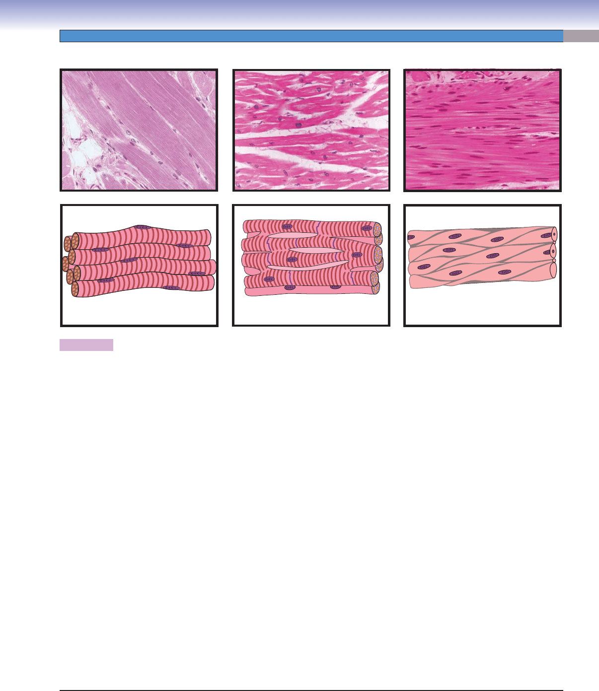
Figure 6-1. Overview of muscle types.
The three major types of contractile tissues in the body, skeletal muscle, cardiac muscle, and smooth muscle, have many properties
in common, but differ in many other ways. Skeletal muscle is usually, but not always, attached to the bones of the skeleton and
is specialized to execute rapid voluntary movements of the limbs, digits, head, etc., in response to signals from the central nervous sys-
tem (CNS). Skeletal muscle cells are long, thin, tubular structures with multiple nuclei clustered just under the cell membrane. Motor
neuron axons form synapses (motor endplates) on each skeletal muscle fi ber. Contraction in skeletal muscle is produced by a calcium-
mediated interaction between myofi laments that are composed primarily of the proteins actin and myosin. The actin and myosin
fi laments are arranged into highly organized, repeating units called sarcomeres, which give skeletal muscle a striped (“striated”)
appearance when viewed at higher magnifi cations in light microscopy. The calcium necessary to initiate the actin-myosin reaction
is stored in modifi ed endoplasmic reticulum structures called the sarcoplasmic reticulum. Calcium is released when electrical charges
fl ow down the transverse tubule system, which is formed by invaginations of the cell membrane, and is located adjacent to parts
of the sarcoplasmic reticulum within the muscle cells. Cardiac muscle, in contrast, is specialized for repeated, rhythmic, automatic
contractions over many years without ceasing. The contractile mechanism is similar to that of skeletal muscle: actin and myosin
myofi laments are arranged in sarcomeres and their interaction is mediated by calcium release. However, cardiac muscle cells are
short and split into two or three branches; these branching cells are joined end to end by intercalated disks. The overall structure of
cardiac muscle is, therefore, one of a meshwork of contractile tissues, instead of being a collection of independent, parallel units such
as is found in skeletal muscle. The autonomic axons that innervate cardiac muscle release their neurotransmitters into the intracel-
lular space rather than onto individual cells at motor endplates as in skeletal muscle. The nervous system, therefore, modulates the
rhythm of contraction of cardiac muscle, but does not command individual contractions. Smooth muscle is found in many organ
systems including the circulatory, respiratory, gastrointestinal, reproductive, and urinary systems. For the most part, smooth muscle
is specialized for automatic, slow, rhythmic contraction, although a few muscles such as the ciliary muscle of the eye are exceptions.
Like skeletal and cardiac muscles, smooth muscle uses actin and myosin fi laments to produce contraction, but the myofi laments
are not organized into sarcomeres. Instead, actin fi laments are anchored to dense plaques in the smooth muscle sarcolemma, and
a myosin fi lament contacts several individual actin fi laments at both of its ends. These actin-myosin combinations are arranged in
a random, crisscross pattern in some muscles and in parallel patterns in other muscles. As in skeletal and cardiac muscles, calcium
is a critical factor in initiating a contraction, but in smooth muscle the calcium is stored in the intercellular space rather than in a
sarcoplasmic reticulum. As in cardiac muscle, autonomic motor nerves release neurotransmitters into the intercellular space rather
than into motor endplates. The nervous system, therefore, modulates the inherent contractile rhythm of smooth muscle. This rhythm
can also be infl uenced by hormones in the bloodstream and by mechanical stretching of the muscle. Electrical excitation and, hence,
muscle contraction, can also spread directly from cell to cell via gap junctions between the membranes of adjacent cells.
D. Cui
J.Lynch
Skeletal muscle Cardiac muscle Smooth muscle
CUI_Chap06.indd 101 6/2/2010 4:08:31 PM
102
UNIT 2
■
Basic Tissues
Skeletal Muscle
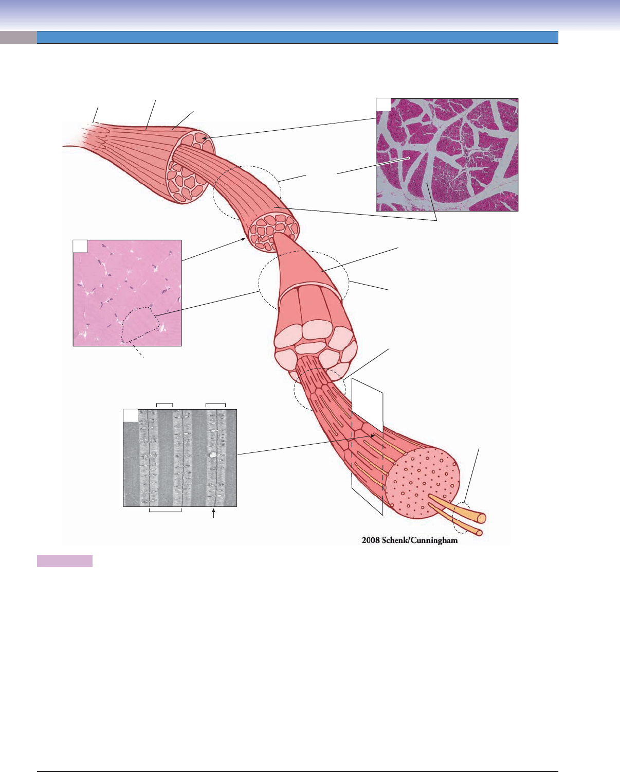
Figure 6-2. Organization of skeletal muscle.
A single skeletal muscle (e.g., the biceps) is composed of numerous fascicles (“small bundles”). The muscle as a whole is enveloped
in a strong layer of dense connective tissue, the epimysium. Each fascicle consists of a large number of muscle fi bers (cells) and is
surrounded by a sheet of less dense connective tissue, the perimysium (Fig. 6-2A). Muscle fi bers are unusual among the cells of
the body in that each contains a large number of nuclei (see Fig. 6-3A), and the nuclei are located around the periphery of the cell
(Figs. 6-2B and 6-3B). Each muscle fi ber is enveloped by a thin layer of delicate connective tissue, the endomysium. An individual
muscle fi ber contains many myofi brils, which, in turn, consist of an array of regularly organized thick and thin myofi laments, the
contractile elements of the muscle (Fig. 6-2D). The myofi laments are visible only with the electron microscope (Figs. 6-2C and 6-4B).
Thick myofi laments are composed of clusters of myosin molecules, and the thin myofi laments are predominantly actin molecules
but contain some additional auxiliary molecules that are important for the contraction process. In cross section, the myofi laments
are arranged in a repeating pattern so that each threadlike cluster of myosin molecules is surrounded by six actin molecules in a
hexagonal array, and the myosin molecule clusters themselves are arranged in a hexagonal array (Fig. 6-4B). Longitudinally, the
actin and myosin molecules form repeating units called sarcomeres (Fig. 6-2C). Actin fi laments are anchored at one end in the Z line,
a transverse membrane-like structure. Myosin molecules lie parallel to the actin molecules and partially overlap the actin molecules
that are attached to two adjacent Z lines (Fig. 6-4B). The region in which the myosin and actin overlap is designated the A band,
and the region in which only actin molecules are present is designated the I band (Figs. 6-2C and 6-4A,B). Muscle contraction is the
result of chemical interactions between the myosin and actin molecules.
Myofibril
Tendon
Muscle
Epimysium
(surrounds entire muscle)
Fascicle
(bundle of
muscle fibers)
Perimysium
(separates fascicles)
Endomysium
(envelops single muscle cell)
Muscle fiber
(muscle cell)
Myofilaments
Myosin
Actin
Z line
Sarcomere
I bandA band
Muscle fiber
(muscle cell)
B
C
A
D
CUI_Chap06.indd 102 6/2/2010 4:08:33 PM
CHAPTER 6
■
Muscle
103
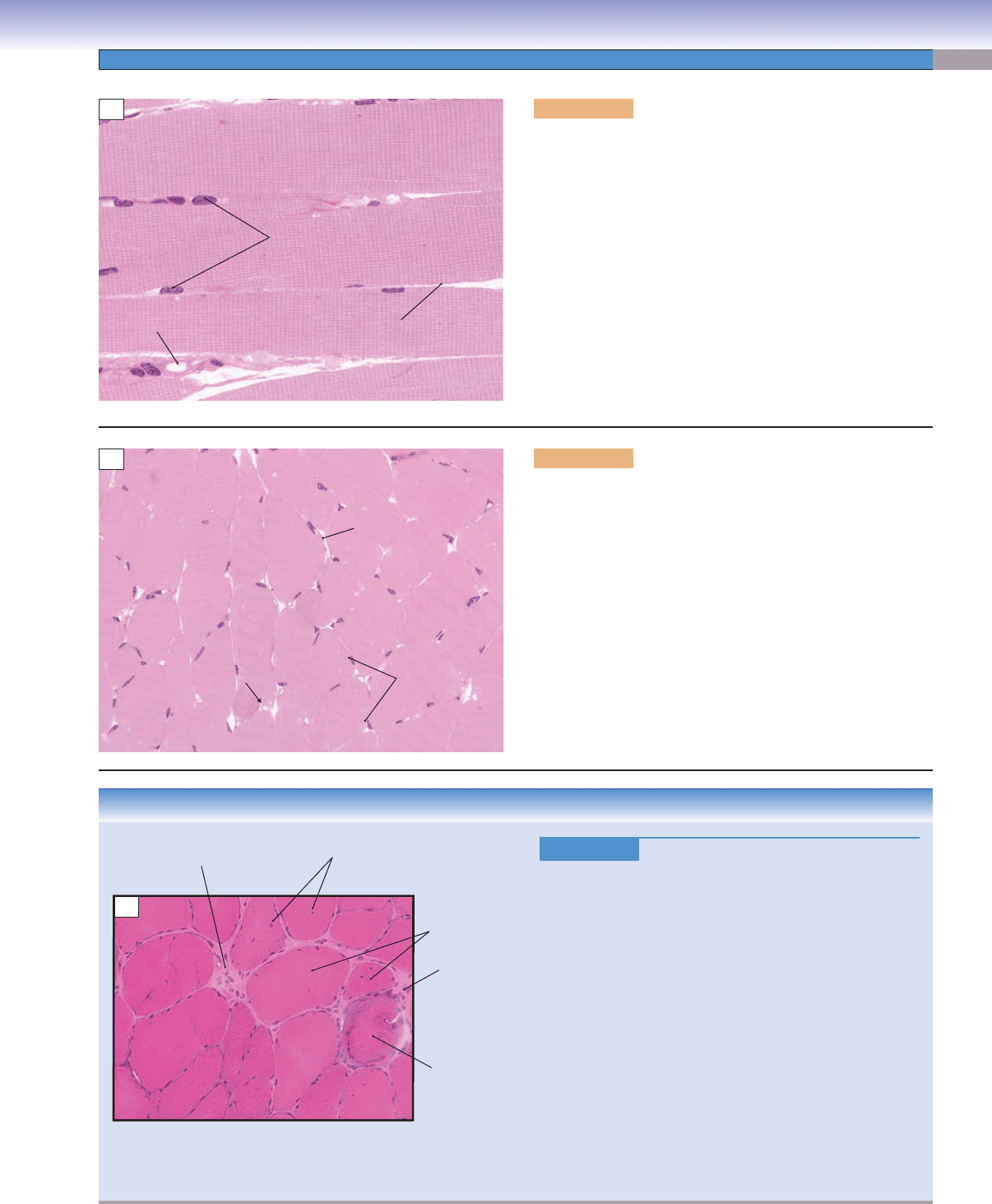
Figure 6-3A. Longitudinal section of striated muscle. H&E,
400
The cellular units of skeletal muscle are called muscle fi bers. Each
fi ber is a long, roughly cylindrical cell bounded by a plasma mem-
brane, the sarcolemma. Muscle fi bers range from 10 to 100 μm in
diameter and may be many centimeters in length in mature mus-
cles. This large size presents a problem for a single cell nucleus
serving far distant cytoplasm and cell membrane. In skeletal
muscle, this problem is solved by the formation of a syncytium,
resulting from the fusion of several myoblasts, during develop-
ment. A single muscle fi ber will therefore have many nuclei.
A distinctive feature of skeletal muscle, visible in this section, is a
repeating pattern of dark and light bands oriented at right angles
to the length of the fi ber. These bands are designated A bands and
I bands (see Fig. 6-4A). Capillaries and myelinated nerve fi bers
are often observed in sections of skeletal muscle tissue.
Nuclei
Sarcolemma
Capillary
A
Nuclei
Sarcolemma
Capillary
B
Figure 6-3B. Transverse section of skeletal muscle (ton -
gue). H&E, 272
Muscle fi bers in the tongue run in several different directions,
so, although most fi bers in this section are cut transversely
(in cross section), some are cut diagonally. Skeletal muscle
fi bers are round or polygonal in cross section, and, in a nor-
mal muscle, the fi ber diameter is relatively uniform. The nuclei
are fl attened and lie peripherally in each fi ber, just beneath the
sarcolemma.
CLINICAL CORRELATION
Figure 6-3C.
Muscular Dystrophy. H&E, 136
The muscular dystrophies are a group of inherited myogenic
disorders characterized by progressive degeneration and
weakness of skeletal muscle without associated abnormality
of the nervous system. They can be subdivided into various
groups based on the distribution and severity of muscle weak-
ness and genetic fi ndings. Duchenne muscular dystrophy
([DMD] illustrated here) is the most common and severe
form of the disease. It is carried by mutation of an X-linked
recessive gene, the dystrophin gene. The lack of the dystro-
phin protein impairs the transfer of force from actin fi la-
ments to the cell wall and causes the progressive weakness.
Pathological changes include large variations in muscle fi ber
diameter, extensive endomysial fi brosis between the fi bers,
degeneration and regeneration of fi bers with necrosis and
phagocytosis, centrally displaced nuclei, and replacement of
muscle by fat and connective tissue. Steroids are the primary
drugs used to treat DMD. Gene therapy using a functioning
dystrophin protein has not yet been successful.
Centrally displaced
nuclei
Abnormally large
range of fiber
diameters
Endomysial fibrosis
Necrotic fiber
Inflammatory
cells
C
CUI_Chap06.indd 103 6/2/2010 4:08:36 PM
104
UNIT 2
■
Basic Tissues
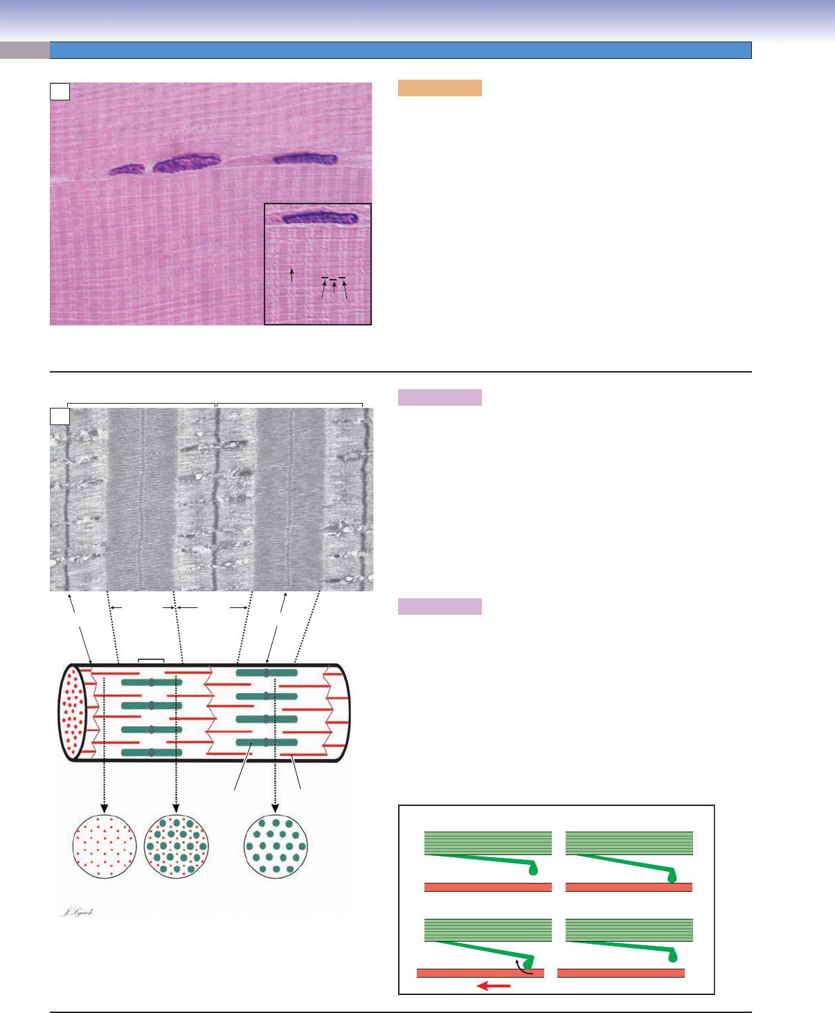
Figure 6-4A. Skeletal muscle, striations. H&E, 1,480; inset
1,800
A pattern of light and dark stripes is readily apparent in higher
magnifi cations of skeletal muscle. This pattern gives skeletal mus-
cle its alternate name, striated (“striped”) muscle. The names of
the striations are based on their behavior under polarized light.
The dark bands are named A bands because they are anisotropic
(rotate polarized light strongly), whereas the I bands are isotropic
(rotate polarized light only slightly). The A bands correspond to
regions in which myosin molecules and actin molecules overlap
to a large extent; I bands correspond to regions in which actin
molecules predominate. In the center of each I band is a thin dark
line, the Z line, which corresponds to a membrane-like structure
to which the ends of actin molecules are attached. This striated
pattern was recognized from the early history of light microscopy,
but its structural signifi cance was not understood until the advent
of practical electron microscope techniques in the 1950s.
A
I
Z
I
A
J.Lynch
Z line M line
H band
Sarcomere Sarcomere
I bandA band
Actin
myofilaments
Actin
Myosin
myofilaments
Myosin
B
C
Actin
1
2
34
Myosin Myosin
Actin Actin
MyosinMyosin
Actin
Figure 6-4B. Skeletal muscle—sarcomeres, myofi laments. EM,
17,600
A sarcomere is defi ned as the portion of a myofi bril between two
adjacent Z lines. The electron micrograph at left illustrates two sar-
comeres. The basic correspondence between the features on the elec-
tron micrograph and the constituent molecules is illustrated in the
diagram at left below. Actin myofi laments (each consisting of many
actin molecules and other accessory molecules) are anchored at the
Z lines. Myosin myofi laments (each consisting of hundreds of myo-
sin molecules) partially overlap the actin fi laments. In cross section,
both actin and myosin fi laments are arrayed hexagonally.
Figure 6-4C. Muscle contraction.
Myosin and actin fi laments (1) are not in contact with each other in
the resting muscle. (2) When a contraction is initiated, myosin mol-
ecules undergo a conformational change and contact adjacent actin
fi laments. (3) An energy (adenosine triphosphate [ATP]) consum-
ing reaction causes a further conformational change in the “head”
of the myosin molecule, which produces a translational movement
between the myosin and actin fi laments. (4) The myosin molecule
is released from the actin fi lament and the conformational changes
are reversed. The process is repeated millions of times in a fraction
of a second to produce contraction of the whole muscle.
CUI_Chap06.indd 104 6/2/2010 4:08:39 PM
