Cui Dongmei. Atlas of Histology: with functional and clinical correlations. 1st ed
Подождите немного. Документ загружается.

CHAPTER 5
■
Cartilage and Bone
85
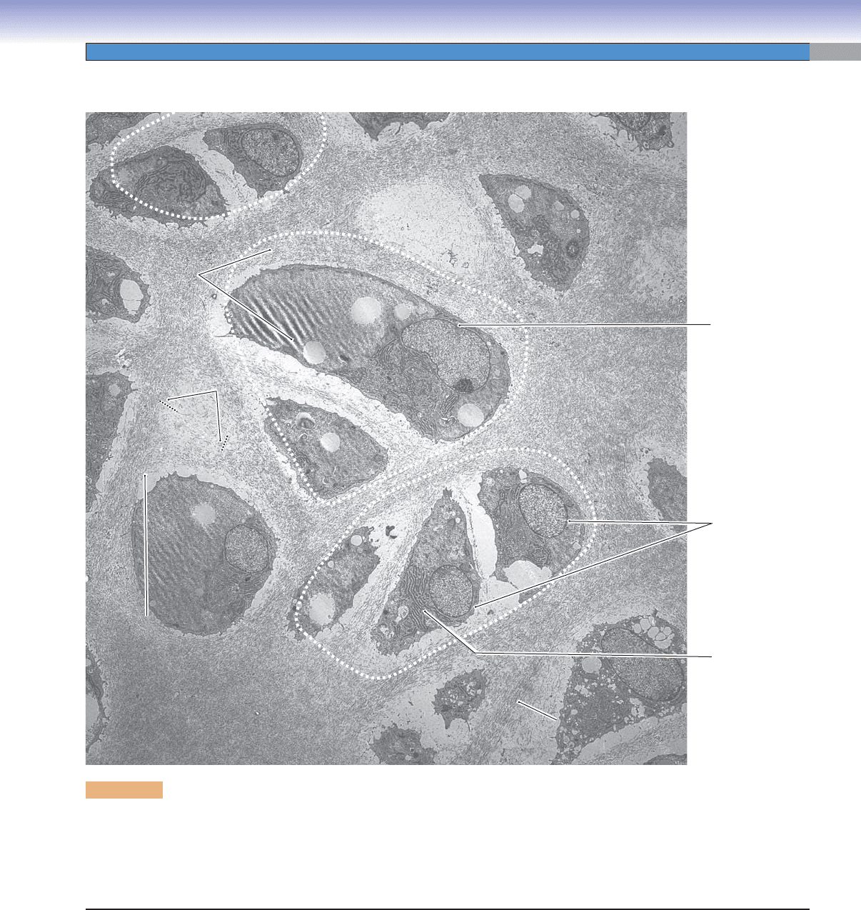
Figure 5-4. Hyaline cartilage and chondrocytes. EM, 6,300
When chondroblasts of the perichondrium have surrounded themselves with matrix and become embedded in a hyaline cartilage,
they are called chondrocytes, the cells seen in this electron micrograph. These chondrocytes are still active in synthesizing matrix
proteins as indicated by their abundant rough endoplasmic reticulum (RER) and by the presence of nucleoli and euchromatin in
their nuclei. Evidence of recent cell divisions is seen in the form of isogenous groups, three of which are circumscribed by dotted
lines. The meshwork of fi laments in the matrix is type II collagen, which does not aggregate to form fi bers.
Interterritorial
Interterritorial
matrix
matrix
Type II
Type II
collagen fibrils
collagen fibrils
Type II
collagen fibrils
Interterritorial
matrix
Lacuna
Lacuna
Territorial
Territorial
matrix
matrix
Lacuna
Territorial
matrix
Territorial
Territorial
matrix
matrix
Territorial
matrix
Interterritorial
Interterritorial
matrix
matrix
Interterritorial
matrix
Euchromatic
nucleus
Rough
endoplasmic
reticulum (RER)
Nuclei
CUI_Chap05.indd 85 6/2/2010 6:29:59 PM
86
UNIT 2
■
Basic Tissues
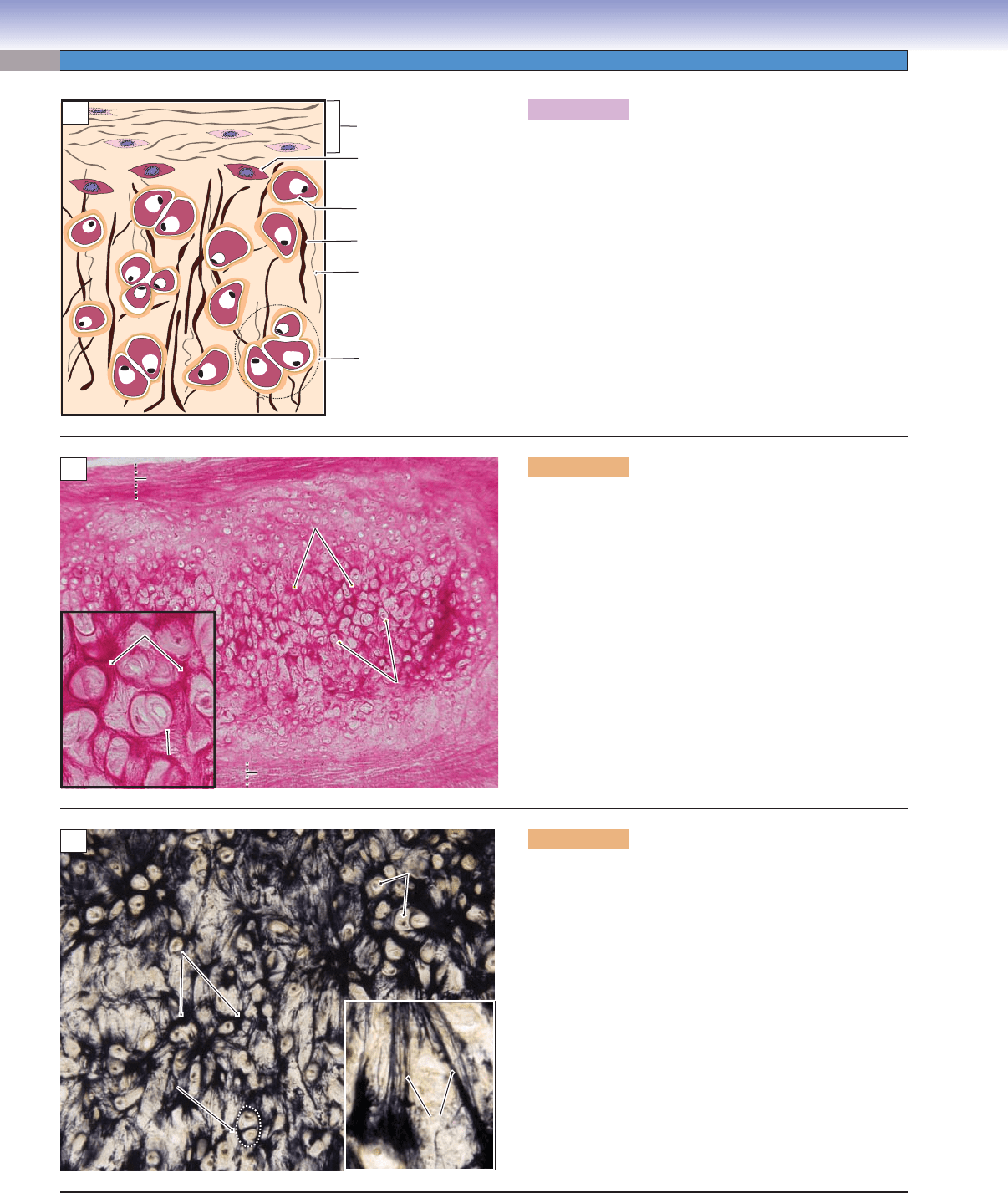
Figure 5-5A. A representation of elastic cartilage.
Elastic cartilage has a rich network of elastic fi bers, which
gives its matrix a rough appearance. It also contains deli-
cate collagen type II fi bers and ground substance in the
matrix, as do other types of cartilage. In general, chondro-
cytes are more abundant in elastic cartilage than in hya-
line cartilage and fi brocartilage. Cartilage growth in elastic
cartilage includes appositional growth, which requires a
perichondrium, and interstitial growth, indicated by isog-
enous groups (see Fig. 5-7). Elastic cartilage provides fl ex-
ible support for tissue and is located in the areas where
fl exible stretching is required, such as the epiglottis, larynx,
pinna of the ear, and the auditory canal and tube.
D. Cui /T. Yang
Chondrocyte in lacuna
Elastic fiber
Type II collagen fiber
Isogenous group
Chondroblast
Perichondrium
A
B
Elastic fibers
Elastic fibers
Elastic fibers
Perichondrium
Perichondrium
Perichondrium
Chondrocyte
Chondrocyte
Chondrocyte
Elastic fibers
Elastic fibers
Elastic fibers
Isogenous
Isogenous
group
group
Isogenous
group
Perichondrium
Perichondrium
Perichondrium
C
Chondrocytes
Chondrocytes
Chondrocytes
Elastic fibers
Elastic fibers
Elastic fibers
Isogenous
Isogenous
group
group
Isogenous
group
Elastic fibers
Elastic fibers
Elastic fibers
Figure 5-5B. Elastic cartilage, epiglottis. H&E, 68;
inset 218
An example of elastic cartilage in the epiglottis is shown.
Elastic cartilage has a perichondrium surrounding it as
does most hyaline cartilage. The perichondrium protects
and provides blood supply for the cartilage tissue. Chon-
drogenic cells and chondroblasts in the perichondrium
layer are responsible for appositional growth of the matrix.
There are abundant elastic fi bers and type II collagen fi bers
in the extracellular matrix. Isogenous groups are created
by the division of existing cells. The resulting daughter cells
that are derived from a single progenitor cell stay in the
same lacuna. Elastic cartilage has both interstitial growth,
which is indicated by the presence of isogenous groups,
and appositional growth, for which a perichondrium is
required (Fig. 5-7).
Figure 5-5C. Elastic cartilage, epiglottis. Elastic fi ber
stain, 68; inset 208
An example of elastic cartilage in the epiglottis is shown.
Elastic cartilage is composed of thick, branching elastic
fi bers with a slight network of collagen fi bers and chon-
drocytes fi lling the interstitial space. Elastic cartilage can
be found in the epiglottis and pinna of the ear. Elastic fi bers
presented here with a special stain are seen as thick, dark,
elongated profi les. Chondrocytes are arranged in individ-
ual and isogenous groups among the elastic fi bers in the
matrix.
CUI_Chap05.indd 86 6/2/2010 6:30:01 PM
CHAPTER 5
■
Cartilage and Bone
87
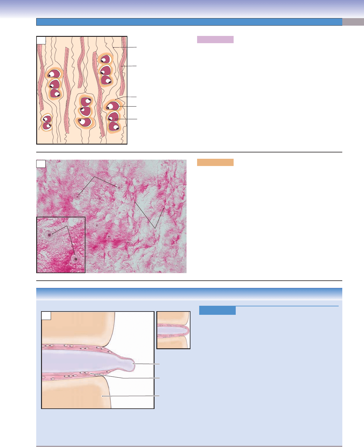
Figure 5-6A. A representation of fi brocartilage.
Fibrocartilage lacks a perichondrium, so no appositional
growth takes place. Chondrocytes in lacunae are often arranged
in small groups in parallel columns or rows, which correlate
with their method of interstitial growth. The chondrocytes are
smaller and fewer in number in fi brocartilage than in the other
two types of cartilage. Because type I collagen fi bers are pres-
ent in its matrix, the matrix has a dense and coarse appear-
ance. Fibrocartilage is less fl exible than the other two types
of cartilage; it provides fi rm support, cushioning, and tensile
strength.
CLINICAL CORRELATION
Figure 5-6C.
Disk Degeneration and Herniation.
Herniation of an intervertebral disk is a common cause
of pain in the lower back and neck. It is most common
in people in their 30s and 40s. Risk factors include age,
occupation, lifestyle, and genetic propensity
. Degenera-
tion of the intervertebral disk is because of a combination
of factors that may result in changes in hydration of the
nucleus pulposus (composed of mucous connective tissue)
and in the strength of collagen, leading to weakening of
the anulus fi brosus (fi brocartilage). The degenerated disk
nucleus pulposus loses its cushioning ability and exerts
uneven pressure on the surrounding anulus; extrusion of the
nucleus pulposus through the weakened annulus is called
herniation. It happens most often at the L4–L5 (lumbar)
and L5–S1 (sacral) vertebral levels, causing back pain and
other neurologic symptoms because of compression of the
nerve roots. Magnetic resonance imaging is widely used to
visualize the herniated disk. Treatment includes bed rest,
the McKenzie exercise, steroid injections, open discectomy,
and minimally invasive endoscopic discectomy.
A
Nucleus pulposus
T. Yang
T. Yang
C
Normal disk
Extrusion of
nucleus
pulposus
Vertebra
Anulus fibrosus
(fibrocartilage)
D. Cui /T. Yang
Chondrocytes
Type I collagen
Type II collagen
Territorial matrix
Lacuna
A
B
Chondrocytes
Type I
collagen
fibers
Chondrocytes
Figure 5-6B. Fibrocartilage, intervertebral disk. H&E,
136; inset 292
Fibrocartilage in the intervertebral disk is shown. Fibrocarti-
lage contains type II and type I collagen fi ber bundles in the
matrix, which makes the matrix look rough, like an oil paint-
ing. Chondrocytes are small and housed in lacunae, which are
widely scattered in the matrix. There is no perichondrium asso-
ciated with fi brocartilage; therefore, cartilage growth proceeds
by interstitial growth only. Fibrocartilage has a fi rm, dense
matrix, and it can be found in the pubic symphysis, interverte-
bral disks, and insertions of tendons and ligaments.
CUI_Chap05.indd 87 6/2/2010 6:30:06 PM
88
UNIT 2
■
Basic Tissues
D. Cui /T. Yang
D. Cui /T. Yang
Interstitial Growth
Chondrocyte in lacuna
Territorial matrix
Interterritorial matrix
Isogenous group
Chondroblast
Chondrogenic cell
Inner cellular layer
of the perichondrium
Outer fibrous layer
of the perichondrium
Appositional Growth
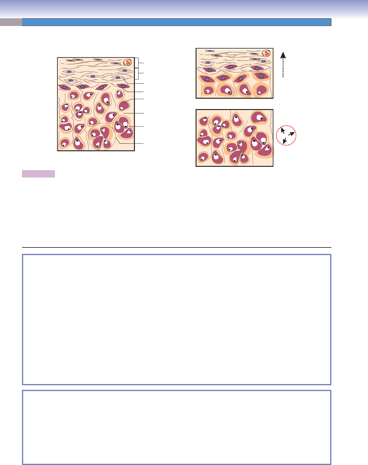
Figure 5-7. A representation of cartilage growth.
Cartilage grows by either appositional or interstitial growth or by both. The growth process is continuous and involves mitosis and the
deposition of additional matrix. Appositional growth begins with the chondrogenic cells in the perichondrium. These chondrogenic cells
differentiate into chondroblasts, which are also called young chondrocytes. Chondroblasts start to elaborate a new layer of matrix at the
surface (periphery) region of the cartilage near the perichondrium. Cartilage grows mostly by appositional growth. Interstitial growth
occurs during the early stages of cartilage formation. The growth begins with the cell division of preexisting chondrocytes (mature chon-
droblasts which are surrounded by territorial matrix). Interstitial growth increases the tissue size by expanding the cartilage matrix from
within the cartilage mass. This type of growth is indicated by the presence of isogenous groups in most cartilage, though sometimes the
chondrocytes are arranged in small groups in parallel columns and rows. Articular cartilage lacks a perichondrium, so it enlarges only by
interstitial growth. Interstitial growth serves to lengthen the bone such as in the epiphyseal plates of long bones (see Fig. 5-12B).
SYNOPSIS 5-1 Functions of Cartilage
Hyaline cartilage
Serves as the
■ cartilage model for the formation of bones during bone development.
Participates in bone-lengthening growth by increasing chondrocyte size and numbers during bone development
■
( endochondral ossifi cation).
Enables free movement by forming smooth surfaces that work with lubricating fl uid (
■ synovial fl uid) in articular cartilage
of the joints.
Provides support and framework for airways in the respiratory tract.
■
Elastic cartilage
Provides elastic but stiff framework for pinna and allows it to return to its former shape after stretching.
■
Provides elastic support for auditory canals and tube; helps to maintain structural shape. ■
Provides a fi rm and elastic support for the epiglottis and larynx; helps to maintain rigid structure and fl exibility. ■
Fibrocartilage
Provides tensile strength for connections between the bones such as the pubic symphysis.
■
Provides cushioning and resistance between vertebrae, enabling the spinal column to endure great pressure. ■
SYNOPSIS 5-2 Special Features of Cartilage
The ■ function is to provide fi rm support with variable fl exibility depending on its location.
The extracellular matrix is
■ nonmineralized and consists of fi brillar proteins (collagen) and ground substance (GAGs,
proteoglycans, and glycoproteins).
The extracellular matrix is produced by
■ chondroblasts and chondrocytes.
Cartilage grows by both
■ interstitial and appositional mechanisms.
It is an
■ avascular tissue; the nutrients are supplied through matrix diffusion.
The
■ perichondrium provides the nearest blood supply to the cartilage.
Vitamins A
■ , D, and C are necessary for cartilage growth and matrix formation.
Cartilage Growth
CUI_Chap05.indd 88 6/2/2010 6:30:09 PM

CHAPTER 5
■
Cartilage and Bone
89
Bone
Introduction and Key
Concepts for Bone
Bone is a special type of supporting connective tissue, which
has a hard, mineralized, extracellular matrix containing osteo-
cytes embedded in the matrix. It is different from cartilage in
that bone is calcifi ed and, hence, is harder and stronger than
cartilage. In addition, it has many blood vessels penetrating the
tissue. Bone protects internal organs, provides support for soft
tissues, serves as a calcium reserve for the body, provides an
environment for blood cell production, detoxifi es certain chem-
icals in the body, and aids in the movement of the body. In gen-
eral, the external surface of the bone is covered by periosteum,
a layer of connective tissue containing small blood vessels,
osteogenic cells, and nerve fi bers conveying pain information.
The inner surface of the bone is covered by endosteum, a thin
connective tissue layer composed of a single layer of osteo-
progenitor cells and osteoblasts that lines all internal cavities
within bone; this lining represents the boundary between the
bone matrix and the marrow cavities. Bone cells include osteo-
genic cells, osteoblasts, osteocytes, and osteoclasts. These cells
contribute to bone growth, remodeling, and repair.
Bone Matrix
Bone is primarily characterized by a hard matrix, which contains
calcium, phosphate, other organic and inorganic materials, and
type I collagen fi bers. Compared to cartilage, bone contains
only about 25% water in the matrix, whereas cartilage matrix
contains about 75% water. This combination makes bone
hard, fi rm, and very strong. Bone matrix has organic and inor-
ganic components. (1) Organic (noncalcifi ed) matrix is mainly
type I collagen with nonmineralized ground substance (chon-
droitin sulfate and keratin sulfate). It is found in the freshly
produced bone matrix, osteoid (also called prebone), which is
produced by osteoblasts. This matrix stains light pink in H&E
preparations (Fig. 5-11A). (2) Inorganic (calcifi ed) matrix,
mainly in the form of hydroxyapatite, contains crystalline min-
eral salts, mostly of calcium and phosphorus. After osteoid is
produced, this fresh matrix undergoes a mineralization process
to become the calcifi ed matrix (Fig. 5-11B).
Bone Cells
The main types of cells in bone are osteoprogenitor cells, osteo-
blasts, osteocytes, and osteoclasts: (1) Osteoprogenitor cells
are located in the periosteum on the surface of the growing
bone and can differentiate into osteoblasts. (2) Osteoblasts
produce the bone matrix. They are cuboidal or low columnar
in shape and have a well-developed Golgi complex and RER,
which correlates with their protein-secreting function (Fig.
5-11). The overall process of mineralization relies on the eleva-
tion of calcium and phosphate within the matrix and the func-
tion of hydroxyapatite crystals. This is brought about by com-
plex functions of the osteoblast. (3) Osteocytes are small, have
cytoplasmic processes, and are unable to divide. These cells
originate from osteoblasts and are embedded in the bone
matrix. Osteoblasts deposit the matrix around themselves and
end up inside the matrix, where they are called “osteocytes.”
Each osteocyte has many long, thin processes that extend into
small narrow spaces called canaliculi. The nucleus and sur-
rounding cytoplasm of each osteocyte occupy a space in the
bone matrix called a lacuna. Thin processes of the osteocyte
course through thin channels (canaliculi) that radiate from
each lacuna and connect neighboring lacunae (Fig. 5-9B,C).
(4) Osteoclasts are large, multinucleated cells, which derive
from monocytes, absorb the bone matrix, and play an essential
role in bone remodeling (Fig. 5-14A,B).
Types of Bone
There are several ways to classify bone tissues. Microscopically,
bone can be classifi ed as primary bone (immature, or “woven”
bone) and secondary bone (mature, or lamellar bone). Bones
can also be classifi ed by their shapes as follows: long bones,
short bones, fl at bones, and irregular bones (Table 5-2). Mature
bone can be classifi ed as compact bone and cancellous bone
based on gross appearance and density of the bone. Compact
bone, also called cortical bone, has a much higher density and
a well-organized osteon system. It does not have trabeculae
and usually forms the external aspect (outside portion) of the
bone (Figs. 5-8 to 5-10B). Cancellous bone, also called spongy
bone, has a much lower density and contains bony trabecu-
lae or spicules with intervening bone marrow (Fig. 5-8A,C). It
can be found between the inner and the outer tables of the
skull, at the ends of long bones, and in the inner core of other
bones.
Bone Development
Bone development can be classifi ed as intramembranous ossifi -
cation and endochondral ossifi cation, according to the mecha-
nism of its initial formation. (1) Intramembranous ossifi cation
is the process by which a condensed mesenchyme tissue is
transformed into bone. A cartilage precursor is not involved;
instead, mesenchymal cells serve as osteoprogenitor cells, which
then differentiate into osteoblasts. Osteoblasts begin to deposit
the bone matrix (Fig. 5-11A,B). (2) Endochondral ossifi cation
is the process by which hyaline cartilage serves as a cartilage
model precursor. This hyaline cartilage proliferates, calci-
fi es, and is gradually replaced by bone. Osteoprogenitor cells
migrate along with blood vessels into the region of the calcifi ed
cartilage. These cells become osteoblasts, which then begin to
deposit the bone matrix on the surface of the calcifi ed cartilage
matrix plate. Endochondral ossifi cation involves several events
(see Figs. 5-12 and 5-13A for a summary of these processes). The
development of long bone is a good example of endochondral
formation. In this particular case, the hyaline cartilage under-
goes proliferation and calcifi cation in the epiphyseal plates. This
epiphyseal cartilage can be divided into fi ve recognizable zones:
reserve zone, proliferation zone, hypertrophy zone, calcifi cation
zone, and ossifi cation zone (see Fig. 5-12B).
CUI_Chap05.indd 89 6/2/2010 6:30:09 PM
90
UNIT 2
■
Basic Tissues
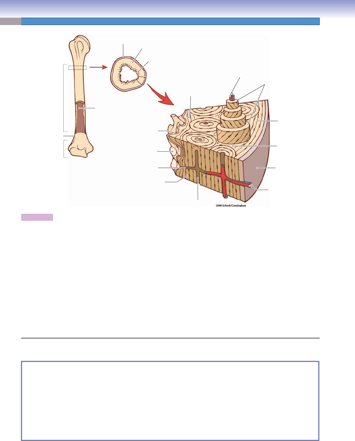
Figure 5-8. Overview of bone structure, long bone.
Bones can be classifi ed as long bones, short bones, fl at bones, and irregular bones according to their shape. Long bones are longer
than they are wide and consist of a long shaft (diaphysis) and two ends (epiphyses). Short bones are roughly cube shaped, such
as wrist and ankle bones. Bone also can be classifi ed as compact bone and cancellous bone based on gross appearance and bone
density. The diaphysis of a long bone is composed primarily of compact bone and an inner medullary cavity, which is fi lled with
bone marrow. The epiphyses of long bones are composed mainly of cancellous (spongy) bone, and the articular surfaces are cov-
ered by articular cartilage, providing a smooth joint surface for articulation with the next bone. The metaphysis is a transitional
zone between the diaphysis and epiphysis; it represents the level that cancellous bone ends and the bone marrow cavity begins.
The external surfaces of compact bone are covered by periosteum, a thick layer of dense connective tissue, which contains blood
vessels. Endosteum, a thin layer of connective tissue with a single layer of osteoprogenitor cells and osteoblasts, forms a boundary
between the bone and the medullary cavity (this layer may be continuous with the trabeculae of the cancellous bone). The general
structure of compact bone includes (1) the osteon, a canal surrounded by layers of concentric lamellae; (2) interstitial lamellae,
lamellae layers in between the osteons; (3) outer circumferential lamellae, outer layers of lamellae located beneath the periosteum
and surrounding the outside of the entire compact bone; and (4) inner circumferential lamellae, layers of lamellae located beneath
the endosteum and forming the innermost layer of compact bone. The Haversian canal is a central space through which blood
vessels pass; the Volkmann canal is the space that sits perpendicularly to the Haversian canals and forms the connection between
two Haversian canals.
Diaphysis
Metaphysis
Epiphysis
Blood vessels
Blood vessels
Osteon
Haversian canal
Volkmann
canal
Endosteum
Cancellous
bone
Compact
bone
Periosteum
Compact bone
Cancellous bone
Inner
circumferential
lamellae
Periosteum
Interstitial
lamellae
Outer
circumferential
lamellae
Bone
marrow
cavity
SYNOPSIS 5-3 Functions of Bone
Provides ■ protection for internal organs, such as the brain, heart, lung, bladder, and reproductive organs.
Provides
■ supporting framework for the body (e.g., long bones for limbs and skull for the support of brain and framework
for facial features).
Enables body
■ movements in conjunction with the muscles and nervous system.
Produces blood cells (
■ hematopoiesis) within the medullary cavity of long bones and cancellous bone.
Provides a
■ calcium and phosphorus reserve for the body.
Provides
■ detoxifi cation for stored heavy metals in the bone tissues. Removes these toxic materials from blood, thereby
reducing damage to other organs and tissues.
Provides
■ sound transduction in the middle ear (auditory ossicles: malleus, incus, and stapes).
CUI_Chap05.indd 90 6/2/2010 6:30:09 PM
CHAPTER 5
■
Cartilage and Bone
91
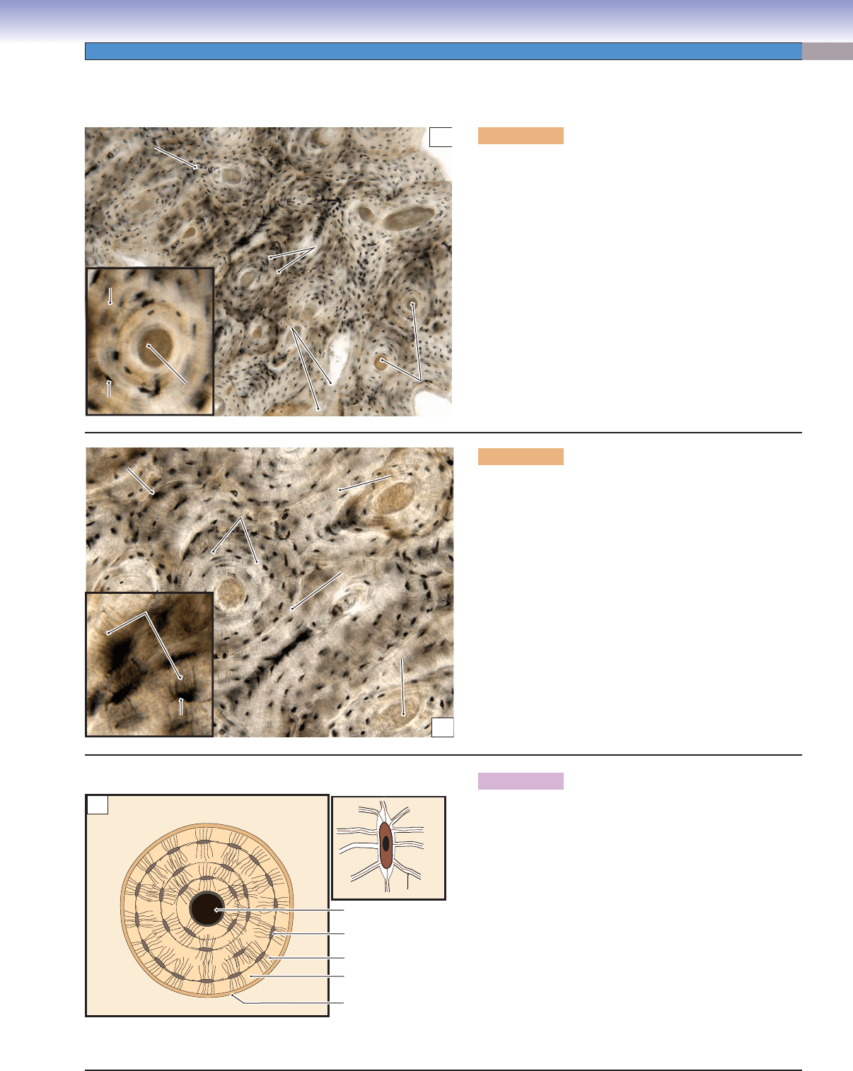
Figure 5-9A. Compact bone. Ground specimen (un-
stained), 68; inset 212
A cross section of compact bone in a ground specimen (with-
out decalcifi cation of tissue) is shown. Haversian canals are
round central spaces in the cross-sectional view; a Volk-
mann canal is shown in the longitudinal view. Volkmann
canals run perpendicularly to and connect Haversian canals
with each other (Fig. 5-8). The inset photomicrograph
shows an osteon (Haversian system), the basic structural
unit of compact bone, which includes a Haversian canal,
lacunae with housed osteocytes, and concentric lamellae
(Fig. 5-9C). Bone matrices located between the osteons are
called interstitial lamellae.
Interstitial
Interstitial
lamellae
lamellae
Concentric
Concentric
lamellae
lamellae
Interstitial
lamellae
Concentric
lamellae
Haversian
Haversian
canals
canals
Lamella
Lamella
Lacuna
Lacuna
Volkmann
Volkmann
canal
canal
Haversian
Haversian
canal
canal
Volkmann
canal
Haversian
canals
Lacuna
Lamella
Haversian
canal
A
lamella
lamella
Haversian
Haversian
canal
canal
Lacuna
Lacuna
Lacuna
Lacuna
Cement line
Cement line
Canaliculi
Canaliculi
Concentric
Concentric
lamellae
lamellae
Interstitial
Interstitial
lamella
Cement line
Haversian
canal
Lacuna
Lacuna
Canaliculi
Concentric
lamellae
Interstitial
B
D. Cui
An osteocyte
within a lacuna
Haversian canal
Canaliculus
Concentric
lamella
Cement line
Lacuna
Canaliculus
C
Figure 5-9B. Compact bone. Ground specimen (un-
stained), 136; inset 388
A higher power view of compact bone in ground specimen
is shown. Concentric lamellae and lacunae are arranged in
rings, which surround the Haversian canal. Each lacuna has
an osteocyte in it. Tiny canals called canaliculi contain pro-
cesses of osteocytes and link the lacunae with each other.
The canaliculi permit the osteocytes to communicate via
gap junctions where the processes of adjacent osteocytes
touch each other inside the canaliculi. A cement line forms
a boundary between adjacent osteons. Compact bone forms
the hard external portion of bone and provides strong sup-
port and protection.
Figure 5-9C. A representation of an osteon of the com-
pact bone.
The osteon, also called a Haversian system, is the basic unit
of the compact bone structure. It has concentrically arranged
laminae (concentric lamellae) surrounding a centrally
located Haversian canal. The Haversian system consists of
(1) a Haversian canal through which blood vessels pass, (2)
concentric lamellae, (3) lacunae, each one of which contains
an osteocyte, (4) canaliculi, which are small narrow spaces
containing osteocyte processes, and (5) a cement line, the
thin dense, external bony layer that surrounds each osteon.
A schematic drawing illustrates an osteocyte occupy-
ing a lacuna (a space in the bone matrix that houses an
osteocyte) and its thin processes within the canaliculi. The
hairlike processes of the osteocyte are in contact with the
processes of adjacent osteocytes and provide a means of
communication between osteocytes.
Types of Bone
CUI_Chap05.indd 91 6/2/2010 6:30:10 PM
92
UNIT 2
■
Basic Tissues
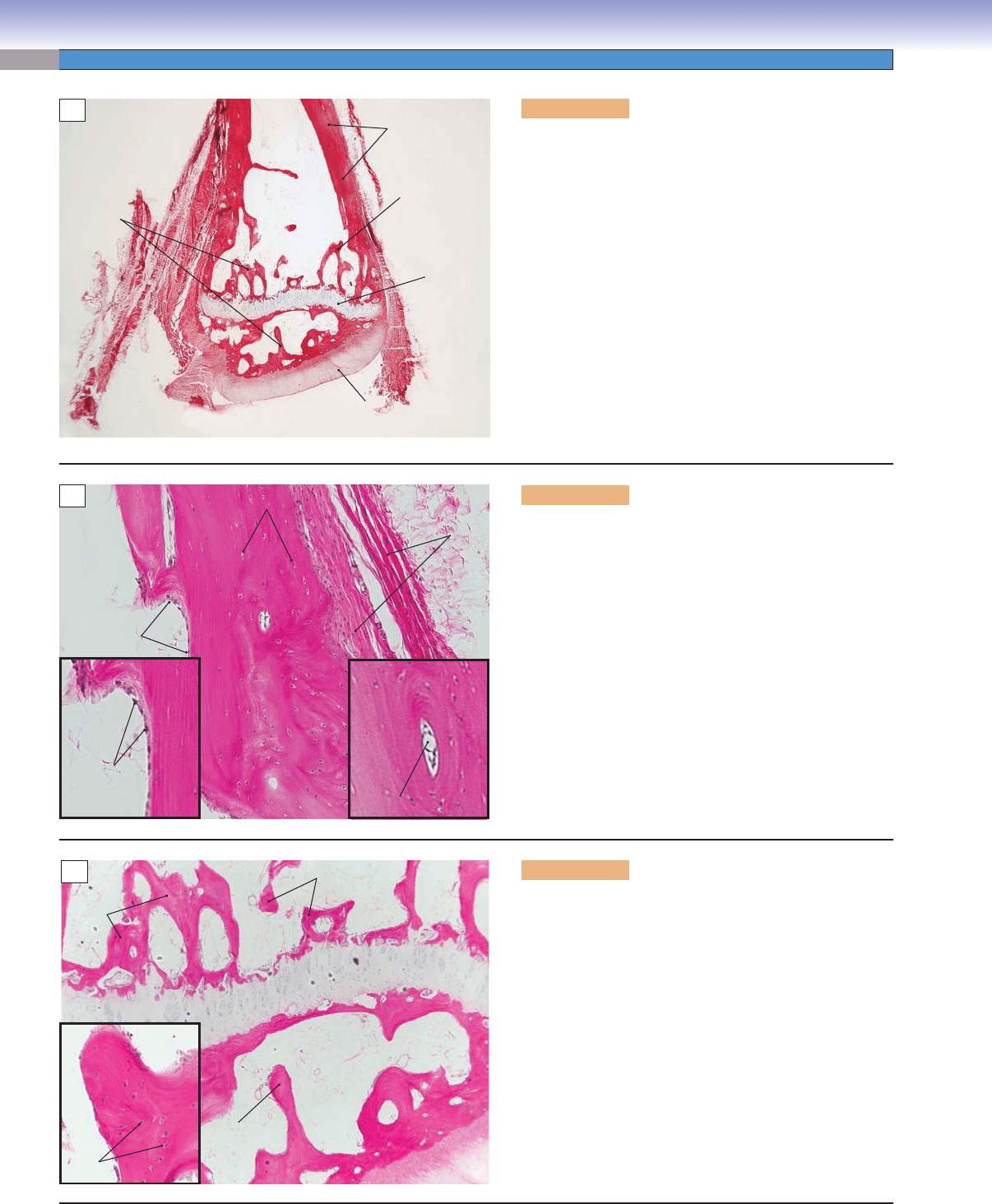
Figure 5-10A. Compact bone and cancellous bone,
fi nger. Decalcifi ed bone, H&E, 11
Bone has a calcifi ed extracellular matrix that is very diffi -
cult to cut into thin sections. In order to have thin sections
with H&E stain, these bone specimens have to go through
a decalcifi cation process that removes calcium compounds
from the specimen. Bone can be classifi ed as compact
bone (cortical bone) and cancellous bone (spongy bone),
based on its gross appearance. Compact bone has a very
high density and a well-organized osteon system (Figs. 5-8
and 5-9A–C). It has no trabeculae and usually forms the
external aspect of a bone. Cancellous bone (spongy bone)
has a much lower density and contains bony trabeculae or
spicules with intervening bone marrow. It usually forms
the inner part of a bone, also called medullary bone, and is
commonly found between the inner and the outer tables of
the skull, at the ends of long bones (limbs and fi ngers), and
in the cores of other bones.
A
Cancellous
bone
Compact
bone
Bony
trabeculae
Epiphyseal
plate
Articular
cartilage
Bone
marrow
B
Osteocytes
Compact
bone
Endosteum
Endosteum
Periosteum
Osteon
Haversian canal
C
Spongy bone
Spongy
bone
Bony
trabeculae
Bone
marrow space
Hyaline cartilage
Osteocytes
Figure 5-10B. Compact bone, fi nger. Decalcifi ed bone,
H&E, 105; inset (left) 154; inset (right) 127
An example of compact bone from the diaphysis of the long
bone (fi nger) is shown. The internal surface is covered by
a single layer of connective tissue cells forming the endos-
teum. It contains osteoprogenitor cells, which are capable
of differentiating into osteoblasts. The external surface is
covered by a thicker layer, the periosteum, which contains
blood vessels, nerves, and osteoprogenitor cells. Osteopro-
genitor cells can differentiate into osteoblasts, which have
the ability to produce bone matrix, osteoid (prebone) (Fig.
5-11A). Blood vessels branch to supply bone through a
system of interconnected Volkmann canals and Haversian
canals (Fig. 5-8). Osteocytes are arranged uniformly in
compact bone. Each osteocyte occupies one lacuna, which
has no isogenous group as it does in cartilage (Fig. 5-9C).
Figure 5-10C. Cancellous bone (spongy bone), nasal.
Decalcifi ed bone, H&E, 34; inset 128
Cancellous bone is also called spongy bone. It has a lower
density than compact bone and consists of bony trabecu-
lae, or spicules, within a marrow-fi lled cavity. Osteoblasts
line the surface of the bony trabeculae. Cancellous bone
displays irregular shapes in the trabecular network. Bone
marrow fi lls the space between the bony trabeculae (Fig.
5-11A). Most osteocytes in the matrix are arranged in an
irregular pattern rather than in circular rings (Fig. 5-8).
Cancellous bone mainly forms the inner core of bone and
provides (1) a meshwork frame that supports and reduces
the overall weight of bone and (2) room for blood vessels
to pass through and a place for marrow to function as a
hemopoietic compartment, housing and producing blood
cells (Fig. 5-11A).
CUI_Chap05.indd 92 6/2/2010 6:30:15 PM
CHAPTER 5
■
Cartilage and Bone
93
Bone Development and Growth
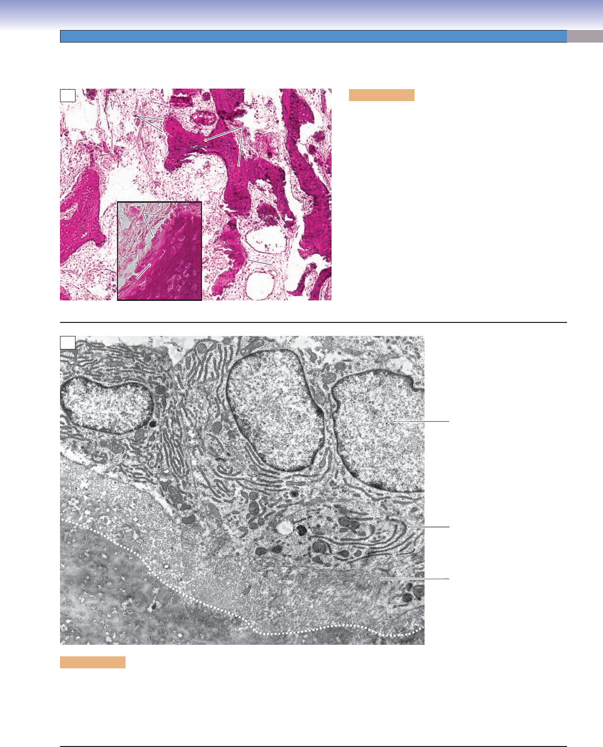
Figure 5-11A. Intramembranous ossifi cation, fetal
head. H&E, 84; inset 210
Intramembranous ossifi cation is a process of bone
formation involving the transformation of condensed
mesenchymal tissue into bone tissue by differentiation
of mesenchymal cells into osteoblasts and deposition of
osteoid (prebone). Osteoid is unmineralized new bone,
which contains organic components. Soon after the new
bone is deposited, it becomes calcifi ed bone, which is
largely composed of calcium and phosphate. Osteoblasts
often line up on the surface of the bone matrix. They are
cuboidal and low columnar in shape, and each osteoblast
contains a large round nucleus and basophilic cytoplasm
containing rich RER and Golgi complexes, indicating
their activity in producing protein and organic compo-
nents (Fig. 5-11B). Mature osteoblasts are trapped inside
the bone matrix to become osteocytes. Osteoid appears
pink in H&E stain, in contrast to mineralized bone
matrix that appears dark red-purple in H&E stain.
Osteoid
Osteoid
(prebone)
(prebone)
Osteoid
(prebone)
Blood
vessel
Mineralized
Mineralized
bone matrix
bone matrix
Mineralized
bone matrix
Osteoblasts
Osteoblasts
Osteoblasts
Bone marrow
Bone marrow
Osteoblasts
Osteoblasts
Osteoblasts
Bone marrow
Osteoid
Osteoid
(prebone)
(prebone)
Osteoid
(prebone)
A
Euchromatic
Euchromatic
nucleus
nucleus
Mineralized
Mineralized
bone matrix
bone matrix
Osteoid (prebone)
Osteoid (prebone)
Mineralized
bone matrix
Euchromatin
Rough
endoplasmic
reticulum (RER)
Type I
collagen
fibrils
Osteoid (prebone)
B
Figure 5-11B. Osteoblasts. EM, 19,600
The three osteoblasts in this electron micrograph are clearly active in the synthesis and secretion of type I collagen and other proteins
of bone matrix. Note the high content of euchromatin in the nuclei and the predominance of RER in the cytoplasm. Minute collagen
fi brils (type I collagen) are just discernible in the layer of matrix adjacent to the cells (prebone or osteoid). The deeper, mineralized
bone matrix has a homogeneous appearance that masks the presence of the collagen fi brils. The dotted white line indicates the inter-
face between the osteoid above and the mineralized bone matrix below.
CUI_Chap05.indd 93 6/2/2010 6:30:21 PM
94
UNIT 2
■
Basic Tissues
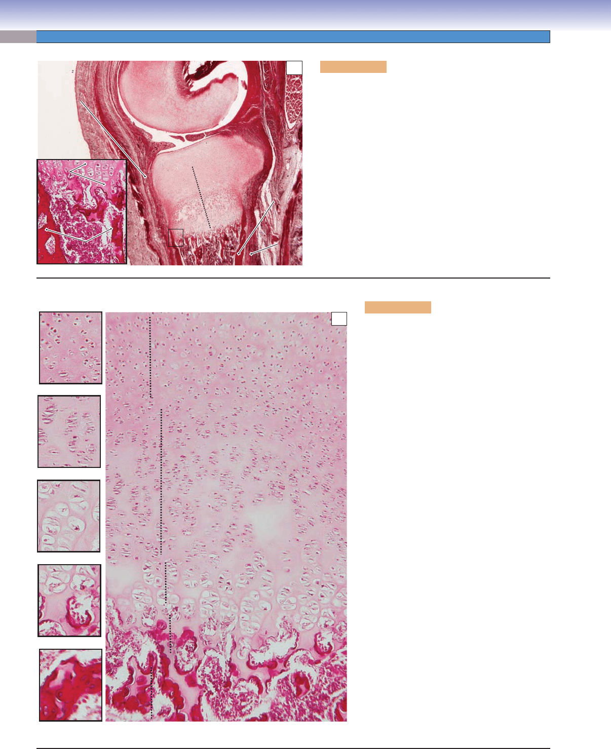
Figure 5-12A. Endochondral ossifi cation, fi nger. H&E,
20; inset 68
Endochondral ossifi cation is a process of bone formation
in which hyaline cartilage serves as a cartilage model (pre-
cursor). Cartilage proliferation occurs, then calcifi cation,
and gradually the cartilage is replaced by bone (see Figs.
5-12B and 5-13A). This is an example of a long bone (fi n-
ger), showing the epiphyseal plate (cartilage plate) with
the primary ossifi cation center (primary marrow cavity).
There is a thick layer of dense connective tissue covering
the peripheral region of the cartilage, called the perichon-
drium. The connective tissue layer that covers the outer
surface of the bone is called periosteum. The primary ossi-
fi cation center contains blood vessels, newly formed bone
tissue, osteoblasts, osteoclasts, calcifi ed cartilage matrix,
and dead chondrocytes. (PC, primary ossifi cation center.)
Bone
Bone
matrix
matrix
Periosteum
Periosteum
Bone
matrix
Epiphyseal
plate
Periosteum
Perichondrium
Perichondrium
Cartilage
Cartilage
matrix
matrix
Bone
Bone
matrix
matrix
Perichondrium
Cartilage
matrix
Bone
matrix
Articular
cartilage
PC
PC
PC
A
1. Reserve zone
2. Proliferation zone
3. Hypertrophy zone
4. Calcification zone
5. Ossification zone
Reserve zone
Proliferation zone
Hypertrophy zone
Calcification zone
Ossification zone
B
Figure 5-12B. Epiphyseal plate, fi nger. H&E,
71; small images 96
The epiphyseal plate is a region of hyaline
cartilage at the ends (epiphyses) of the shafts of
long bones. Its chondrocytes are undergoing the
process of proliferation, hypertrophy, and cal-
cifi cation, during the process of endochondral
ossifi cation. The epiphyseal plate can be divided
into fi ve functionally distinct zones beginning
at the epiphyseal end: (1) In the reserve zone,
cartilage chondrocytes are inactive and individ-
ual cells are not arranged in isogenous groups.
These cells are small and randomly scattered
in the matrix. (2) In the proliferation zone,
chondrocytes undergo frequent mitosis and are
arranged in groups of columns (indicative of
interstitial growth of cartilage) in this region.
Chondrocytes are fl at, and their size is increased
leading to increased length of the cartilage. (3)
In the hypertrophy zone, chondrocytes become
mature, and their size increases markedly (big
and fat cells). Isogenous groups are clearly evi-
denced and cells actively deposit matrix (type
X and XI collagen). (4) In the calcifi cation
zone, cartilage matrix becomes calcifi ed, and
chondrocytes die because nutrients and oxygen
cannot diffuse through the calcifi ed cartilage
matrix. The matrix in this region is fi lled with
hydroxyapatite (a complex phosphate of cal-
cium). (5) In the ossifi cation zone, blood vessels
invade and create primary marrow; osteopro-
genitor cells arrive in this region and differen-
tiate into osteoblasts to start depositing bone
matrix (osteoid or new bone) on the surface
of the calcifi ed cartilage. Osteoclasts are also
present and function as phagocytes to remove
unwanted calcifi ed cartilage matrix and dead
chondrocytes.
CUI_Chap05.indd 94 6/2/2010 6:30:25 PM
