Cui Dongmei. Atlas of Histology: with functional and clinical correlations. 1st ed
Подождите немного. Документ загружается.

CHAPTER 4
■
Connective Tissue
75
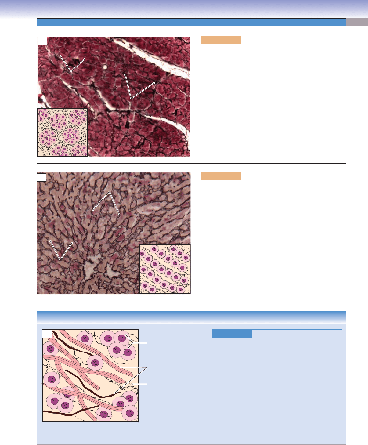
Figure 4-19A. Reticular connective tissue, pancreas. Silver
stain, 136
Reticular tissue is a specialized loose connective tissue that
provides a delicate supporting framework for many highly
cellular organs, such as endocrine glands, lymphoid organs,
the spleen, and the liver. Reticular fi bers are shown in black
with a silver stain. These fi bers are small in diameter and do
not form large bundles. They are arranged in a netlike frame-
work to support parenchymal cells, in this example, pancre-
atic cells. The inset drawing represents the organization of
reticular fi bers and pancreatic cells.
Figure 4-19B. Reticular connective tissue, liver. Silver
stain, 312
The reticular fi bers can be selectively visualized with a silver
stain, that is, they are argyrophilic. These fi bers consist of col-
lagen type III, which forms a meshlike network that supports
the liver cells and holds these cells together. The liver cells’
cytoplasm is unstained in this preparation, and the structure
of the cells is not easy to distinguish here. The inset draw-
ing represents the organization of reticular fi bers and hepato-
cytes. There is a sinusoid running between the reticular fi bers,
which appears as empty space here.
D. Cui
Reticular fibers
Reticular fibers
Reticular fibers
Pancreatic cells
Pancreatic cells
Pancreatic cells
A
Hepatocytes
Hepatocytes
Hepatocytes
Reticular fibers
Reticular fibers
Reticular fibers
B
CLINICAL CORRELATION
Figure 4-19C.
Cirrhosis.
Cirrhosis is a liver disorder caused by chronic injury
to the hepatic parenchyma. The major causes of cir
-
rhosis include alcoholism and chronic infection with
hepatitis B or hepatitis C virus. Pathologic changes are
characterized by the collapse of the delicate support-
ing reticular connective tissue with increased numbers
of collagen and elastic fi bers. There is disruption of
the liver architecture and vascular bed. Regenerating
hepatocytes form nodules rather than the characteristic
columnar plates. Symptoms include jaundice, edema,
and coagulopathy (a defect of blood coagulation). The
resulting damage to the liver tissue impedes drain-
age of the portal venous system, a condition known
as portal hypertension, which may eventually lead to
gastroesophageal varices, splenomegaly, and ascites.
D. Cui
Damaged and reduced
reticular fibers
Collagen bundles and
elastic fibers have replaced
normal reticular fibers
Nodule of regenerated
liver cells
C
CUI_Chap04.indd 75 6/2/2010 7:48:17 AM
76
UNIT 2
■
Basic Tissues
CLINICAL CORRELATION
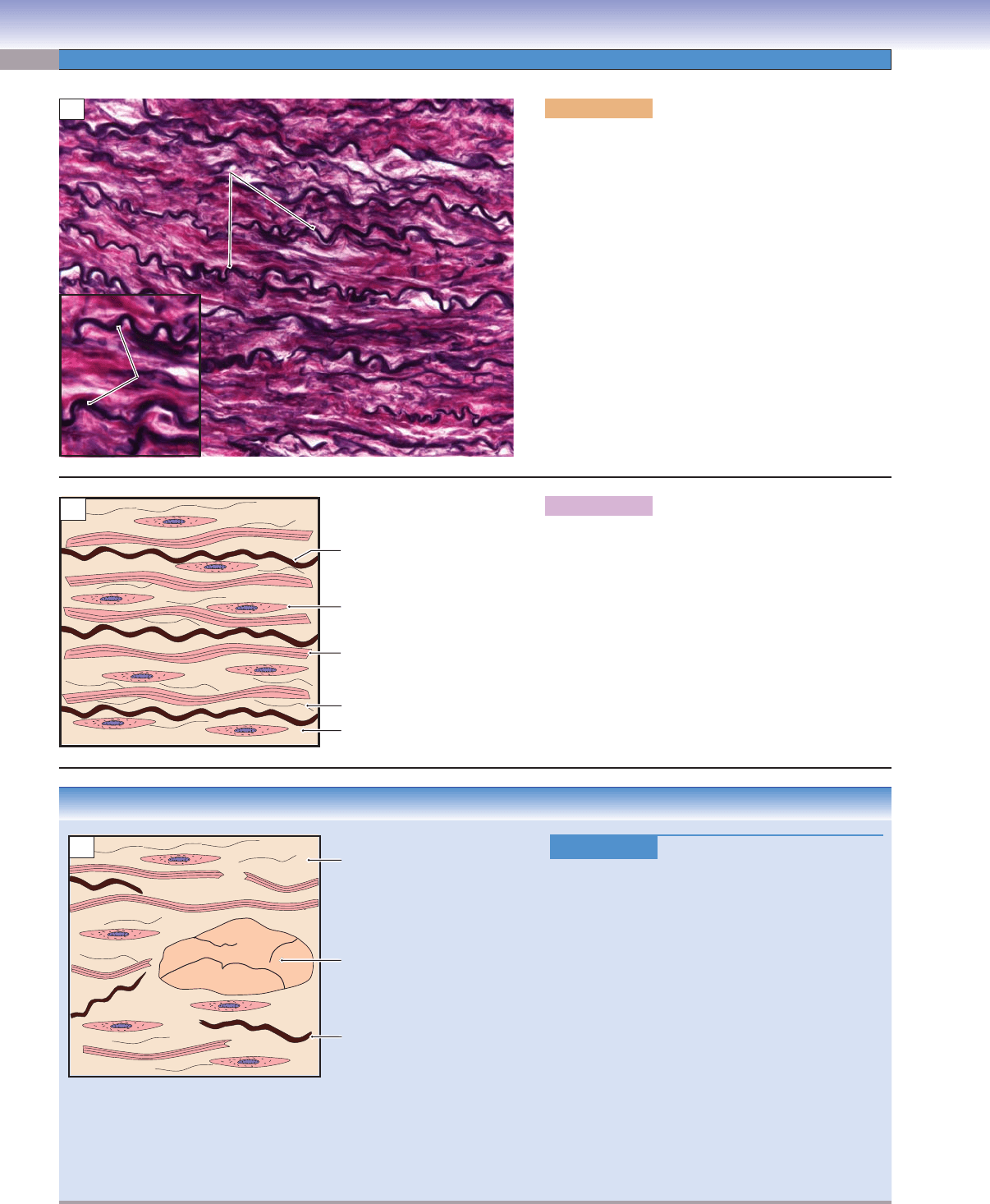
Figure 4-20C.
Marfan Syndrome—Cystic Medial
Degeneration.
Marfan syndrome is an autosomal dominant disorder
caused by an FBN1 gene mutation, which affects the
formation of elastic fi bers, particularly those found in
the aorta, heart, eye, and skin. Signs and symptoms
include tall stature with long limbs and long, thin fi n-
gers and enlargement of the base of the aorta accom-
panied by aortic regurgitation. There is increased
probability of dissecting aortic aneurysms and pro-
lapse of the mitral valve. Treatment includes pharma-
cologic or surgical intervention to prevent potentially
fatal or long-term complications, but no permanent
cure is yet available. This illustration depicts cystic
medial degeneration (cystic medionecrosis) of the
aorta, including disruption and fragmentation of
elastic lamellae in the tunica media of the aorta, loss
of elastic fi bers, and increase in ground substance
causing formation of cystic space.
D. Cui
Loss of elastic lamellae and
increased ground substance
Fragmentation of elastic lamellae
Cystic space filled with
amorphous extracellular matrix
C
Figure 4-20A. Elastic connective tissue, carotid
artery. Elastic stain (Verhoeff), 275; inset 516
This is an example of elastic connective tissue in the
tunica media of a carotid artery. The wavy elastic
lamellae are distributed among collagen and smooth
muscle cells in the tunica media layer of a large artery.
The smooth muscle cells are not visible here because
of the type of stain. In general, the elastic material (as
either elastic fi bers or elastic lamellae) and other con-
nective tissue fi bers are produced by fi broblasts in the
connective tissue, but in blood vessels, smooth muscle
cells are the principal cells that produce elastic material
and other connective tissue fi bers. Elastic connective
tissue consists predominately of elastic material, and
this allows distension and recoil of the structure. This
tissue can be found in some vertebral ligaments, arte-
rial walls, and in the bronchial tree.
Figure 4-20B. A representation of elastic connective
tissue in the tunica media of a large artery.
Thick bundles of elastic lamellae are arranged in parallel
wavy sheets, with the smooth muscle cells and collagen
fi bers insinuated between alternating lamellae. The elas-
tic fi bers are formed by elastin and fi brillin microfi brils.
Elastic connective tissue is able to recoil after stretch-
ing. This property in large arteries helps to moderate
the extremes of pressure associated with the cardiac
cycle. Abnormal expression of the fi brillin (FBN1) gene
is associated with abnormal elastic tissue disease.
Elastic
Elastic
lamellae
lamellae
Elastic
lamellae
Elastic lamellae
Elastic lamellae
Elastic lamellae
A
D. Cui
Ground substance
Collagen fiber
Reticular fiber
Smooth muscle
cell
Elastic lamellae
B
CUI_Chap04.indd 76 6/2/2010 7:48:24 AM
CHAPTER 4
■
Connective Tissue
77
Types of Connective Tissue: Embryonic Connective Tissues
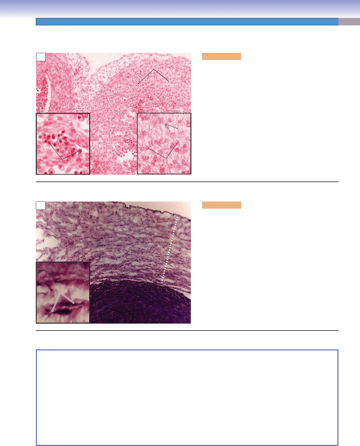
Figure 4-21A. Mesenchyme, embryo. H&E, 136; inset
408 (left) and 438 (right)
Mesenchyme (mesenchymal connective tissue) is found in
the developing structures in the embryo. It contains scat-
tered reticular fi bers and mesenchymal cells, which have
irregular, star or spindle shapes and pale-stained cytoplasm.
These cells exhibit cytoplasmic processes, which often give
the cells a stellate appearance. Mesenchymal cells are rela-
tively unspecialized and are capable of differentiating into
different cell types in mature tissue cells, such as cartilages,
bones, and muscles. Embryonic red blood cells can be seen
in this specimen. These blood cells contain a nucleus in each
cell; this is characteristic of their immature state (anucle-
ated red blood cells are characteristic of the mature state
and are found in adult tissues). Interestingly enough, some
vertebrates, such as frogs and chickens, have nucleated red
blood cells in the adult state.
Figure 4-21B. Mucous connective tissue, umbilical cord.
Toluidine blue stain, 68; inset 178
An example of mucous connective tissue that has an abun-
dance of a jellylike matrix with some fi ne aggregates of col-
lagen fi bers and stellate-shaped fi broblasts is shown. It is
found in the umbilical cord and subdermal connective tis-
sue of the embryo. Mucous tissue is a major constituent of
the umbilical cord, where it is referred to as Wharton jelly.
This type of connective tissue does not differentiate beyond
this stage. In this example, the viscous ground substance has
been stained with a special stain to reveal jellylike mucin,
which contains hyaluronic acid and glycoproteins. Colla-
gen fi bers and large stellate-shaped fi broblasts (not mesen-
chymal cells) predominate in the mucous tissue.
Mesenchymal
cells
Embryonic red blood
cells
Mesenchymal
connective
tissue
Cytoplasmic
processes
Mesenchymal
cells
A
Mucous
Mucous
connective tissue
connective tissue
Smooth muscle
Smooth muscle
Mucous
connective tissue
Smooth muscle
Fibroblasts
Fibroblasts
Fibroblasts
B
SYNOPSIS 4-3 Pathological Terms for Connective Tissue
Urticaria ■ : An itchy skin eruption, also known as hives, characterized by wheals with pale interiors and well-defi ned red
margins, often the result of an allergic response to insect bites, foods, or drugs (Fig. 4-3C).
Pruritis
■ : Itching of the skin due to a variety of causes including hyperbilirubinemia and allergic and irritant contact condi-
tions (Fig. 4-3C).
Cirrhosis
■ : An abnormal liver condition characterized by diffuse nodularity, due to fi brosis and regenerative nodules of
hepatocytes; frequent causes are alcohol abuse and viral hepatitis (Fig. 4-19C).
Jaundice
■ : Yellow staining of the skin, mucous membranes, or conjunctiva of the eyes caused by elevated blood levels of
the bile pigment bilirubin (Fig. 4-19C).
Coagulopathy
■ : A disorder that prevents the normal clotting process of blood; causes may be acquired, such as hepatic dys-
function, or congenital, such as decreased clotting factors, as seen in inherited conditions like hemophilia (Fig. 4-19C).
Necrosis
■ : Irreversible cell changes that occur as a result of cell death (Fig. 4-20C).
CUI_Chap04.indd 77 6/2/2010 7:48:37 AM

78
UNIT 2
■
Basic Tissues
Type Connective Tissue
Cells
Connective
Tissue Fibers
Organization of
Fibers and Cells
Main Locations Main Functions
Connective Tissue Proper
Dense irregular
connective tissue
Predominantly
fi broblasts; other
connective tissue
cells occasionally
present
Collagen fi bers,
elastic fi bers,
reticular fi bers
Fewer cells and
more fi bers; fi bers
arranged randomly
without a defi nite
orientation in
relatively less ground
substance
Dermis of the skin,
capsules of many
organs
Resists stress
from all
directions;
protects organs
Dense regular
connective tissue
Predominantly
fi broblasts; other
connective tissue
cells occasionally
present
Collagen fi bers,
elastic fi bers,
reticular fi bers
Fewer cells and
more fi bers; fi bers
arranged in uniform
parallel bundles
Tendons, ligaments Provides
resistance to
traction forces
Loose
connective tissue
Fibroblasts,
macrophages,
adipocytes, mast
cells, plasma cells,
leukocytes
Collagen fi bers
predominate;
elastic and
reticular fi bers
also present
More cells and
fewer fi bers; fi bers
randomly distributed
in abundant ground
substance
Lamina propria
of gastrointestinal
tract; around the
nerves and vessels
(in adventitia layer)
Provides
protection,
suspension, and
support; conduit
for vessels and
nerves; environ-
ment for immune
defense function
Specialized Connective Tissues
Adipose
connective tissue
Predominantly
adipocytes (fat cells);
fi broblasts and other
connective tissue
cells occasionally
present
Collagen fi bers
and reticular
fi bers
Fibers form fi ne
meshwork that
separates adjacent
adipocytes
Hypodermis of the
skin, mammary
glands, and around
many organs
Provides
energy storage,
insulation;
cushioning of
organs; hormone
secretion
Reticular
connective tissue
Fibroblasts,
reticular cells,
hepatocytes,
smooth muscle
cells, Schwann cells
depending on the
location
Reticular fi bers Fibers form delicate
meshlike network;
cells with process
attached to the fi bers
Liver, pancreas,
lymph nodes,
spleen, and bone
marrow
Provides support-
ive framework
for hematopoietic
and parenchymal
organs
Elastic
connective tissue
Predominantly
fi broblasts or
smooth muscle cells;
other connective
tissue cells occasion-
ally present
Elastic fi bers
predominate;
collagen and
reticular fi bers
also present
Fibers arranged
in parallel wavy
bundles
Vertebral ligaments,
walls of the large
arteries
Provides fl exible
support for the
tissue; reduces
pressure on the
walls of the
arteries
Embryonic Connective Tissues
Mesenchymal
connective tissue
Mesenchymal cells Reticular fi bers
and collagen
fi bers
Scattered fi bers
with spindle-
shaped cells having
long cytoplasmic
processes;
mesenchymal cells
uniformly distributed
Embryonic
mesoderm
Gives rise to all
connective tissue
types
Mucous
connective tissue
Spindle-shaped
fi broblasts
Collagen fi bers
predominate; few
elastic and reticu-
lar fi bers
Fibers and
fi broblasts randomly
displayed in jellylike
matrix (Wharton
jelly)
Umbilical cord,
subdermal layer
of the fetus, dental
pulp of the devel-
oping teeth, nucleus
pulposus of the disk
Provides cushion
to protect the
blood vessels in
the umbilical cord
TABLE 4-3 Connective Tissue Types
CUI_Chap04.indd 78 6/2/2010 7:48:42 AM
79
5
Cartilage and Bone
Cartilage
Introduction and Key Concepts for Cartilage
Types of Cartilage
Figure 5-1 Overview of Cartilage Types
Table 5-1 Cartilage
Figure 5-2A A Representation of Hyaline Cartilage
Figure 5-2B,C Hyaline Cartilage, Bronchus
Figure 5-3A Hyaline Cartilage, Trachea
Figure 5-3B Hyaline Cartilage, Finger Bone
Figure 5-3C Clinical Correlation: Osteoarthritis
Figure 5-4 Hyaline Cartilage and Chondrocytes
Figure 5-5A–C Elastic Cartilage
Figure 5-6A,B Fibrocartilage
Figure 5-6C Clinical Correlation: Disk Degeneration and Herniation
Cartilage Growth
Figure 5-7 A Representation of Cartilage Growth
Synopsis 5-1 Functions of Cartilage
Synopsis 5-2 Special Features of Cartilage
Bone
Introduction and Key Concepts for Bone
Figure 5-8 Overview of Bone Structure, Long Bone
Synopsis 5-3 Functions of Bone
Types of Bone
Figure 5-9A,B Compact Bone
Figure 5-9C A Representation of an Osteon of the Compact Bone
Figure 5-10A–C Compact and Cancellous Bone
CUI_Chap05.indd 79 6/2/2010 6:29:39 PM

80
UNIT 2
■
Basic Tissues
Bone Development and Growth
Figure 5-11A Intramembranous Ossifi cation, Fetal Head
Figure 5-11B Osteoblasts
Figure 5-12A Endochondral Ossifi cation, Finger
Figure 5-12B Epiphyseal Plate, Finger
Figure 5-13A A Representation of the Development of the Long Bone
Figure 5-13B Clinical Correlation: Osteosarcoma
Figure 5-14A Bone Remodeling, Nasal
Figure 5-14B Osteoclast
Table 5-2 Bone
Synopsis 5-4 Pathological and Clinical Terms for Cartilage and Bone
Cartilage
Introduction and Key Concepts
for Cartilage
Cartilage and bone are two types of supporting connective
tissues. Cartilage is an avascular specialized form of connective
tissue whose support function is a result of a fi rm extracellular
matrix that has variable fl exibility depending on its location.
This type of tissue is able to bear mechanical stress without
permanent deformation. Cartilage has features that are differ-
ent from other types of connective tissues but, like bone, has
the characteristic of isolated cells embedded in extensive matrix.
Most cartilage is covered by a layer of dense irregular connective
tissue called perichondrium, which contains a rich blood supply
and is innervated by nerve fi bers conveying pain. The excep-
tions are fi brocartilage and articular cartilage of the joint, which
do not have perichondrium. Perichondrium is important for the
growth (appositional growth) and maintenance of cartilage; it
has two layers. The outer fi brous layer of the perichondrium
contains connective tissue fi bers, fi broblasts, and blood vessels.
These perichondrial vessels represent an essential blood supply
for cartilage. Because cartilage itself is avascular, these vessels
are the route through which nutrients access the matrix by dif-
fusion. The inner cellular layer of the perichondrium consists of
chondrogenic cells, which are able to differentiate into chondro-
blasts (Fig. 5-2). The functions of cartilage include the support
of soft tissues, the facilitation of smooth movement of bones at
joints, and the mediation of growth of the length of bones dur-
ing bone development.
Cartilage Cells
The main types of cells in cartilage are chondrogenic cells, chon-
droblasts, and chondrocytes. (1) Chondrogenic cells are located
in the perichondrium and differentiate into chondroblasts to par-
ticipate in appositional growth of cartilage (Fig. 5-7). These cells
are diffi cult to identify under the light microscope with H&E
stain. (2) Chondroblasts are young chondrocytes, which derive
from chondrogenic cells, and are able to actively manufacture
the matrix of cartilage. The chondroblasts have ribosome-rich
basophilic cytoplasm. They synthesize and deposit cartilage
matrix around themselves. As the matrix accumulates and
separates the chondroblasts from one another, the cells become
entrapped in small individual compartments called lacunae and
are then referred to as “chondrocytes.” (3) Chondrocytes are
mature chondroblasts that are embedded in the lacunae of the
matrix. Chondrocytes retain the ability to divide and often pres-
ent as an isogenous group, two or more chondrocytes arranged
in a group that was derived from a single progenitor cell
(Fig. 5-2). The isogenous group represents the active division
of cells, which contribute to interstitial growth (see below,
Cartilage Growth). In most cartilage, chondrocytes are arranged
in an isogenous group. However, in some locations such as in
fi brocartilage, chondrocytes are more likely to be arranged in
groups of small columns or rows instead of isogenous groups.
This is also a sign of interstitial growth.
Cartilage Matrix
The matrix of cartilage is nonmineralized and consists of fi bers
and ground substance. Collagen fi bers are mainly type II in
the matrix, although some cartilage may also contain type I
or elastic fi bers. The major components of ground substance
include glycosaminoglycans (GAGs), proteoglycans, and glyco-
proteins. The matrix of cartilage surrounding each chondrocyte,
or immediately adjacent to chondrocytes of isogenous groups, is
called territorial matrix. This newly produced matrix has abun-
dant proteoglycans and less collagen and stains more intensely
in routine H&E preparations. Another type of matrix, which
surrounds the regions of territorial matrix and fi lls the rest of the
space, is called interterritorial matrix. This type of matrix stains
more lightly than does the territorial matrix (Figs. 5-2 and 5-4).
Types of Cartilage
Cartilage can be classifi ed into three types based on the charac-
teristics of the matrix. All three types of cartilage contain type II
collagen; in addition, some types contain type I collagen or elas-
tic fi bers in the extracellular matrix. Types of cartilage include
hyaline cartilage, elastic cartilage, and fi brocartilage.
HYALINE CARTILAGE is characterized by the presence of a
glassy, homogeneous matrix that contains type II collagen, which
is evenly dispersed within the ground substance. Most hyaline
cartilage is covered by perichondrium, except at the articular
surfaces of joints. Hyaline cartilage is the most common type
of cartilage; is found in the articular ends of long bones, nose,
larynx, trachea, bronchi, and the distal ends of ribs; and is the
template for endochondral bone formation (Figs. 5-2 to 5-4 and
5-12). Hyaline cartilage covers the smooth surface of joints,
CUI_Chap05.indd 80 6/2/2010 6:29:46 PM

CHAPTER 5
■
Cartilage and Bone
81
providing for free movement, and is also involved in bone
formation and long bone growth (Figs. 5-3B and 5-12).
ELASTIC CARTILAGE is similar to hyaline cartilage except
for its rich network of elastic fi bers, arranged in thick bundles
in the matrix. This type of cartilage has a perichondrium, as
does hyaline cartilage, and it also contains type II collagen
in the matrix. The chondrocytes of elastic cartilage are more
abundant and larger than those of hyaline cartilage. Elastic car-
tilage is located in areas where elasticity and fi rm support are
required, such as the epiglottis and larynx, auditory canal and
tube, and the pinna of the ear, which is able to recover its shape
after deformation (Fig. 5-5).
FIBROCARTILAGE does not have a perichondrium. It has
type II collagen, as do the other two types of cartilage. It is
characterized by thick, coarse bundles of type I collagen fi bers
that alternate with parallel groups of columns (or rows) of
chondrocytes within the matrix. The chondrocytes of fi brocar-
tilage are smaller and much less numerous than in the other
two types of cartilage and are often arranged in columns or
rows. Because fi brocartilage has no perichondrium, its growth
depends on interstitial growth. Fibrocartilage is resistant to
tearing and compression, can accommodate great pressure,
and is often found at connections between bones that do not
have an articular surface. It is found in areas where support and
tensile strength are required, such as intervertebral disks, the
pubic symphysis, and the insertions of tendons and ligaments
(Fig. 5-6).
Cartilage Growth
Cartilage grows by either appositional or interstitial growth or
both. The growth process is prolonged and involves mitosis and
the deposition of additional matrix (Fig. 5-7). (1) Appositional
growth begins with the chondrogenic cells located in the per-
ichondrium. These chondrogenic cells differentiate into chon-
droblasts, also called young chondrocytes, and these cells start
to elaborate a new layer of matrix at the surface ( periphery)
region of the cartilage near the perichondrium. Most carti-
lage growth in the body is appositional growth. (2) Interstitial
growth occurs during the early stages of cartilage formation
in most types of cartilage. It begins with the cell division of
preexisting chondrocytes (mature chondroblasts surrounded by
territorial matrix). Interstitial growth increases the tissue size
by expanding the cartilage matrix from within. Fibrocartilage
lacks a perichondrium, so it grows only by interstitial growth.
In the epiphyseal plates of long bones, interstitial growth serves
to lengthen the bone.
CUI_Chap05.indd 81 6/2/2010 6:29:47 PM
82
UNIT 2
■
Basic Tissues
Types of Cartilage
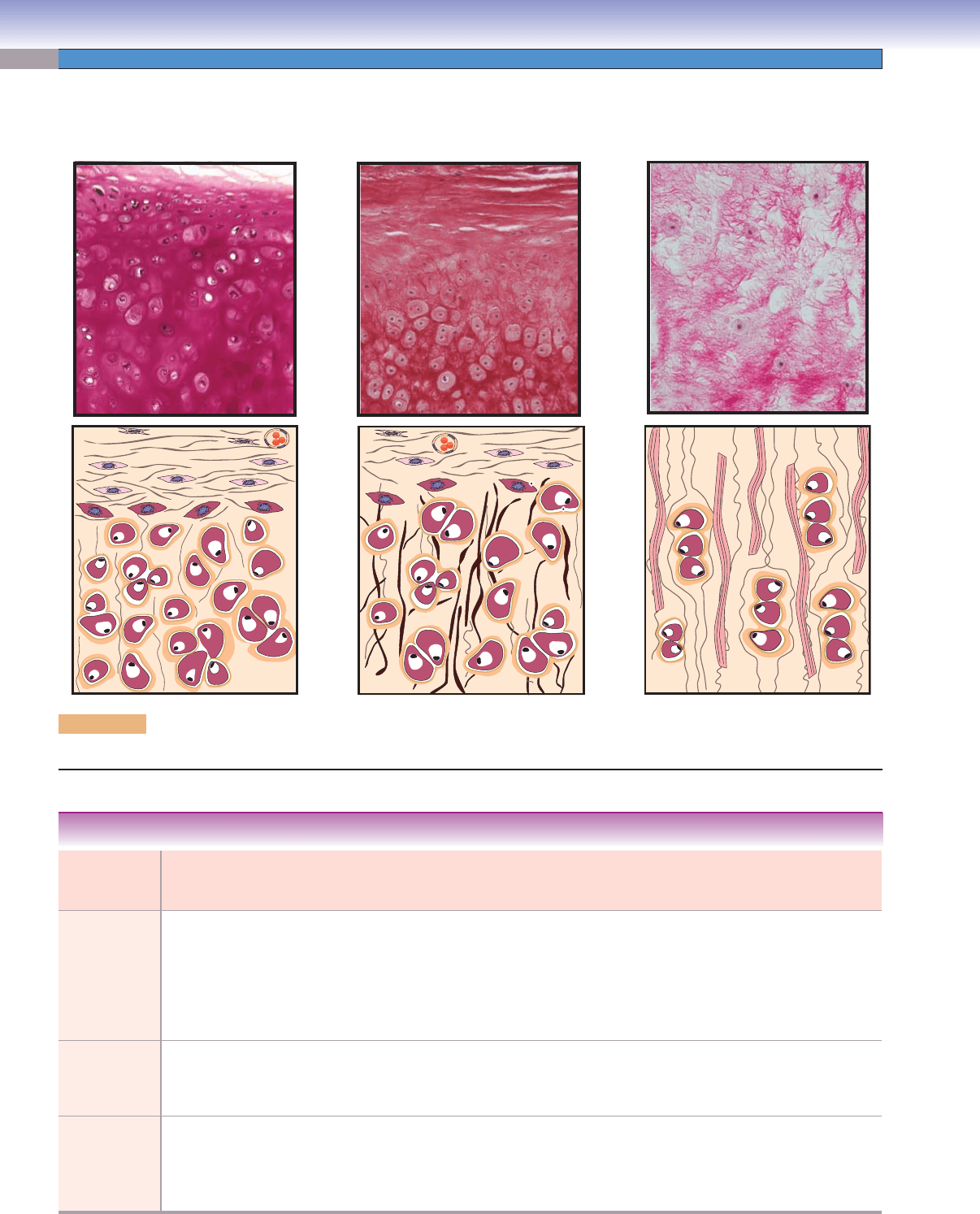
Figure 5-1. Overview of cartilage types. Cartilage can be classifi ed into three types (hyaline, elastic, and fi brocartilage) based on
the characteristics of the matrix.
D. Cui /T. Yang
D. Cui /T. Yang
Hyaline cartilage
Elastic cartilage
Fibrocartilage
Types of
Cartilage
Characteristics of
the Extracellular
Matrix
Chondrocyte
Arrangement
Perichondrium
Coverage
Main Locations Main Functions
Hyaline
cartilage
Type II collagen Mostly in groups
(isogenous groups)
Yes, except
articular
cartilage surface
Trachea, bronchi,
ventral ends
of ribs, nose,
articular ends and
epiphyseal plates
of long bones
Confers shape and
fl exibility (respiratory
tract); forms cartilage
model for bone growth in
the fetus; forms smooth
surface to provide free
movement in joints
Elastic
cartilage
Type II collagen
and elastic fi bers
Mostly in groups
(isogenous groups)
Yes Epiglottis, larynx,
pinna of the ear,
and auditory canal
and tube
Confers shape and
elasticity
Fibrocartilage Type II and type I
collagen
Most are small and
sparsely arranged
in parallel columns
or rows
No Articular disks,
intervertebral disks,
pubic symphysis,
and the insertion of
tendons
Provides resistance to
compression, cushioning,
and tensile strength
TABLE 5-1 Cartilage
CUI_Chap05.indd 82 6/2/2010 6:29:47 PM
CHAPTER 5
■
Cartilage and Bone
83
D. Cui /T. Yang
Chondrocyte
Territorial matrix
Type II collagen
Interterritorial matrix
Isogenous group
Chondroblast
Chondrogenic cell
Inner cellular layer
of the perichondrium
Outer fibrous layer
of the perichondrium
A
Chondrocytes
Chondrocytes
Isogenous
Isogenous
group
group
Chondrocytes
Chondroblasts
Isogenous
group
Perichondrium
Chondroblasts
B
Chondrocytes
Chondrocytes
Interterritorial
Interterritorial
matrix
matrix
Chondrocytes
Interterritorial
matrix
Territorial
Territorial
matrix
matrix
Territorial
matrix
C
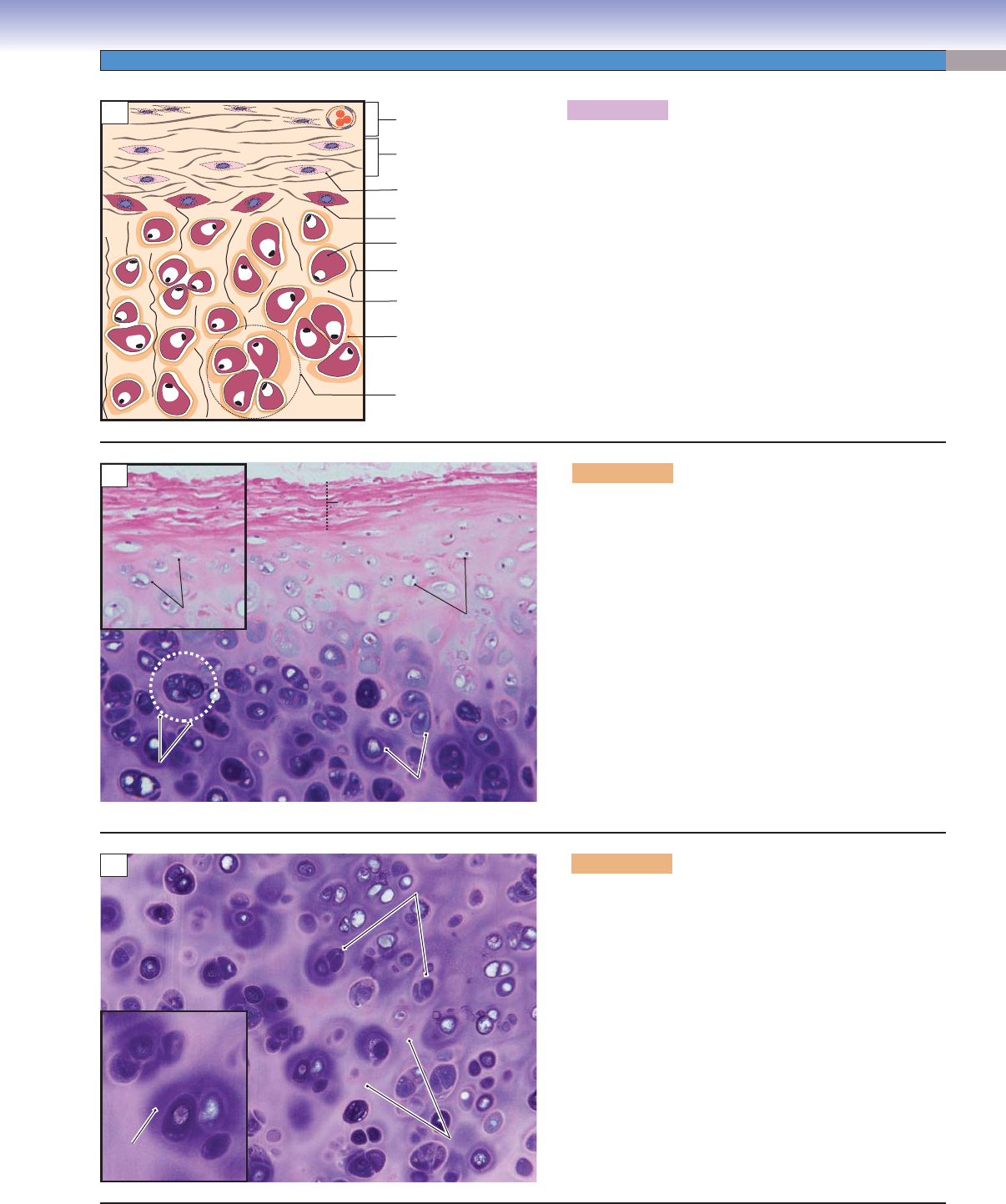
Figure 5-2A. A representation of hyaline cartilage.
Hyaline cartilage is the most common of the three types of
cartilage (hyaline cartilage, elastic cartilage, and fi brocar-
tilage). It can be found in the trachea, bronchi, distal ends
of ribs, and articular ends and epiphyseal plates of long
bones. Most hyaline cartilage is covered by perichondrium,
a dense irregular connective tissue sheath. However, the
hyaline cartilage in the articular joint surfaces of long bones
is an exception (Fig. 5-3B). Hyaline cartilage is composed
of chondroblasts, chondrocytes, delicate collagen (type II
collagen), and a homogenous ground substance (matrix),
which makes it glassy in appearance. The matrices include
the territorial matrix and the interterritorial matrix.
Figure 5-2B. Hyaline cartilage, bronchus. H&E,
139; inset 167
This is an example of hyaline cartilage in the bronchus.
Cartilage is an avascular tissue; nutrients are supplied
through matrix diffusion. The perichondrium, a dense
irregular connective tissue sheath surrounding the surface
of the hyaline cartilage, provides the nearest blood sup-
ply to the cartilage. The perichondrium consists of (1) an
outer fi brous layer, which is composed of type I collagen,
fi broblasts, and blood vessels and (2) an inner cellular
layer, which contains chondrogenic cells that give rise to
new chondroblasts. These cells are fl attened cells, which
actively secrete matrix and often are located beneath the
perichondrium (Fig. 5-2A). Chondrogenic cells are dif-
fi cult to identify under the light microscope with H&E
stain. The chondrocytes are often arranged in small clus-
ters called isogenous groups, which contribute to intersti-
tial growth (Figs. 5-2A and 5-3A).
Figure 5-2C. Hyaline cartilage, bronchus. H&E, 136;
inset 251
The chondrocytes have small, round nuclei and shrunken,
pale-staining cytoplasm, which contains large Golgi appa-
ratuses and lipid droplets. The matrix that surrounds each
chondrocyte or isogenous group is called the territorial
matrix. The matrix that fi lls in the space between isoge-
nous groups and chondrocytes is called the interterritorial
matrix (Fig. 5-2A). In general, the territorial matrix stains
darker than the interterritorial matrix in H&E.
CUI_Chap05.indd 83 6/2/2010 6:29:51 PM
84
UNIT 2
■
Basic Tissues
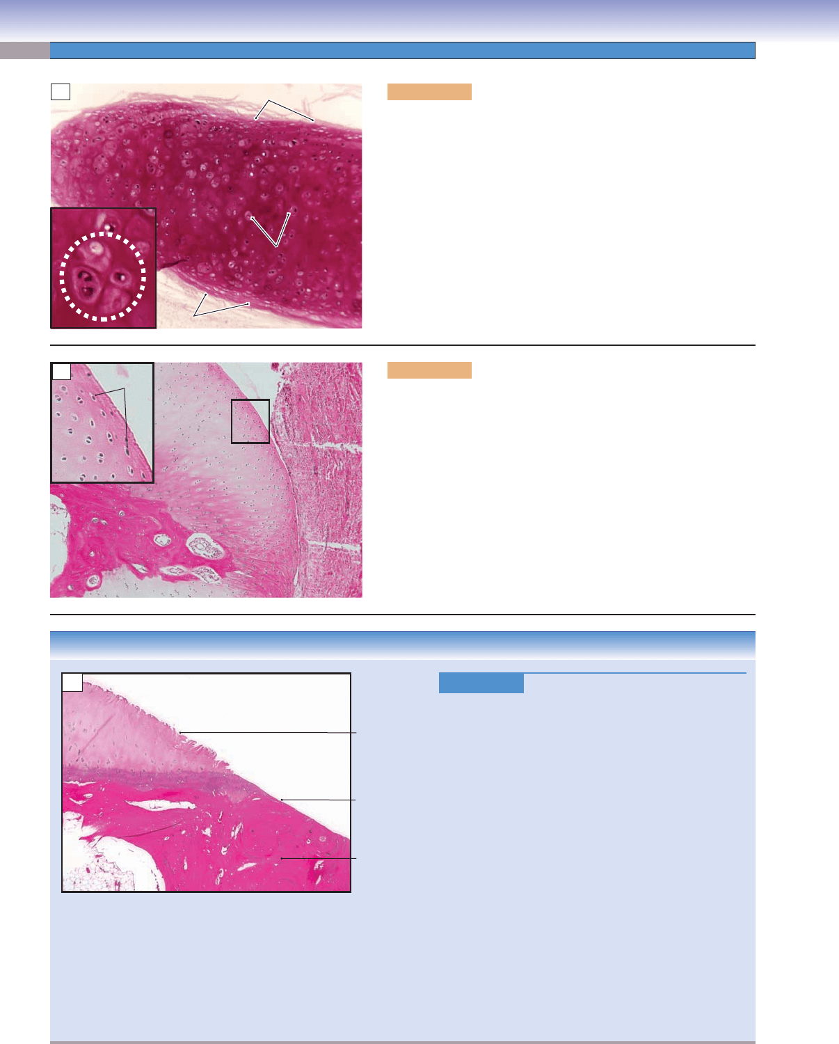
Figure 5-3A. Hyaline cartilage, trachea. H&E, 68; inset 276
This is an example of hyaline cartilage in the trachea. The hyaline
cartilage forms a structural framework to support tissues such as
the larynx, trachea, and bronchi in the respiratory tract. The chon-
drocytes are mature chondroblasts that are embedded in the matrix.
Each chondrocyte is contained within a small cavity in the matrix
called a lacuna; sometimes, one lacuna may contain two cells (Fig.
5-4).
Isogenous
Isogenous
group
group
Isogenous
group
Chondrocytes
Chondrocytes
Chondrocytes
Perichondrium
Perichondrium
Perichondrium
Perichondrium
Perichondrium
Perichondrium
A
Bone
Bone
Hyaline cartilage
Small, flattened
chondrocytes
Joint
cavity
Bone
B
CLINICAL CORRELATION
Figure 5-3C.
Osteoarthritis. H&E, 29
Osteoarthritis is a chronic condition that is charac-
terized by a gradual loss of hyaline cartilage from the
joints. It commonly affects the hand, knee, hip, spine,
and other weight-supporting joints. Risk factors include
genetic factors, aging, obesity
, female gender, injury,
and wear and tear of the joints. Symptoms and signs
include joint pain that is worsened by physical activ-
ity and relieved by rest, morning stiffness, and changes
in the shape of affected joints. There are two types of
osteoarthritides: idiopathic and secondary. Idiopathic
has no obvious cause, whereas secondary has an identi-
fi able cause. Monocyte-derived peptides cause chondro-
cytes to proliferate. Increased numbers of chondrocytes
release degradative enzymes, which cause inadequate
repair responses and subsequent infl ammation in car-
tilage, bone, and synovium. Cartilage fragments and
soluble proteoglycan and type II collagen can be found
in the synovial fl uid. This illustration shows the rough
surface of the hyaline cartilage with fi brillations and
eburnation as a result of softening, thinning, and loss of
the articular cartilage and exposure of the subchondral
bone, which becomes worn and polished.
Fibrillation
of articular
cartilage
Eburnation
Subchondral
bone
C
Figure 5-3B. Hyaline cartilage, fi nger bone. H&E, 68; inset
189
This is an example of the hyaline cartilage in the articular ends of a
long bone (fi nger bone). The cartilage that covers the articular sur-
face of the bone is called articular cartilage. In this particular region,
the cartilage is exposed without perichondrium present. The surface
area of the articular cartilage is composed of small, dense, fl attened
chondrocytes, which enable it to resist pressure and form a smooth
surface to provide free movement in the presence of a lubricating
fl uid (synovial fl uid).
CUI_Chap05.indd 84 6/2/2010 6:29:55 PM
