Cui Dongmei. Atlas of Histology: with functional and clinical correlations. 1st ed
Подождите немного. Документ загружается.

CHAPTER 3
■
Epithelium and Glands
45
D. Cui
Connective tissue
Basal cuboidal cell
Dome-shaped cell
Basement membrane
B
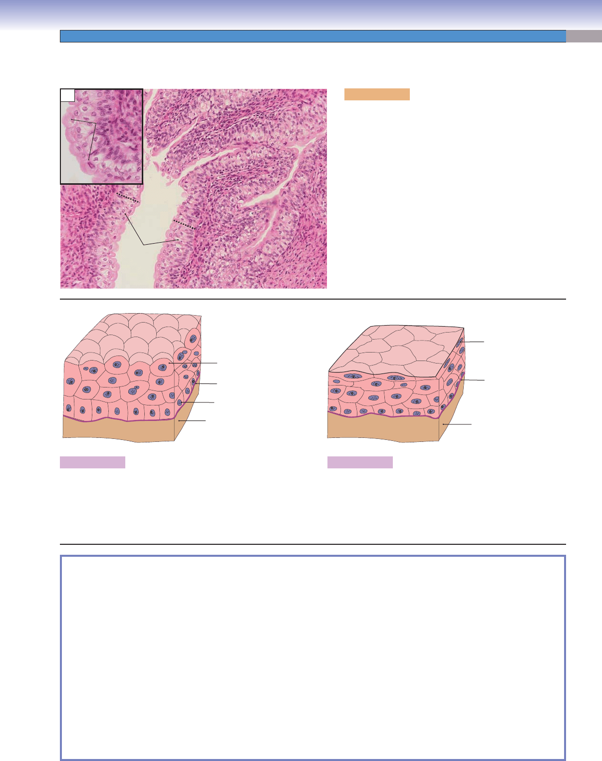
Figure 3-17B. Transitional epithelium (relaxed).
The relaxed, normal transitional epithelium is composed of
four to six layers of cells. The cells located basally are smaller,
low columnar or cuboidal cells. By contrast, the cells located
in the most superfi cial layer are larger and exhibit a rounded,
dome shape that bulges into the lumen.
D. Cui
Connective tissue
Basement membrane
Flattened top cell
C
Figure 3-17C. Transitional epithelium (distended).
A presentation of transitional epithelium in the distended state
is shown. These cells change shape according to the degree
of distention of the bladder. When the transitional epithe-
lium is stretched, the top dome-shaped cells become fl attened
squamous cells and the epithelium becomes thinner.
SYNOPSIS 3-2 Pathological Terms for Epithelial Tissue
Metastasis ■ : The spread of a malignant neoplasm from its site of origin to a remote site, usually through blood and
lymphatic vessels (Fig. 3-2C).
Dyslipidemia
■ : A general term describing a disorder of lipoprotein metabolism causing abnormal amounts of lipids and
lipoproteins in the blood; certain dyslipidemias constitute a major risk factor for the development of atherosclerosis, such
as hypercholesterolemia (Fig. 3-3C).
Osteomalacia
■ : Abnormal bone mineralization producing weak, soft bones; may be caused by vitamin D defi ciency or
kidney disorders, including renal Fanconi syndrome (Fig. 3-6C).
Metaplasia
■ : The reversible process by which one mature cell type changes into another mature cell type, as in squamous
metaplasia of respiratory or glandular epithelia (Figs. 3-9C and 3-14C).
Microabscess
■ : Collection of neutrophils and neutrophil debris within the parakeratotic scale in the skin disease psoriasis
(Fig. 3-13C).
Micropustule
■ : Collection of neutrophils within the epidermis, abutting the parakeratotic scale in the skin disease psoriasis
(Fig. 3-13C).
Parakeratosis
■ : Persistence of the nuclei of keratinocytes into the stratum corneum of the skin or mucous membranes;
parakeratotic scales containing neutrophils are seen in the skin disease psoriasis (Fig. 3-13C).
Connective
Connective
tissue
tissue
Connective
Connective
tissue
tissue
Transitional
epithelium
Lumen
Dome shaped
Dome shaped
surface cells
surface cells
Connective
tissue
Connective
tissue
Dome shaped
surface cells
A
Figure 3-17A. Transitional epithelium, urinary blad-
der. H&E, 155; inset 250
Transitional epithelium has the special characteristic of
being able to change shape to accommodate a volume
change in the organ it lines. In the relaxed state, tran-
sitional epithelium contains four to six cell layers, and
each surface cell appears dome shaped, often contain-
ing two nuclei (these cells are “binucleate”). This picture
illustrates transitional epithelium in a relaxed state (in
most slides, this tissue is unstretched and the surface cells
appear dome shaped). The lumen of the bladder appears
as a white space. The transitional epithelium lining the uri-
nary tract including the bladder, ureter, and major calyces
of the kidney is also referred to as urothelium. The black
dashed line indicates the thickness of the epithelium.
Transitional Epithelium (Stratifi ed Epithelium)
CUI_Chap03.indd 45 6/2/2010 4:57:20 PM

46
UNIT 2
■
Basic Tissues
Types of
Epithelia
Number of
Layers
Type of Cells in the
Epithelium
Apical
Surface
Main Locations
(Lining)
Main Functions
Simple
squamous
epithelium
One Flattened, squamous
epithelial cells
Smooth Cornea, blood, and
lymphatic vessels—
endothelium; surface
of body cavities—
mesothelium (pleural,
pericardial, peritoneal);
alveoli in the lung
Fluid transport,
lubrication, and
exchange
Simple
cuboidal
epithelium
One Cuboidal epithelial cells
(height equal to width)
Smooth/short
microvilli;
long microvilli
depending on
location
Kidney tubules, thyroid
follicles; small ducts
of exocrine glands and
surface of ovary
Absorption,
secretion, and
transportation
Simple
columnar
epithelium
One Absorptive columnar cells
and secretary cells, such as
goblet cells
Mostly
microvilli;
cilia in some
locations
Most of digestive
tract and gallbladder;
oviducts and ductuli
efferentes
Secretion,
absorption,
protection, and
transportation
Pseudostratifi ed
columnar
epithelium
One Ciliated columnar cells,
goblet cells, and short basal
cells not reaching lumen; all
cells rest on the basement
membrane
Mostly cilia;
stereocilia
in some
locations
Most of respiratory
tract; ductus deferens
and epididymis
Secretion,
transportation,
and absorption
Stratifi ed
squamous
epithelium
Several Flattened surface cells,
polygonal cells in the
middle layers, and cuboidal
cells in basal layer
Keratinized
or nonkerati-
nized surface
layer
Epidermis of the skin;
oral cavity, epiglottis,
and esophagus; vagina
Protection
(barrier)
Stratifi ed
cuboidal
epithelium
Two to three Cuboidal cells Mostly
smooth
Large ducts of exocrine
glands and ducts
of sweat gland (not
common type)
Transportation
Stratifi ed
columnar
epithelium
Two to three Low columnar surface cells
and cuboidal basal cells
Smooth Large ducts of exocrine
glands; conjunctiva of
the eye (not common
type)
Transportation
and protection
Transitional
epithelium
Four to six layers
(relaxed); two
to three layers
(distended)
Dome-shaped surface cells
(relaxed), polygonal in the
middle layer, cuboidal cells
in the basal layer
Smooth Urinary tract
Transportation
and protection
(distensible
property)
TABLE 3-2 Epithelium
SYNOPSIS 3-3 Functions of Epithelial Tissue
Promotes gliding between two surfaces (mesothelium of pleural cavity [Fig. 3-2A,B]). ■
Senses changes in blood pressure, oxygen tension, and blood fl ow and controls blood coagulation (endothelium of blood ■
vessels [Fig. 3-3A,B]).
Pumps the excess fl uid out of the stroma and keeps the cornea clear (simple squamous epithelium in cornea).
■
Mediates gas exchange (type 1 pneumocytes and simple squamous epithelium in the alveoli of the lung). ■
Absorbs material from a lumen (simple cuboidal epithelium in kidney and simple columnar epithelium in small and large ■
intestines [Figs. 3-6A,B and 3-7A,B]).
Transports material along a surface (pseudostratifi ed ciliated columnar epithelium in the respiratory tract [Fig. 3-9A,B]).
■
Provides conduit for fl uid (simple and stratifi ed cuboidal and columnar epithelia forming ducts of some large exocrine ■
glands [Fig. 3-15A,B]).
Protects the body from abrasion and injury (stratifi ed squamous epithelium in the skin and esophagus [Figs. 3-13A,B and
■
3-14A,B]).
Becomes highly distensible when the bladder is fi lled with urine and the tissue is stretched (transitional epithelium in
■
bladder [Fig. 3-17A–C]).
Secretes mucus, hormones, and proteins (secretory epithelium, glands [Figs. 3-18 and 3-19]).
■
CUI_Chap03.indd 46 6/2/2010 4:57:23 PM

CHAPTER 3
■
Epithelium and Glands
47
Glands
Introduction and Key Concepts for Glands
Glands are composed of epithelial tissue and can be classifi ed
as endocrine and exocrine according to how the secretory prod-
uct leaves the gland. Endocrine glands release their products into
interstitial fl uid or directly into the bloodstream (see Chapter 17,
“Endocrine System”). Exocrine glands are discussed in this chap-
ter; these glands secrete their products either through ducts into
the lumen of an organ or directly onto the body surfaces. Exo-
crine glands can be classifi ed into several categories according to
various criteria.
Exocrine Glands Classifi ed by Product
Exocrine glands can be classifi ed as serous glands, mucous glands,
mixed glands (seromucous), and sebaceous glands, depending on
what type of secretion is produced. (1) Serous glands secrete a watery
proteinaceous fl uid (Figs. 3-18A and 3-19A). The parotid gland, the
gland of von Ebner of the tongue, the pancreas, and sweat glands are
examples of this type of gland. (2) Mucous glands secrete mucus, a
viscous mixture of glycoprotein and water (Figs. 3-18B and 3-19B).
Goblet cells in the small and large intestines, respiratory epithelium
(Fig. 3-20), some glands in the hard and soft palates, and stomach
epithelium are examples of mucous glands. (3) Mixed glands have
both serous and mucous secretions (Fig. 3-18C) and include the
submandibular gland, sublingual gland, and glands in the trachea
and esophagus. (4) Sebaceous glands produce lipids (Fig. 3-18D).
The sebaceous glands in the skin are good examples.
Exocrine Glands Classifi ed by Mechanisms
of Secretion
Exocrine glands can be classifi ed into merocrine secretion,
apocrine secretion, and holocrine secretion based on the path-
way by which the secretory products are released from the cell.
(1) In merocrine secretion, the secretory product is released from
the cell by exocytosis without the loss of cell material (cyto-
plasm). The release of secretory zymogen granules by pancre-
atic acinar cells is an example of merocrine secretion. Merocrine
mechanism is the most common mode. (2) In apocrine secretion,
the secretory product is released together with part of the apical
cytoplasm of the secretory cell. The lipid secretion by epithelial
cells of the mammary gland belongs to this mode of secretion.
(3) In holocrine secretion, the secretory product is released by
disintegration of the entire cell. The secretory cell dies and a
new secretory cell is formed from a nearby basal cell. The fatty
lubricant secretory product, sebum, is released by the cells of
sebaceous glands by holocrine secretion.
Exocrine Glands Classifi ed by Morphology
The exocrine glands also can be classifi ed into unicellular and
multicellular glands depending upon the number of cells that
form the gland.
UNICELLULAR GLANDS are composed of only single cells.
The secretory products are released directly onto the surface of
an epithelium. Goblet cells are an example of this type of gland
(Fig. 3-20A,B).
MULTICELLULAR GLANDS consist of numbers of secretory
cells arranged in different organizations. The multicellular glands
can be further classifi ed into several subtypes according to their
morphology. In general, the terms simple and compound are tied
to their duct shape. Simple glands have unbranched ducts or
lack ducts. Compound glands have branched ducts. The cells of
the multicellular glands are arranged into secretory units in the
form of acini or tubules.
The multicellular glands also can be classifi ed using a com-
bination of duct shape and the shape of secretory units. (1)
Simple tubular glands have no ducts. The secretory cells are
arranged in straight tubules (Fig. 3-21A,B). This type of gland
can be found in small and large intestines. (2) Simple branched
tubular glands do not have ducts, and their secretory cells are
split into two or more tubules (Fig. 3-22A,B). This type of
gland can be found in the stomach. (3) Simple coiled tubular
glands have a long duct, and secretory cells are formed by coiled
tubules (Fig. 3-23A,B). Sweat glands are examples of this type of
gland. (4) Simple acinar glands have a short, unbranched duct;
the secretory cells are arranged in acini form (Fig. 3-24A,B). The
mucus-secreting glands in the submucosa of the penile urethra
are examples of this type of gland. (5) Simple branched acinar
glands have a short, unbranched duct, and their secretory cells
are formed into branched acini (Fig. 3-25A,B). The sebaceous
glands of the skin belong to this type. (6) Compound tubular
glands have branched ducts. Their secretory cells are formed
into branched tubules as can be found in the Brunner glands
of the duodenum (Fig. 3-26A,B). (7) Compound acinar glands
have branched ducts, and the secretory units are branched acini
(Fig. 3-27A,B). The pancreas and mammary glands are exam-
ples of this type of gland. (8) Compound tubuloacinar glands
have branched ducts, and the secretory units are formed by both
an acinar component and a tubular component (Fig. 3–28A,B).
The submandibular and sublingual glands are good examples of
this type of gland.
Duct System
The compound glands often have complex duct systems. The
secretory acini or tubules are arranged in lobules. The secretory
cells empty their products into small ducts called small intral-
obular ducts, which are often referred to as intercalated ducts.
The small intralobular ducts drain secretory products into larger
intralobular ducts, which in salivary glands are called striated
ducts. The striated ducts are so named because the basal cyto-
plasm of these cells often appears “striped” under the micro-
scope. The striped appearance is a result of the arrangement of
the basal cytoplasm into deep folds packed with mitochondria.
This organization provides the large surface area and genera-
tion of energy needed for extensive pumping of ions across the
basolateral membrane of the cell. Some glands, such as the pan-
creas, have intercalated ducts but not striated ducts. In general,
the ducts located inside of lobules are called intralobular ducts;
and ducts located between lobules are called interlobular ducts.
The large intralobular ducts feed into the interlobular ducts; the
interlobular ducts course through the connective tissue (septa)
between the lobules. The secretory products then pass through
the major ducts, the lobar ducts. Finally, lobar ducts feed into
the main duct of the gland and the secretory products exit the
organ.
CUI_Chap03.indd 47 6/2/2010 4:57:23 PM
48
UNIT 2
■
Basic Tissues
Exocrine Glands Classifi ed by Product
The exocrine glands can be classifi ed as serous glands, mucous glands, mixed glands (seromucous), and sebaceous glands, depending
on what type of secretion is produced.
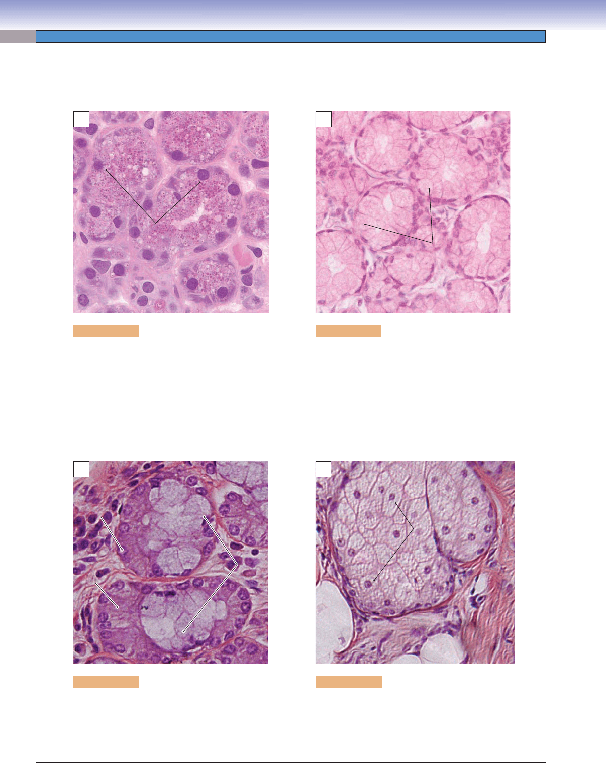
Figure 3-18A. Parotid gland. H&E, 668
An example of a serous gland from the parotid
gland is shown. Each serous secretory cell has a
spherical nucleus, the cytoplasm is basophilic, and
the secretory vesicles (granules) are located in the
apical part of the cytoplasm. These serous cells are
organized in acini and produce a watery proteina-
ceous secretion.
A
Serous
Serous
cells
cells
Serous
cells
B
Mucous
Mucous
cells
cells
Mucous
cells
Figure 3-18B. Duodenum. H&E, 396
An example of a mucous gland in the duodenum
is shown. The mucous secretory cell has a fl attened
nucleus at the base of the cell and an empty vacuo-
lated appearance of the apical cytoplasm. This refl ects
the removal of the mucus from the secretory gran-
ules during processing of the specimen. These cells
are arranged in tubules and produce gellike mucin
(glycoprotein and water mixture) secretions that
usually protect or lubricate epithelial cell surfaces.
C
Serous
Serous
cells
cells
Serous
cells
Serous
Serous
demilune
demilune
Serous
demilune
Mucous
Mucous
cells
cells
Mucous
cells
Figure 3-18C. Sublingual gland. H&E, 609
An example of a mixed gland, the sublingual
gland, containing both mucous secretory por-
tions and serous secretory portions is shown. The
serous cells forming a moon-shaped cap on top of
the mucus are called a serous demilune.
D
Lipid secretary
Lipid secretary
cells
cells
Lipid secretory
cells
Figure 3-18D. Skin (scalp). H&E, 306
An example of a sebaceous gland in the scalp is
shown. The sebaceous gland cells are tightly packed
together in groups. This type of gland produces
sebum, an oily substance that is a mixture of lipids
and debris of dead lipid-producing cells.
CUI_Chap03.indd 48 6/2/2010 4:57:23 PM
CHAPTER 3
■
Epithelium and Glands
49
SYNOPSIS 3-4 Glands Classifi ed by Product
Serous glands ■ : Generate serous product, which is a thin, watery fl uid containing proteins, glycoproteins, and water.
Mucous glands
■ : Produce mucin, which is a thick, viscous material containing high concentration of glycosylated glyco-
proteins and water.
Mixed glands
■ : Consist of both serous and mucous secretary cells and produce serous and mucous materials.
Sebaceous
■ glands: Produce lipids (sebum), which contains an oily substance.
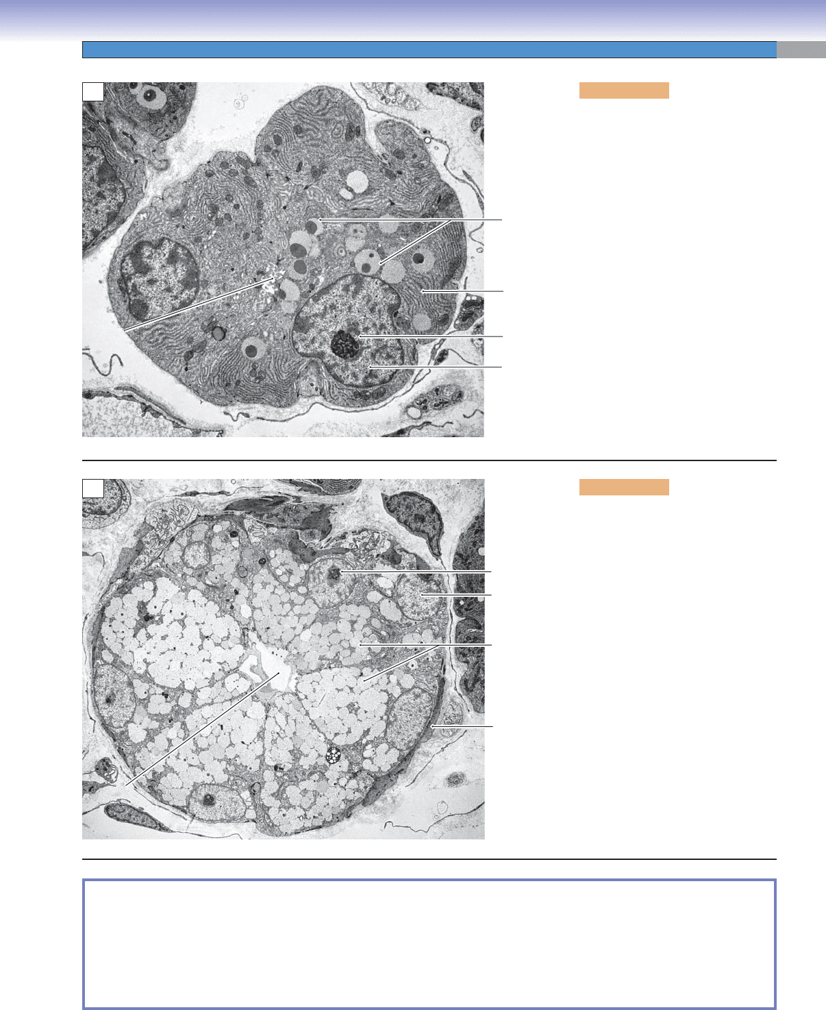
Figure 3-19A. Serous acinus from
the parotid gland. EM, 5,200
Cells that produce serous (thin,
proteinaceous fl uid) secretions have
features in common whether they
are from one of the salivary glands
or from the pancreas. The nuclei are
relatively large with considerable
euchromatin and one or more promi-
nent nucleoli. The cytoplasm in the
basal region is fi lled with RER. The
apical cytoplasm contains secretory
vesicles (granules), which vary in
their staining characteristics.
Nucleous
RER
Secretory
granules
(vesicles)
Nucleus
Lumen
Lumen
Lumen
A
B
Nucleous
Secretory
granules
(vesicles)
Nucleus
Myoepithelial
cell
Lumen
Lumen
Lumen
Figure 3-19B. Mucous acinus
from the submandibular gland.
EM, 6,300
Although mucus-secreting cells have
the same general organization as cells
that produce serous secretions, there
are some distinctions. The mucous
acinus cell has a smaller nucleus,
which is located against the basal
edge of the cell. In addition, the secre-
tory granules are generally more elec-
tron lucent than secretory granules of
serous secreting cells.
CUI_Chap03.indd 49 6/2/2010 4:57:28 PM
50
UNIT 2
■
Basic Tissues
Exocrine Glands Classifi ed by Morphology
The exocrine glands also can be classifi ed into unicellular and multicellular glands depending upon the number of cells that form
the gland.
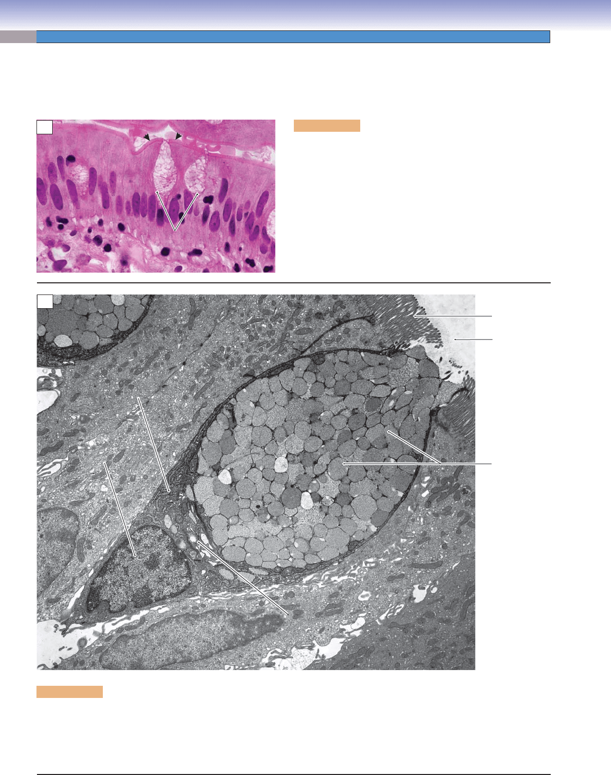
Figure 3-20A. Unicellular gland, small intestine. H&E, 962
Unicellular glands are composed of only a single cell. The secre-
tory products are released directly onto the surface of an epithe-
lium. Goblet cells are an example of this type of gland. Microvilli
with glycocalyx coating form a brush border (arrows). Note that
goblet cells themselves do not have microvilli on their apical
surfaces (see Fig. 3-20B).
Goblet
Goblet
cell
cell
Goblet
cell
B
ru
ru
sh
b
b
o
rd
rd
er
Brush border
Lumen
Lumen
A
Secretory
granules
(mucous granules)
Rough endoplasmic
Rough endoplasmic
reticulum (RER)
reticulum (RER)
Rough endoplasmic
reticulum (RER)
Nucleus
Nucleus
Nucleus
Golgi
Golgi
complex
complex
Golgi
complex
Microvilli
Lumen
B
Figure 3-20B. Goblet cell, unicellular glands (single-cell gland). EM, 5,100
The goblet cell can be considered a single-cell gland because it is commonly inserted into an epithelium among nonsecretory cells. In
this example, the goblet cell is surrounded by enterocytes, absorptive cells of the small intestine. Goblet cells are also found among
ciliated cells in the respiratory epithelium. Typical features of goblet cells are illustrated here. The relatively small heterochromatic
nucleus is packed into the narrow base of the cell along with some RER. A Golgi complex is barely visible adjacent to the apical end
of the nucleus. Most of the cytoplasm is fi lled with secretory vesicles.
CUI_Chap03.indd 50 6/2/2010 4:57:30 PM
CHAPTER 3
■
Epithelium and Glands
51
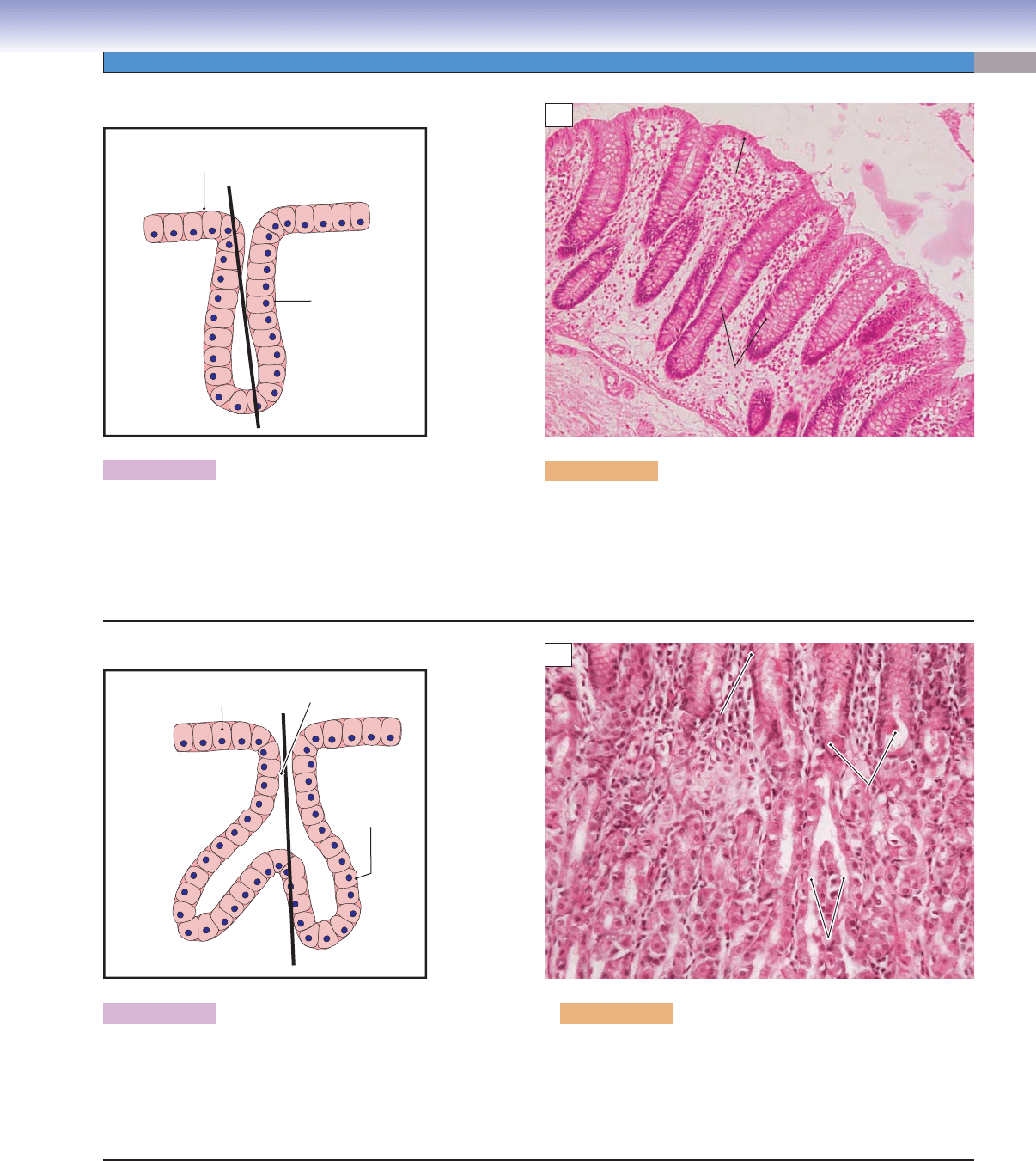
Figure 3-21A. A simple tubular gland.
The secretory cells of this simple tubular gland are
arranged in straight tubules, and the gland has no duct.
The heavy black line represents the approximate plane of
the section in Figure 3-21B.
A
D. Cui
Surface
epithelium
Secretory
cells
B
Surface
Surface
epithelium
epithelium
Secretory
Secretory
cells
cells
Lumen
Surface
epithelium
Secretory
cells
Figure 3-21B. Large intestine. H&E, 99
An example of the simple tubular glands in the large intestine is
shown. The secretory cells (goblet cells) are arranged in straight
tubules, and secretory products are released directly into the
lumen of the intestine. This type of gland also can be found in
the small intestine.
Figure 3-22A. An example of a simple branched
tubular gland.
This type of gland has no duct, and the secretory cells of
the simple branched tubular gland are arranged in two or
more branched tubules. The heavy black line represents
the approximate plane of the section in Figure 3-22B.
Figure 3-22B. Stomach. H&E, 198
An example of the simple branched tubular glands in the
stomach is shown. The surface epithelium invaginates to form
gastric pits. The secretory cells form branched tubular gastric
glands that empty their secretory products into gastric pits.
D. Cui
Surface
epithelium
Gastric pit
Secretory
cells
A
Gastric pits
Gastric pits
Surface
Surface
epithelium
epithelium
Branched
Branched
secretory tubules
secretory tubules
Gastric pits
Surface
epithelium
Branched
secretory tubules
B
CUI_Chap03.indd 51 6/2/2010 4:57:34 PM

52
UNIT 2
■
Basic Tissues
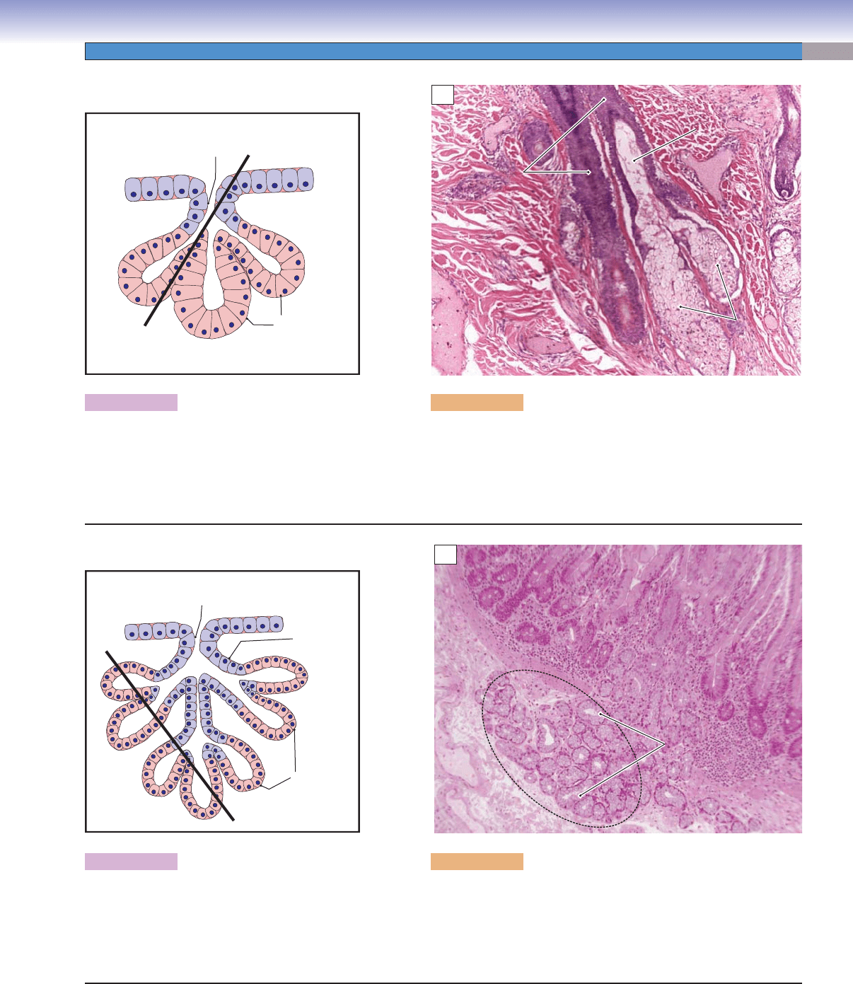
Figure 3-23A. A simple coiled tubular gland.
The simple coiled tubular gland has a long excretory
duct that is unbranched (indicated in blue). The
secretory portions are formed by coiled tubules.
The heavy black line represents the approximate
plane of the section in Figure 3-23B.
Figure 3-23B. Sweat gland of the skin. H&E, 377
Sweat glands in the integument (skin) are examples of simple
coiled tubular glands. The secretory cells form coiled tubules that
are lined by simple cuboidal cells. The excretory ducts are lined
by stratifi ed cuboidal epithelium. The ducts are long, unbranched,
and open at the skin surface.
Figure 3-24A. A simple acinar gland.
The simple acinar gland has a short, unbranched
duct (blue cells). The secretory portion is formed
by secretory cells arranged in an unbranched
acinus or alveolus (a small, grape-shaped secretory
unit). The heavy black line represents the approxi-
mate plane of the section in Figure 3-24B.
Figure 3-24B. Penis. H&E (perfusion), 158
Small mucous glands (Littré glands) in the submucosa of the male
urethra are examples of simple acinar glands. They have very short
excretory ducts that are directly linked to the surface of the epithe-
lium. The mucous secretory cells are arranged in acinar form.
D. Cui
Excretory
duct
Secretory
cells
A
Secretory
Secretory
cells
cells
Duct-forming cells
Duct-forming cells
Secretory
cells
Duct-forming cells
B
D. Cui
Secretory
cells
Excretory
duct
A
Secretory
Secretory
cells
cells
Secretory
cells
B
CUI_Chap03.indd 52 6/2/2010 4:57:37 PM
CHAPTER 3
■
Epithelium and Glands
53
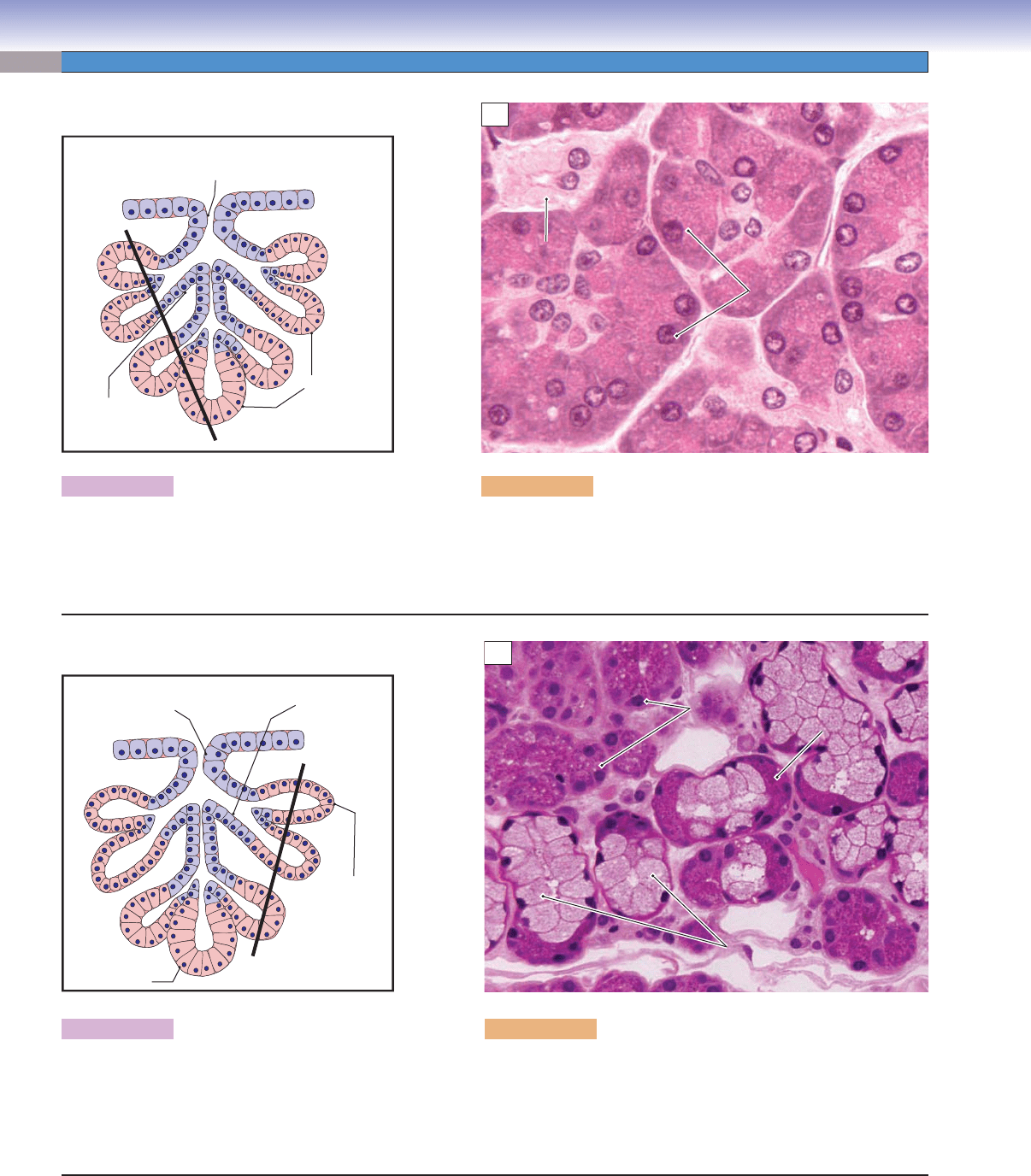
Figure 3-25A. A simple branched acinar gland.
The simple branched acinar gland has a short,
unbranched duct (blue cells). The secretory
portions are branched acini. The heavy black line
represents the approximate plane of the section in
Figure 3-25B.
Figure 3-25B. Skin. H&E, 78
The sebaceous glands in the skin are good examples of a simple
branched acinar gland. The secretory cells are arranged in several
acinar units and open into a short excretory duct. The secretory
product, sebum, is discharged from the acini through a short duct
into the hair follicle.
Figure 3-26A. A compound tubular gland.
The compound tubular gland has branched
ducts (blue cells). The secretory portions are
formed by branched tubules. The heavy black line
represents the approximate plane of the section in
Figure 3-26B.
Figure 3-26B. Duodenum. H&E, 83
The Brunner glands in the duodenum are examples of compound
tubular glands. The mucous secretory cells are arranged in tubular
components. The secretory products exit through branched ducts
into the lumen of the duodenum.
D. Cui
Excretory
duct
Acinar
components
A
Hair
Hair
follicle
follicle
Excretory
Excretory
duct
duct
Acinar
Acinar
components
components
Hair
follicle
Excretory
duct
Acinar
components
B
D. Cui
Excretory
duct
Tubular
components
Branched
duct
A
Secretory tubular
Secretory tubular
portion
portion
Secretory tubular
portion
Brunner
Brunner
glands
glands
Brunner
glands
B
CUI_Chap03.indd 53 6/2/2010 4:57:42 PM
54
UNIT 2
■
Basic Tissues
Figure 3-27A. A compound acinar gland.
The compound acinar gland has branched ducts
(blue cells), and the secretory units are branched
acini. The heavy black line represents the approxi-
mate plane of the section in Figure 3-27B.
Figure 3-27B. Pancreas. H&E, 812
Exocrine glands of the pancreas are examples of compound acinar
glands. The secretory cells form numbers of acinar compounds, and
the secretory products are evacuated into the duodenum through the
duct system of the glands.
D. Cui
Excretory
duct
Acinar
compounds
Branched
duct
A
Serous acinar
Serous acinar
cells
cells
Serous acinar
cells
Small
Small
duct
duct
Small
duct
B
Figure 3-28A. A compound tubuloacinar gland.
The compound tubuloacinar gland has branched
ducts (blue cells) and branched secretory portions
of tubular and acinar components. The heavy black
line represents the approximate plane of the section
in Figure 3-28B.
D. Cui
Interlobular
duct
Intralobular
duct
Tubular
component
Acinar
component
A
Figure 3-28B. Submandibular gland. H&E, 436
The submandibular glands and sublingual glands are good examples
of this category. The acinar components are made up of serous cells;
the tubular components are formed by mucous cells (see Fig. 3-18C).
There are several levels of excretory ducts, including intralobular
ducts and interlobular ducts. A serous demilune is also visible
Serous
Serous
demilune
demilune
Serous
demilune
Mucous
Mucous
cells
cells
(tubular component)
(tubular component)
Mucous
cells
(tubular component)
Serous
Serous
cells
cells
(acinar component)
(acinar component)
Serous
cells
(acinar component)
B
CUI_Chap03.indd 54 6/2/2010 4:57:46 PM
