Cui Dongmei. Atlas of Histology: with functional and clinical correlations. 1st ed
Подождите немного. Документ загружается.

CHAPTER 2
■
Cell Structure and Function
15
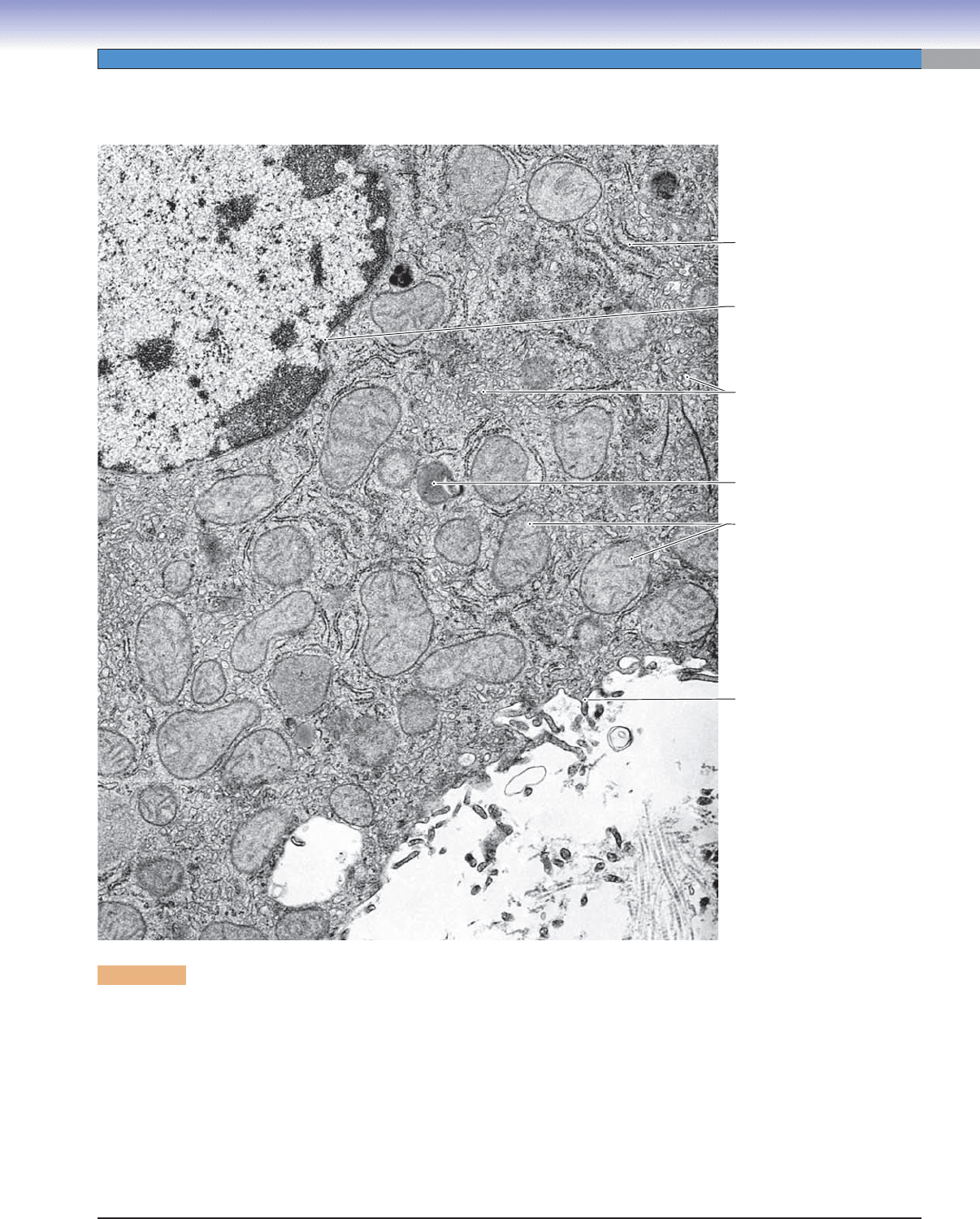
All of the ultrastructural features of cells can be viewed with electron microscopy under optimum conditions of specimen preparation,
orientation, and magnifi cation. The unit membrane that delimits the cell and many of its major components is seen as a thin,
electron-dense line in transmission electron micrographs. Segments of membrane that are oriented vertically to the plane of section
are most sharply defi ned, whereas membrane segments that are oriented at or nearly parallel to the plane of section cannot be readily
discerned. Identifi cation of the major organelles relies, to a great extent, on the characteristic arrangements of the membranes that
defi ne them. Two major structures, the nucleus and the mitochondria, are bounded by double membranes. Identifi cation of mito-
chondria is aided by the infoldings of the inner mitochondrial membrane that form shelfl ike or tubular cristae, extending through the
interior of the organelle. The inner nuclear membrane typically has heterochromatin apposed to it, and the outer nuclear membrane
is often studded with ribosomes. The membrane that forms the RER encloses a cisternal space in the shape of either disks or tubules,
and identifi cation is confi rmed by the presence of polyribosomes on the outer surface of the membrane. SER typically has a branch-
ing tubular form, so that, in sections, patches of circular, ovoid, and Y-shaped profi les of membrane are seen.
Plasmalemma
Rough
endoplasmic
reticulum
Nuclear
envelope
Smooth
endoplasmic
reticulum
Secondary
lysosome
Mitochondria
Figure 2-2. Membranes defi ne the major components and compartments of the cell. EM, 19,000
Cell Ultrastructure
CUI_Chap02.indd 15 6/2/2010 6:25:19 PM

16
UNIT 1
■
Basic Principles of Cell Structure and Function
The outer and inner nuclear membranes and the perinuclear cisternae are readily identifi ed in electron micrographs if there is
adequate magnifi cation and a favorable plane of section. In some cells, the outer nuclear membrane is studded with ribosomes, and
the perinuclear cisternae are continuous with the cisternae of RER. In a cell that is actively synthesizing proteins, the nuclear enve-
lope has numerous nuclear pores that can be identifi ed in electron micrographs as interruptions in the double-membrane arrange-
ment of the nuclear envelope. Some face-on views of nuclear pores can be seen in the inset. It can be seen that the pores are not
simply openings, but rather each has a diaphragm. Chromatin that is highly condensed, or heterochromatin, is much more electron
dense than chromatin that is accessible to transcription, or euchromatin. Clumps of heterochromatin tend to be located adjacent to
the inner nuclear membrane, with gaps that correspond to sites of nuclear pores. Nucleoli appear similar to heterochromatin but can
usually be distinguished by a more complex substructure of granular, fi brous, and nucleolar organizer components.
J. Naftel
J. Naftel
Outer nuclear
membrane
Cisterna of
nuclear envelope
Heterochromatin
Euchromatin
Nucleolus
Rough
endoplasmic
reticulum
Nuclear pore
(vertical section)
Nuclear pores
(face-on view)
Inner nuclear
membrane
Figure 2-3.
The nucleus and its components. EM, 43,000; inset 42,000
CUI_Chap02.indd 16 6/2/2010 6:25:21 PM
CHAPTER 2
■
Cell Structure and Function
17
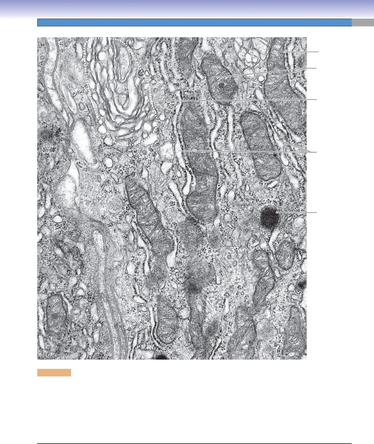
Mitochondria vary both in size and shape and in the appearance of their cristae, yet they are generally the easiest organelles to
identify in transmission electron micrographs. The striking features are the double membrane and the cristae, which are inwardly
directed extensions of the inner mitochondrial membrane. The RER typically consists of fl attened, membrane-delimited sacs. The
distinguishing feature of RER is the presence of ribosomes (polyribosomes) attached to the outer surface of the membrane. SER
also consists of membrane-delimited spaces, but these are usually in the form of a labyrinth of branching tubules. The surface of
the membrane of SER is smooth, with no attached ribosomes. Golgi complexes are composed of stacks of fl attened, membrane-
delimited sacs along with associated vesicles. Shapes of the sacs can vary, but often they are bowl shaped, so that there is a convex
face (the forming face) and a concave face (the maturing face).
Cisternae
of rough
endoplasmic
reticulum
LysosomeLysosome
Golgi complex
Cristae of
mitochondrion
Double
membrane of
mitochondrion
Figure 2-4. Cytoplasmic organelles. EM, 66,000
CUI_Chap02.indd 17 6/2/2010 6:25:28 PM
18
UNIT 1
■
Basic Principles of Cell Structure and Function
Golgi
complex
Cisterna of
rough
endoplasmic
reticulum
Outer
nuclear
membrane
Nuclear
pore
Condensing
(secretory)
vesicle
A
Smooth
endoplasmic
reticulum
Intermediate
filaments
Mitochondrion
Plasmalemma
Nucleus
B
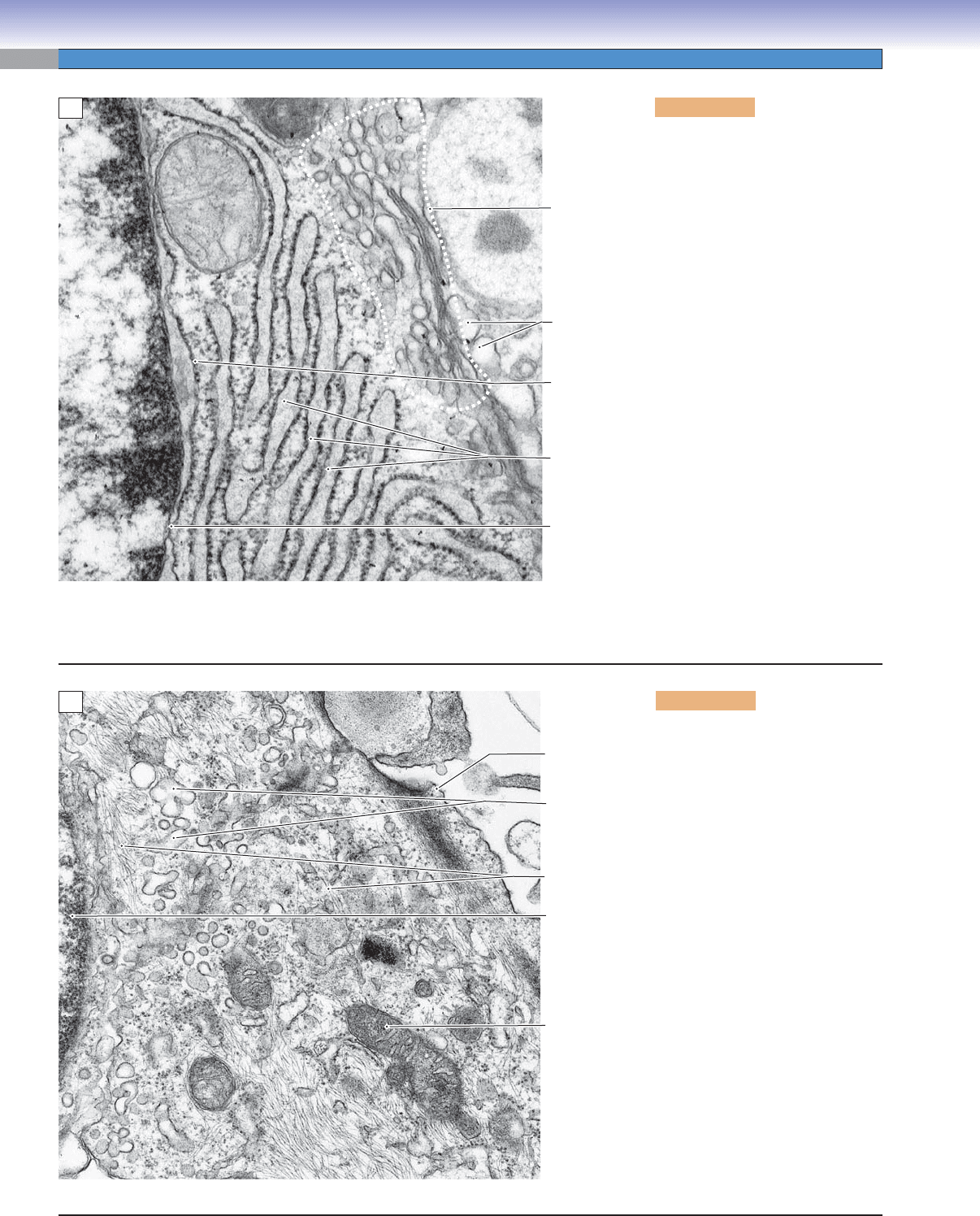
Figure 2-5A. Cytoplasmic organ-
elles: Rough endoplasmic reticulum
and the Golgi complex. EM,49,000
This view includes only a small area
at the edge of the nucleus of a cell
that is actively synthesizing proteins
for secretion. Both euchromatin and
heterochromatin can be seen, but the
nucleolus, although present in the
cell, is not in view here. The nuclear
pore is the gateway for materials
leaving the nucleus, for example,
mRNA, tRNA (transfer RNA), and
preribosomal particles. Entering
the nucleus through nuclear pores
are histones and other proteins of
chromatin, DNA polymerases and
RNA polymerases, and ribosomal
proteins. The outer nuclear mem-
brane is studded with ribosomes, an
indication that the nuclear envelope
is continuous with the RER, which
is abundant in this cell. A part of the
Golgi complex is identifi able as a
stack of fl attened membranous sacs
with smooth surfaces. The small ves-
icles associated with the Golgi com-
plex include transport vesicles that
convey polypeptides from the RER.
Figure 2-5B. Cytoplasmic orga-
nelles: Smooth endoplasmic reticu-
lum. EM, 33,000
Smooth endoplasmic reticulum is
considerably less conspicuous in
appearance than RER with its broad,
fl attened cisternae and arrays of
attached ribosomes. The usual con-
fi guration of SER is a labyrinth of
branching tubules with swellings.
Thus, it presents in sections as pro-
fi les of smooth-surfaced circular or
oval membranes, much the same as
slices through spherical vesicles. The
true distinctive structure of the SER
is revealed by the occasional profi les
that have a Y-shaped or branching
lumen, as can be seen in this image.
The cytoplasm in this view also con-
tains some mitochondria and numer-
ous intermediate fi laments coursing
through the cytosol. Some of the fi la-
ments near the plasmalemma of the
cell are more likely actin fi laments,
which are concentrated in the cortex
(outer layer) of many cells.
CUI_Chap02.indd 18 6/2/2010 6:25:35 PM
CHAPTER 2
■
Cell Structure and Function
19
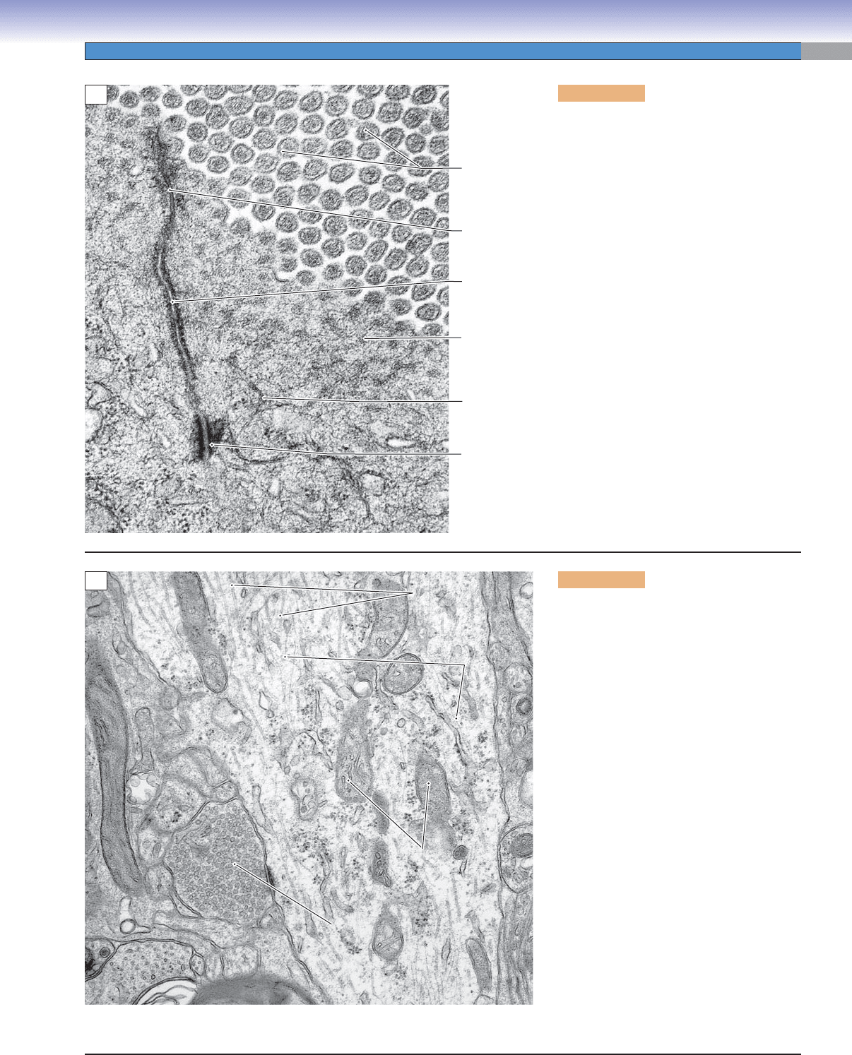
Figure 2-6A. Cell surface and cyto-
skeleton, intestinal absorptive cells. EM,
73,000
This image is restricted to a very small part of
the surfaces of two absorptive cells (entero-
cytes) in the wall of the small intestine. The
plane of section is tangential to the plane of
the surface, so that the right side of the image
shows numerous microvilli that project into
the lumen and function to greatly increase the
surface area exposed to the contents of the
intestine. The electron-dense dots in the cores
of the microvilli are actin fi laments, which,
in this case, provide for stiffness rather than
motility. These actin fi laments extend from
the microvilli into the cytoplasm as part of
the terminal web of actin fi laments near the
cell surface. Components of a junctional
complex provide a seal (zonula occludens)
and adhesion (zonula adherens and macula
adherens) between neighboring enterocytes.
Beneath the terminal web of actin fi laments
are thicker fi laments, intermediate fi laments,
which provide mechanical strength to the
cell. Some of the intermediate fi laments are
anchored in the macula adherens.
Zonula
occludens
Terminal
web
(actin
filaments)
Intermediate
filaments
Macula
adherens
Zonula
adherens
Microvilli
with core
of actin
filaments
A
Microtubules
Microtubules
Microtubules
Intermediate
Intermediate
filaments
filaments
/neuro-
/neuro-
filaments)
filaments)
Intermediate
filaments
/neuro-
filaments)
Mitochondria
Mitochondria
Mitochondria
Synaptic
Synaptic
vesicles
vesicles
Synaptic
vesicles
B
Figure 2-6B. Cytoskeleton, the dendrite
of a neuron. EM, 20,000
The central structure in this view is a
dendrite of a neuron. It is a process extend-
ing from the cell body of the cell. Dendrites
and axons, the other type of neural pro-
cess, require cytoskeletal elements both for
mechanical support and for conveying essen-
tial molecules, particles, and organelles over
distances that can be quite extensive. The
structural support is provided mainly by the
intermediate fi laments, which are termed
neurofi laments in neurons because of their
specialized molecular structure. Intermediate
fi laments appear in electron micrographs as
electron-dense single lines when they course
within the plane of the section or as electron-
dense dots when their orientation is vertical
to the plane of the section. The movement of
molecules and particles along the lengths of
dendrites and axons requires microtubules
as tracks and motor molecules (dyneins
and kinesins) to transport the cargo struc-
tures and molecules along the microtubules.
Because of their tubular structure, microtu-
bules appear as a closely paired set of paral-
lel lines when they course within the plane of
the section or as circles when their orienta-
tion is vertical to the plane of the section.
CUI_Chap02.indd 19 6/2/2010 6:25:38 PM

20
UNIT 1
■
Basic Principles of Cell Structure and Function
Figure 2-7A. Cell components in light micros-
copy. H&E, 1,075
Only the larger cell components can be dis-
tinguished individually by light microscopy.
Examples of such readily identifi able compo-
nents include the nucleus, nucleoli, blocks of
heterochromatin and euchromatin, larger secre-
tory vesicles, and larger secondary lysosomes.
Because of its limited resolution of about 0.2 μm,
light microscopy cannot distinguish smaller cel-
lular components, such as ribosomes, centrioles,
cytoskeletal elements, and most primary lyso-
somes and mitochondria, although the presence
of some of these structures can be inferred by
their infl uence on staining reactions when they
are present in abundance. For example, ribo-
somes, whether free or associated with endoplas-
mic reticulum, impart basophilic staining to a
region of cytoplasm where they are concentrated.
By contrast, mitochondria impart acidophilic
(eosinophilic) staining to regions of cytoplasm
where they are concentrated. In cells with a large
Golgi complex, its presence can sometimes be
distinguished as an unstained region adjacent to
the nucleus.
Bilobed
Bilobed
nucleus
nucleus
Bilobed
nucleus
Secretory granules
Secretory granules
(contents extracted)
(contents extracted)
Secretory granules
(contents extracted)
Secretory granules
Secretory granules
(contents eosinophilic)
(contents eosinophilic)
Secretory granules
(contents eosinophilic)
Hof (Golgi)
Hof (Golgi)
Hof (Golgi)
Heterochromatin
Heterochromatin
Heterochromatin
Euchromatin
Euchromatin
Euchromatin
Nucleolus
Nucleolus
Nucleolus
Inactive nucleus
Inactive nucleus
(heterochromatin)
(heterochromatin)
Inactive nucleus
(heterochromatin)
Basophilic
Basophilic
cytoplasm (RER)
cytoplasm (RER)
Basophilic
cytoplasm (RER)
Granules of
Granules of
eosinophilic
eosinophilic
leukocyte
leukocyte
Granules of
eosinophilic
leukocyte
A
Cytoplasm of neuronal
Cytoplasm of neuronal
cell body with basophilic
cell body with basophilic
patches of RER
patches of RER
Cytoplasm of neuronal
cell body with basophilic
patches of RER
Euchromatin in nucleus
Euchromatin in nucleus
of neuronal cell body
of neuronal cell body
Euchromatin in nucleus
of neuronal cell body
Nucleolus
Nucleolus
Nucleolus
Nucleus of
Nucleus of
satellite cell
satellite cell
Nucleus of
satellite cell
Shrinkage artifact
B
Figure 2-7B. Wide range of cell sizes, spinal
ganglion sensory neuron. H&E, 780
Human cells have a wide range of sizes and shapes.
Some of the largest cell types, such as oocytes and
megakaryocytes, can be as much as 100 μm in diam-
eter. Similar diameters can be seen in cell bodies of
neurons with long axons such as the sensory neurons
in this illustration. The enormity of the cell body in
the center of the image can be appreciated if its size
is compared with that of the satellite cells that sur-
round it. The nucleolus in the nucleus of the neuron
is equal in size to the entire nucleus of the satellite
cell. The large volume of cytoplasm that surrounds
the nucleus of the neuron contains large expanses of
RER and Golgi complexes. As large as the cell body
is, it amounts to less than 1% of the total cell volume
because the axon may be over a meter in length. Cell
types with the smallest volumes include spermato-
zoa, lymphocytes (spherical cells with diameters as
little as 5 μm), and endothelial and alveolar cells
(extremely thin cells that allow exchange of gases
and other materials between compartments).
Cell Structure
CUI_Chap02.indd 20 6/2/2010 6:25:40 PM
CHAPTER 2
■
Cell Structure and Function
21
Basophilic
Basophilic
cytoplasm (RER)
cytoplasm (RER)
Basophilic
cytoplasm (RER)
Nucleus
Nucleus
Nucleus
Site of Golgi
Site of Golgi
complex (hof)
complex (hof)
Site of Golgi
complex (hof)
Nucleolus
Nucleolus
Nucleolus
B
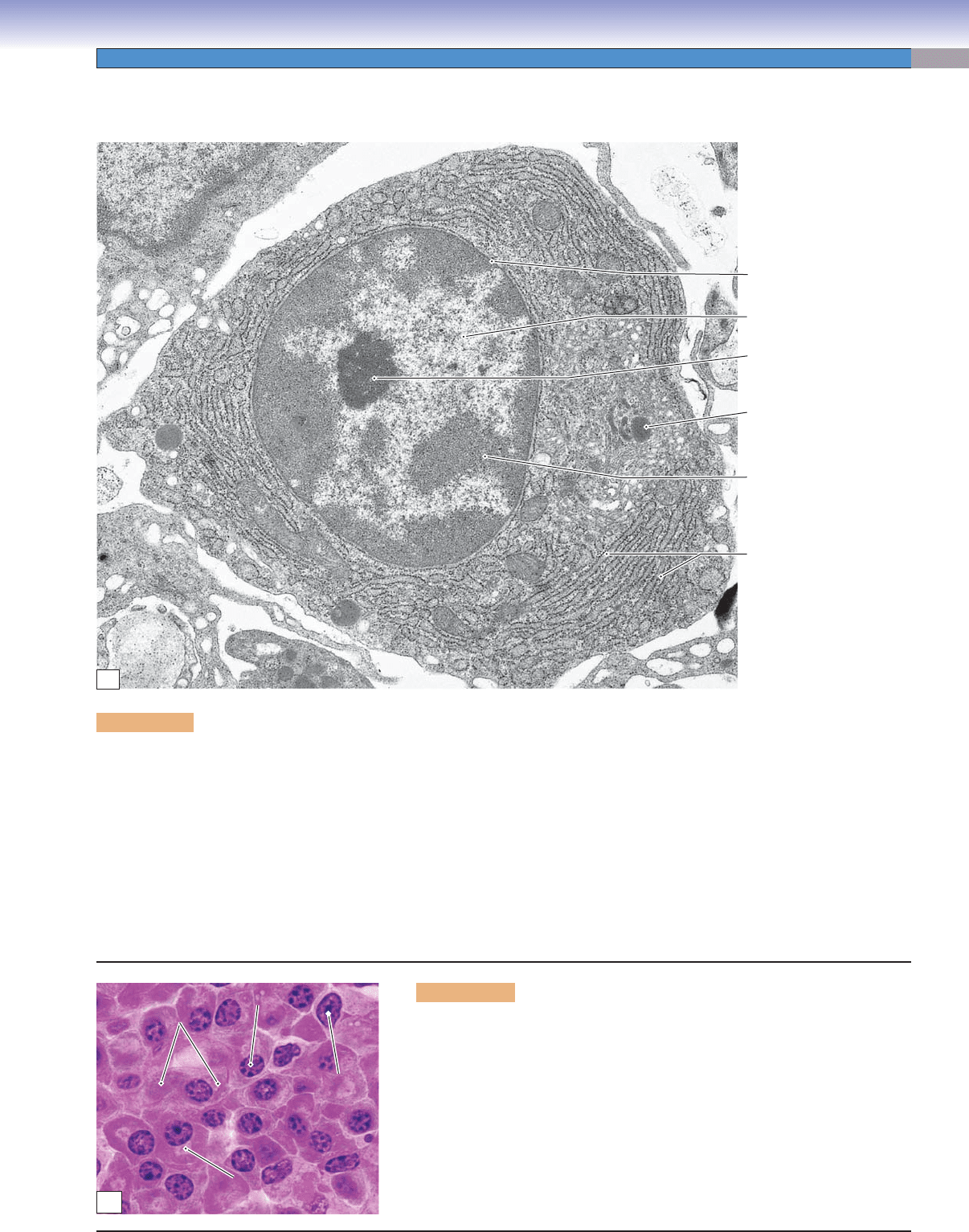
Figure 2-8B. Light microscopic appearance of plasma cells. H&E, 1,200
The appearance of plasma cells in light microscopy is consistent with the
ultrastructure of these antibody-secreting cells. The nucleus has a mixture of
heterochromatin and euchromatin in an arrangement that is variously described
as a “clock face” or checkerboard pattern. A large nucleolus or two occupy the
center of the nucleus. The cytoplasm is basophilic as a result of the ribonucleic
acid associated with the extensive RER. Depending upon the orientation of the
cell in the section, a pale area, called the hof, can be seen adjacent to the nucleus.
This unstained area is the location of the Golgi complex.
Cell Structure Correlates with Function
Plasma cells function to synthesize and secrete immunoglobulin, a glycoprotein. Once these cells have differentiated from stimulated
B lymphocytes, they secrete the antibodies as fast as they are generated for 1 to 2 weeks before they die. The major structures
required for this process are RER, the Golgi complex, and secretory vesicles. The RER is the site of synthesis and sequestering of the
polypeptides of the antibody. Posttranslational modifi cation and packaging occur in the Golgi complex, and the secretory vesicles
convey the product to the cell surface. Because plasma cells do not store the immunoglobulin, few, if any, secretory vesicles are seen
in the cytoplasm. Rather, the cytoplasm is packed with RER, and there is a large Golgi complex (not visible in this section) located
adjacent to the nucleus. The nucleus has one or more well-developed nucleoli, but there is a considerable amount of heterochroma-
tin, considering that this is an active protein-secreting cell. The explanation may be that only one protein, an antibody molecule,
is secreted, and the cell is terminally differentiated, so it will never divide. The immunoglobulin is secreted into the surrounding
interstitial compartment, from which it can enter circulation through walls of small blood or lymph vessels. Even though the cell is
not sharply polarized, the nucleus tends to occupy an eccentric position, with the Golgi complex near the center of the cell.
Figure 2-8A. Protein-secreting cells, plasma cell. EM, 17,000
Rough
endoplasmic
reticulum
Heterochromatin
Nucleolus
Euchromatin
Nuclear
envelope
Secretory
vesicle
A
CUI_Chap02.indd 21 6/2/2010 6:25:48 PM
22
UNIT 1
■
Basic Principles of Cell Structure and Function
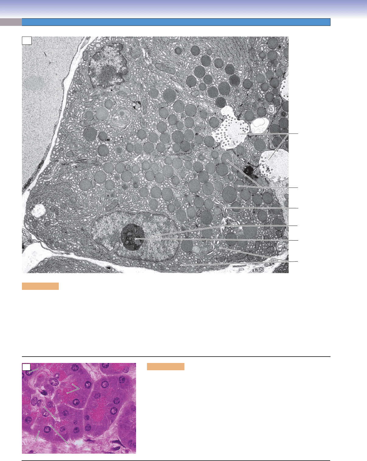
Figure 2-9A. Exocrine protein–secreting cells, pancreatic acinar cell. EM, 7,000
Rough
endoplasmic
reticulum
Golgi
complex
Lumen
Secretory
granules
Euchromatin
in nucleus
Nucleolus
A
Secretory
Secretory
granules
granules
Secretory
granules
Lumen
Lumen
Lumen
Basophilic
Basophilic
cytoplasm
cytoplasm
Basophilic
cytoplasm
Nucleolus
Nucleolus
Nucleolus
Site of Golgi
Site of Golgi
Site of Golgi
B
There are many examples of glandular epithelial cells that function to synthesize and secrete proteins and glycoproteins into a lumen.
All cells have in common the equipment needed for this function, and all cells are distinctly polarized, with a typical arrangement of
cellular constituents. The necessary organelles are the RER, Golgi complex, and secretory vesicles (often called secretory granules).
The RER functions to synthesize polypeptides, sequester them, and initiate posttranslational modifi cations such as glycosylation. The
polypeptides are then shunted by small transport vesicles to the Golgi complex for further modifi cation and packaging into secretory
vesicles, which convey the products to the part of the cell’s plasmalemma that borders a lumen or free surface. The nucleus of an
exocrine protein–secreting cell can vary in shape and position, but it will typically have nucleoli evident and a substantial proportion
of its chromatin in the euchromatin (transcription-capable) form.
Figure 2-9B. Light microscopic appearance of an exocrine protein–
secreting cell. H&E, 750
Light microscopic views of exocrine secretory cells in sections are consistent
with the structures that can be discerned in electron micrographs. The nucleus
usually exhibits nucleoli and a considerable amount of euchromatin. The cyto-
plasm at the basal end of the cell is basophilic owing to the concentration of
RER at this location. The position of the Golgi apparatus may be evident as a
pale, unstained area of cytoplasm at the apical-facing edge of the nucleus. The
sizes and staining characteristics of the secretory granules vary according to
the type of cell and secretory product. In this example, the secretory granules
are acidophilic (eosinophilic).
CUI_Chap02.indd 22 6/2/2010 6:25:51 PM
CHAPTER 2
■
Cell Structure and Function
23
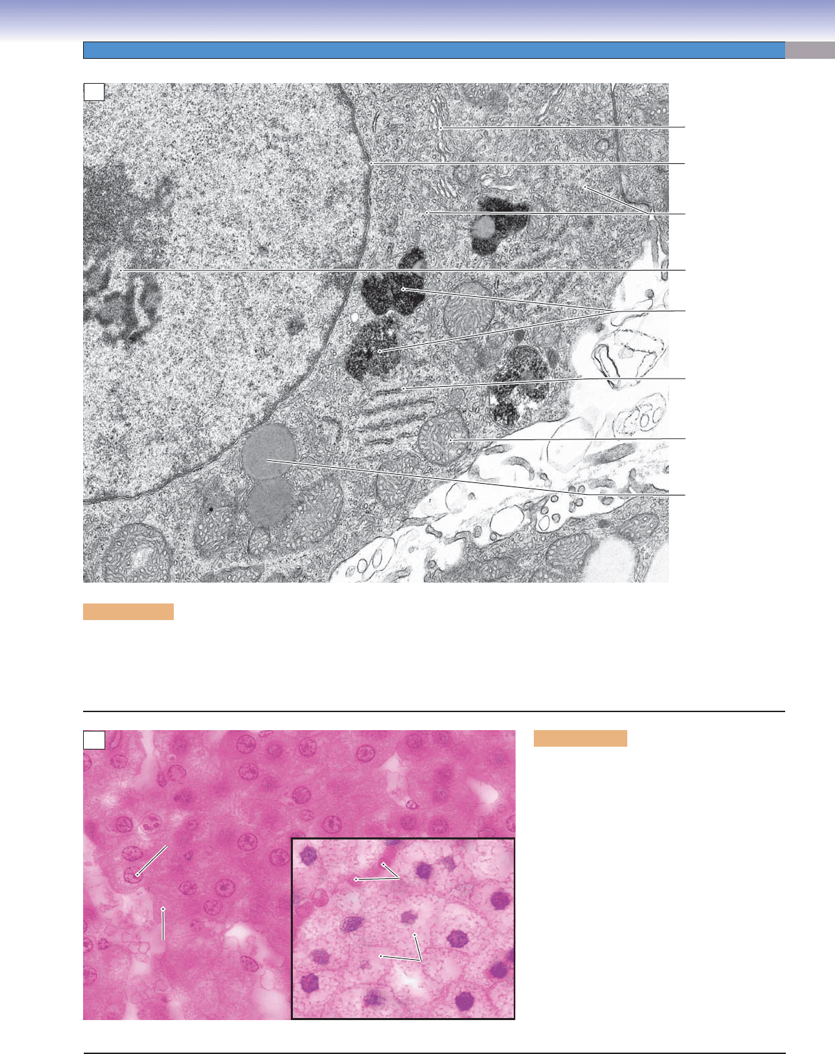
Figure 2-10A. Steroid hormone–secreting cells, adrenal cortex. EM, 21,000
Smooth
endoplasmic
reticulum
Nuclear
envelope
Nucleolus
Mitochondrion
with tubular
cristae
Rough
endoplasmic
reticulum
Lipid
droplet
Secondary
lysosome
Golgi
complex
A
Red cells in
Red cells in
capillary
capillary
Red cells in
capillary
Cytoplasm filled
Cytoplasm filled
with lipid droplets
with lipid droplets
Cytoplasm filled
with lipid droplets
Eosinophilic
Eosinophilic
cytoplasm
cytoplasm
Eosinophilic
cytoplasm
Nucleolus
Nucleolus
in nucleus
in nucleus
Nucleolus
in nucleus
B
Adrenal cortical cells, testicular interstitial cells, and ovarian follicular cells are examples of cell types that function to synthesize and
secrete steroid hormones. Each of these cells have in common the equipment needed for synthesis of steroid hormones. The neces-
sary components include lipid droplets containing cholesterol esters as substrates, SER, and mitochondria that function together in
the synthesis of hormones. The mitochondria characteristically have cristae that are tubular rather than shelfl ike.
Figure 2-10B. Light microscopic appearance
of steroid hormone–secreting cells. H&E, 800
main panel and inset
Light microscopic views of steroid hormone–
synthesizing cells reveal only a hint of the
distinctive set of structures that characterize
the cytoplasm of these cells. The eosinophilia
(acidophilia) of the cytoplasm is largely attribut-
able to the abundance of mitochondria. Steroid
hormone–secreting cells vary in the amount of
cholesterol stored in the form of lipid droplets,
which appear in most preparations as empty
vacuoles because their contents are extracted
during specimen preparation. The cells in the
main panel are in the zona reticularis and have
few droplets, similar to the cell in the electron
micrograph (Fig. 2-10A). The cells in the inset
are from the zona fasciculata and have numer-
ous lipid droplets.
CUI_Chap02.indd 23 6/2/2010 6:25:54 PM
24
UNIT 1
■
Basic Principles of Cell Structure and Function
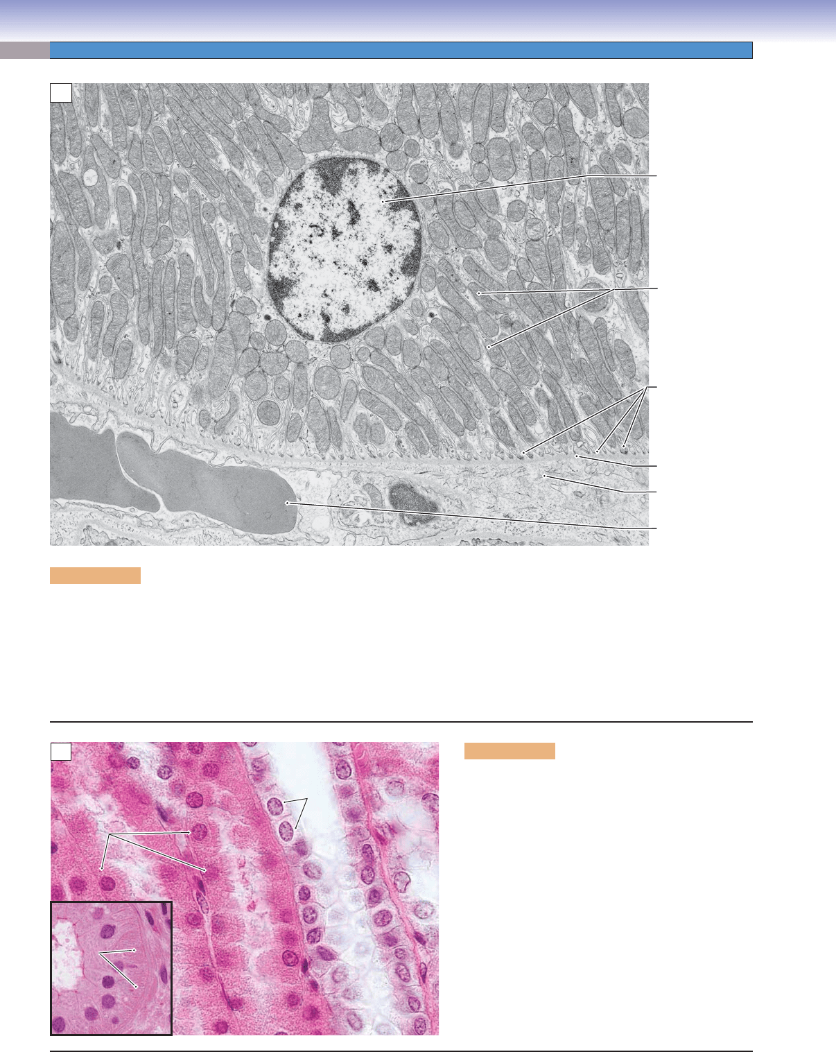
Figure 2-11A. Ion-pumping cells, renal tubule. EM, 9,060
Nucleus
Mitochondria
Basal lamina of
renal tubule cell
Red blood cell
in capillary
Interstitial
compartment
Basolateral folds
of renal tubule
epithelial cell
A
Striations
Striations
Striations
Proximal tubule cells
Proximal tubule cells
with basolateral folds,
with basolateral folds,
ion pumps, and abundant
ion pumps, and abundant
mitochondria
mitochondria
Proximal tubule cells
with basolateral folds,
ion pumps, and abundant
mitochondria
Collecting duct
Collecting duct
cells with channels,
cells with channels,
but without ion pumps
but without ion pumps
Collecting duct
cells with channels,
but without ion pumps
Striated duct
Renal medulla
B
Examples of epithelial cells that function primarily to move ions from a lumen to the interstitial compartment form the walls of
some renal tubules and of some ducts of salivary glands. Perhaps surprisingly, the molecular pumps are located not in the apical
cell membrane but rather in the plasmalemma of the basal and lateral surfaces of the cells. These ATP-requiring pumps transfer
sodium ions from the cytosol of the cell into the interstitial compartment. Sodium ions are pulled from the lumen at the apex of the
cell by the gradient created by the basolateral pumps. The surface area of the membrane containing the pumps is greatly increased
by numerous, deep basolateral folds. The energy required to pump ions against a concentration gradient is supplied by numerous
mitochondria packed into the cytoplasm of the basolateral folds.
Figure 2-11B. Light microscopic appearance of
ion-pumping cells. H&E, 770 main panel; 710
inset
Light microscopic views of cells that pump ions from
one compartment to another are consistent with the
ultrastructural features of these cells. The cytoplasm is
distinctly acidophilic (eosinophilic in an H&E-stained
specimen) owing to the abundance of mitochondria,
particularly in the basal portions of the cells. In paraf-
fi n sections such as the main panel view of the renal
medulla, the basolateral folds are usually diffi cult to
discern. The inset shows a plastic-embedded, 1-μm
thick section of a duct from a submandibular salivary
gland. Evidence of the basolateral folds can be seen
here as vertical stripes in the basal cytoplasm of these
cells, which function to retrieve ions from the secre-
tion (saliva). This appearance is the basis of the term
striated duct for this structure.
CUI_Chap02.indd 24 6/2/2010 6:25:59 PM
