Cui Dongmei. Atlas of Histology: with functional and clinical correlations. 1st ed
Подождите немного. Документ загружается.

CHAPTER 6
■
Muscle
105
B
J.Lynch
Triad
Triad
Mitochondrion
Mitochondrion
Openings into
transverse tubules
Sarcoplasmic reticulum
Transverse tubules
A band I band
I band
Z line
Sarcolemma
Myofibril
Mitochondrion
Nucleus
AA
A band
T-tubule
Triad
Sarcoplasmic reticulum
(terminal cistern)
Voltage-gated
calcium channel
Mitochondrion
Resting state
Muscle action potential opens calcium channels
Calcium ions
C
D
Figure 6-5. Muscle contraction: Transverse tubule system
(T tubules) and the sarcoplasmic reticulum. EM, 40,000
Skeletal muscle contracts very quickly after a nerve action potential
releases acetylcholine (Ach) at the neuromuscular junction (see
Fig. 6-6A,B). The Ach causes Na
+
channels in the sarcolemma to
open and a wave of electrical excitation (depolarization) sweeps
down the length of the muscle fi ber. The depolarization is carried
into the interior of the muscle fi ber by a system of tubules, the
transverse tubules (T tubules) that are themselves extensions of
the cell membrane. The T tubules branch within the muscle fi ber
and encircle each myofi bril. Immediately adjacent to each T tubule
are two enlargements of the sarcoplasmic reticulum called ter-
minal cisterns. The three structures together form a muscle triad
(Fig. 6-5A,B). In mammalian skeletal muscle, these triads lie at
the junction of the A and I bands. The sarcoplasmic reticulum, a
specialized form of endoplasmic reticulum, is a plexus of membra-
nous channels that fi lls much of the space between the myofi brils.
It serves as a reservoir for Ca
++
ions, which are essential to the
process of muscle contraction (Fig. 6-5C). When a muscle action
potential is initiated, the depolarization spreads through the entire
T-tubule system almost instantaneously and causes the voltage-
gated calcium channel proteins to change confi guration, permit-
ting large amounts of Ca
++
to move from the terminal cisterns into
the surrounding cytosol (Fig. 6-5D). Here, the Ca
++
initiates the
reaction between the actin and myosin fi laments that produces
muscle contraction (see Fig. 6-4C). At the end of the contraction,
the Ca
++
is quickly returned to the sarcoplasmic reticulum by an
ATP-dependent pump in its membrane.
CUI_Chap06.indd 105 6/2/2010 4:08:42 PM
106
UNIT 2
■
Basic Tissues
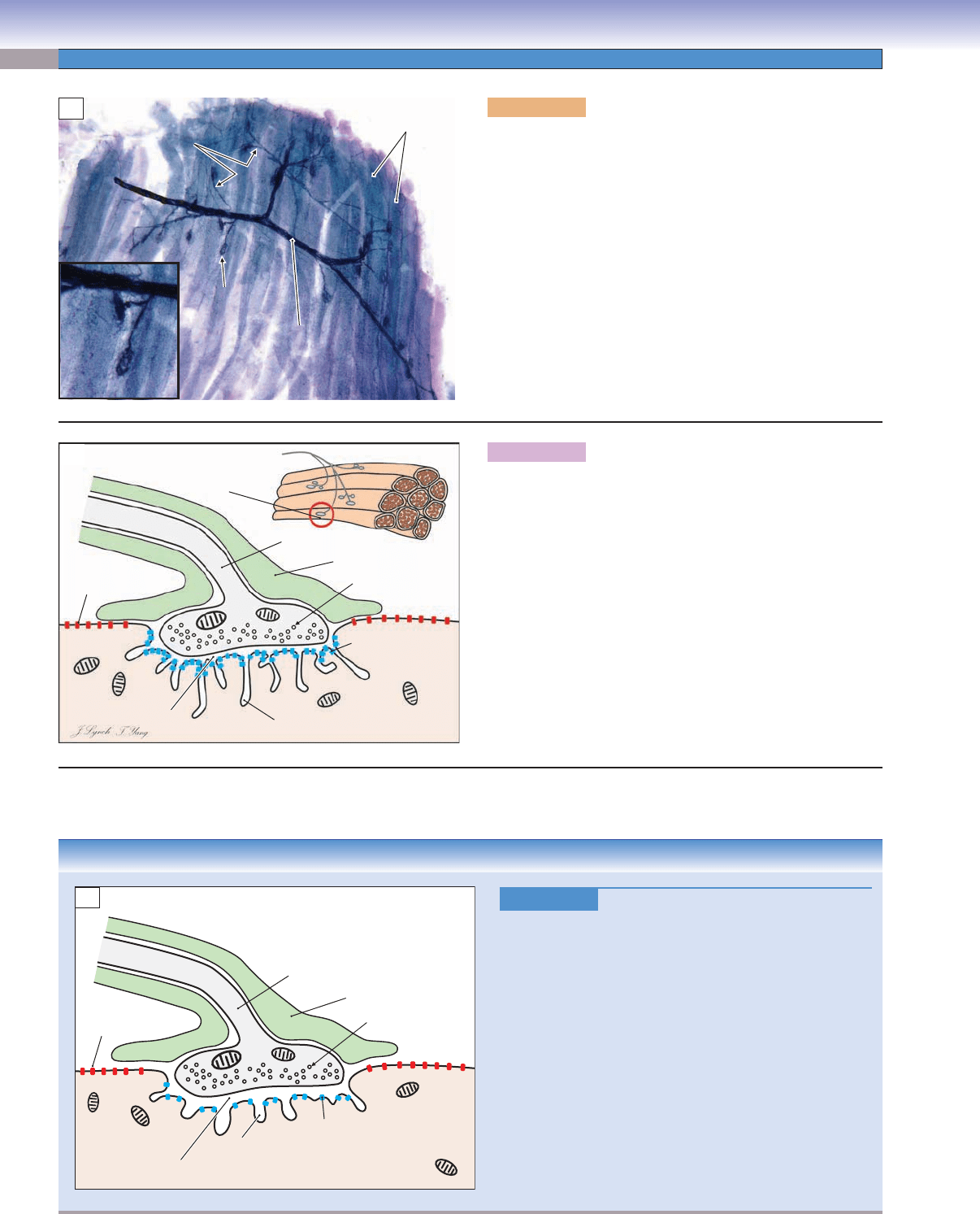
Figure 6-6A. Motor endplates on skeletal muscle. Silver
stain, 83; inset 184
A motor nerve (black) is shown terminating on skeletal muscle
fi bers (violet). The nerve contains several dozen individual
axons, which leave the nerve and form multiple motor end-
plates. These endplates are the sites of the neuromuscular junc-
tions, where the axon makes synaptic contact with individual
muscle fi bers. A single axon contacts numerous muscle fi bers.
The motor neuron, its associated axon, and all of the muscle
fi bers that it contacts are defi ned as a motor unit. Each time an
action potential travels along the axon and causes the release
of Ach at the neuromuscular junction, a contraction is pro-
duced in the muscle fi bers innervated by that axon. In small
muscles involved in fi ne movements, a single axon may contact
10 to 100 muscle fi bers; in large muscles, which produce great
force, the motor units may include 500 to 1,000 muscle fi bers.
Motor nerve
Motor nerve
Motor
Motor
endplate
endplate
Single axons
Single axons
Motor nerve
Muscle fibers
Single axons
Motor
endplate
A
Voltage-gated
channel
Transmitter-gated
channel
Synaptic vesicle
Axon terminal
Schwann cell
Subjunctional fold
Synaptic cleft
Motor
endplate
B
Figure 6-6B. Neuromuscular junction.
A single motor endplate (red circle in inset) is shown in cross
section. The nervous system controls muscle contraction using a
combination of electrical and chemical signals. When an action
potential travels to the end of an axon, the associated electrical
charge causes the synaptic vesicles clustered in the axon terminal to
release a neurotransmitter, ACh, into the synaptic cleft. The ACh
acts upon receptors in transmitter-gated ion channels (blue) in the
postsynaptic membrane. When the channels open, a voltage change
occurs across the membrane, which, in turn, activates voltage-gated
channels (red) in the sarcolemma. This voltage change sweeps rap-
idly along the sarcolemma and invades the T-tubule system, in
which it causes the release of calcium ions and consequent muscle
contraction. The subjunctional folds in the postsynaptic membrane
serve as a reservoir for the enzyme acetylcholinesterase, which rap-
idly inactivates the ACh after each transmitter release.
CLINICAL CORRELATION
Figure 6-6C.
Myasthenia Gravis
Myasthenia gravis is an autoimmune disease that affects
the neuromuscular junction, causing fl uctuating
weakness
and fatigue of skeletal muscles, including ocular, bulbar,
limb, and respiratory muscles. Acetylcholine receptor
antibodies, which block and attack ACh receptors in the
postsynaptic membrane of the neuromuscular junction,
are the most common causes, especially for patients who
develop the disease in adolescence and adulthood. The
mechanism may involve thymic hyperplasia, the binding
of T lymphocytes to ACh receptors to stimulate B cells to
produce autoantibodies, or genetic defects. This illustra-
tion shows fewer ACh receptors than normal, reduction
in subjunctional fold depth, and increased synaptic cleft
width. Treatments include using anticholinesterase agents,
immunosuppressive agents, and thymectomy (surgical
excision of the thymus).
J.Lynch
T. Yang
Voltage-gated
channel
Fewer ACh receptors
(transmitter-gated
channels)
Synaptic vesicle
Axon terminal
Schwann cell
Shallower
subjunctional folds
Wider
synaptic cleft
C
CUI_Chap06.indd 106 6/2/2010 4:08:44 PM
CHAPTER 6
■
Muscle
107
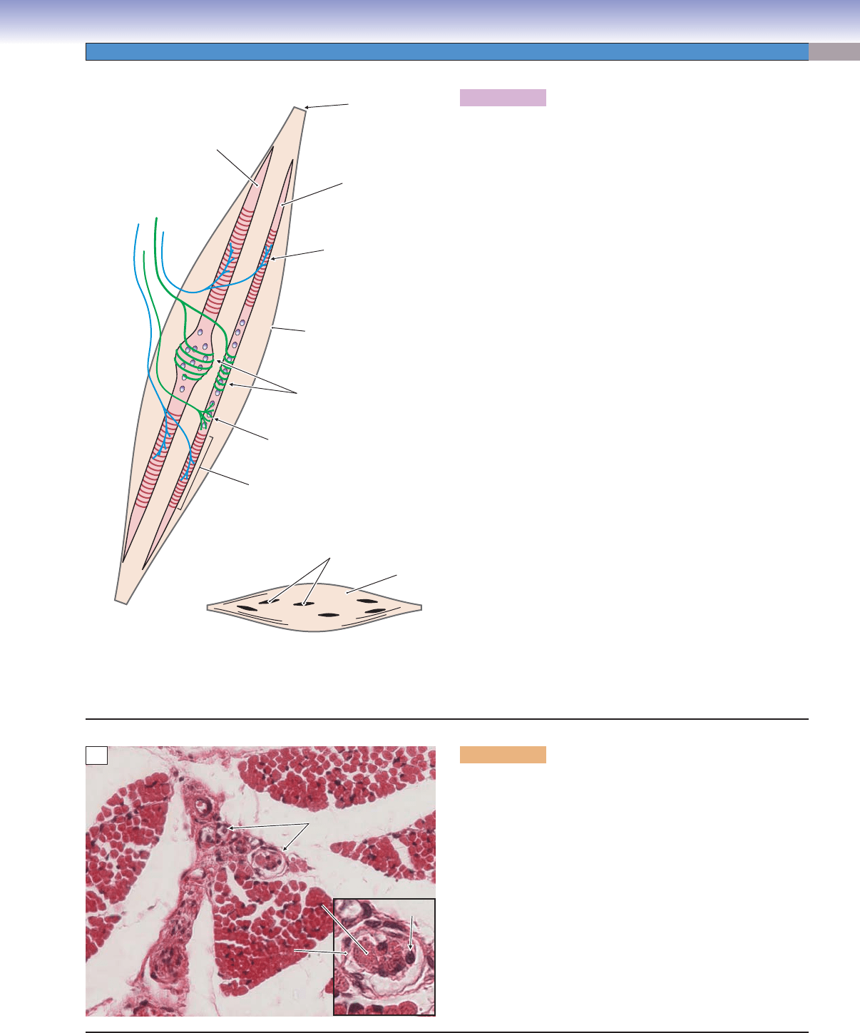
Figure 6-7A. Simplifi ed schematic diagram of the intrafusal
muscle fi bers of a muscle spindle receptor.
Muscle spindles (a type of stretch receptor) play an important
role in the control of voluntary movement, constantly moni-
toring the length of each muscle and the rate of change of that
length. Each spindle contains 10 to 15 specialized muscle fi bers
(intrafusal fi bers) innervated by sensory and motor nerve fi bers
and surrounded by a fl uid-fi lled connective tissue capsule.
Muscle spindles are generally about 1.5 mm in length and are
anchored at each end to connective tissue attached to ordinary
muscle fi bers (extrafusal fi bers). The spindle is stretched when
the muscle lengthens and is shortened when the muscle itself
becomes shorter. A given muscle will contain from a few dozen
to a few hundred spindles distributed throughout the bulk of the
muscle (small drawing). Two general types of muscle fi bers are
included in spindles: nuclear bag fi bers (which have a swelling
in the middle of the fi ber where most of the nuclei are concen-
trated) and nuclear chain fi bers (which are smaller in diameter
and have a single row of nuclei). A typical human muscle spin-
dle contains three to fi ve nuclear bag fi bers and 8 to 10 nuclear
chain fi bers. There are several highly specialized receptors associ-
ated with the sensory nerve endings, which are able to measure
(1) muscle length, (2) change in muscle length, and (3) rate of
change of muscle length. The sensory axons form two types of
endings: (1) primary (or annulospiral) endings (green) in which
the axon wraps around the equator of nuclear bag or nuclear
chain fi bers and (2) secondary (fl ower-spray) endings (green),
which are more common on nuclear chain fi bers. The two ends
of each intrafusal fi ber consist of contractile muscle very similar
to that of the extrafusal fi bers (striated region in drawing). These
contractile portions of the intrafusal fi bers are innervated by
small-diameter myelinated motor axons (gamma motor neurons
or fusimotor neurons [blue]). This innervation causes the intra-
fusal fi bers to shorten when the muscle as a whole shortens and
to relax when the muscle as a whole lengthens, therefore main-
taining the sensitivity of the length-sensitive stretch receptors in
their optimum range and providing accurate information about
the state of the muscle to the motor centers of the CNS.
J.Lynch
Nuclear chain
fiber
Connective tissue
capsule
Gamma motor
ending on
contractile portion
of fiber
Attached to
extrafusal
fibers
Nuclear bag
fiber
Secondary
(flower-spray)
ending
Contractile portion
of intrafusal
muscle fiber
Primary
(annulospiral)
endings
Muscle spindles
Muscle
A
Perimysium
Nucleus
Muscle spindles
Capsule
Capsule
(fibroblast)
(fibroblast)
Intrafusal fiber
Intrafusal fiber
Intrafusal fiber
Capsule
(fibroblast)
B
Figure 6-7B. Skeletal muscle—muscle spindle, cross
section. H&E, 272; inset 680
Fascicles of skeletal muscle separated by perimysium are
illustrated (see Fig. 6-2). Several muscle spindles can be seen
in tangential section in the central fascicle. The fl attened fi bro-
blasts making up the capsule can be seen in the inset, as well as
fi ve or six intrafusal fi bers. In general, muscles that are used in
delicate, highly controlled movements contain the largest num-
bers of muscle spindles. The intrinsic muscles of the hand, for
example, contain a relatively larger number of spindles than do
larger muscles, such as the quadriceps and gluteus maximus,
which are specialized for producing large amounts of force.
CUI_Chap06.indd 107 6/2/2010 4:08:47 PM
108
UNIT 2
■
Basic Tissues
Cardiac Muscle
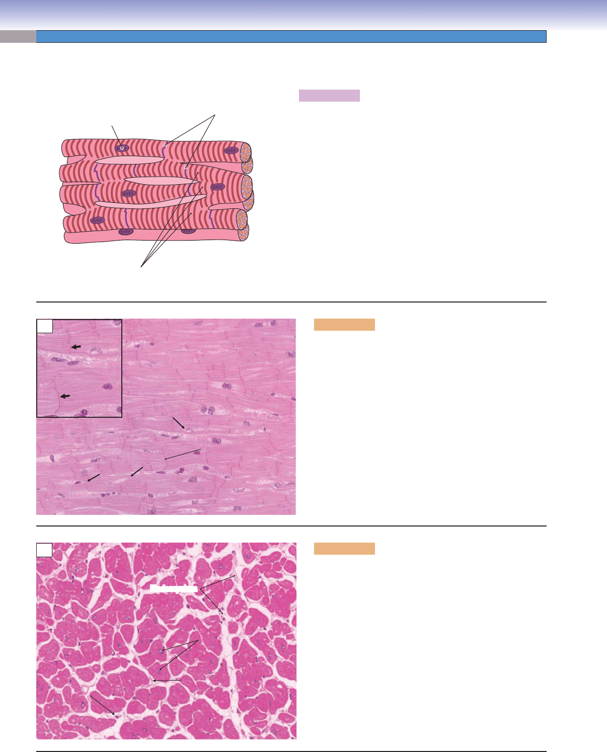
Figure 6-8A. Organization of cardiac muscle—a branching
network of interconnected muscle cells.
Cardiac muscle fi bers split and branch repeatedly and join other
muscle fi bers end to end to form an anastomosing network of
contractile tissues. In contrast to skeletal muscle, cardiac muscle
fi bers contract and relax spontaneously. The intercalated disks
at the boundaries between fi bers contain gap junctions, which
permit electrical depolarization to move directly and rapidly
from one myocyte to the next. The sympathetic and parasym-
pathetic innervation of the heart serves to increase or decrease
the rhythm of contraction rather than to command individual
contractions as the peripheral nervous system does for skeletal
muscle. This modulation of heart rate occurs via a system that
includes the sinoatrial and atrioventricular (AV) nodes and
specialized, highly conductive muscle fi bers (AV bundle and
Purkinje fi bers) that connect the AV node with the contractile
myocytes (see Figs. 9-2 and 9-4A).
D. Cui
J.Lynch
Single nucleus
in each fiber
Intercalated disks
Fibers branch
and anastomose
A
Endomysium
Nuclei
Nuclei
Endomysium
Capillary
Capillary
Fibroblast
Fibroblast
Nuclei
Capillary
Fibroblast
C
Figure 6-8C. Cardiac muscle, transverse section. H&E,
272; inset 418
Cardiac muscle fi bers (myocytes) are elliptical or lobulated
in transverse section. Each fi ber has a single nucleus, which
is irregular in shape and centrally located in the fi ber. Many
capillaries traverse the tissue, and the endomysium is typi-
cally more prominent than in skeletal muscle. The inset
shows nuclei of myocytes and fi broblasts at higher power,
with a capillary in the lower right quadrant (arrow).
Intercalated disks
B
Figure 6-8B. Cardiac muscle, longitudinal section.
H&E, 272; inset 418
Cardiac muscle is like skeletal muscle in that it is striated.
Actin and myosin fi laments are arranged into sarcomeres,
with A bands, I bands, H bands, and Z lines (see Fig. 6-9).
However, cardiac muscle is different in several respects.
Actin and myosin fi laments are not arranged in discrete
myofi brils. Cardiac muscle fi bers are much shorter than
skeletal muscle fi bers and typically split into two or more
branches (thin arrows). The branches are joined, end to
end, by intercalated disks (thick arrows in inset) and form a
meshwork of muscle fi bers. Each fi ber has a single, centrally
located nucleus. Cardiac muscle tissue is highly vascularized
and contains many more mitochondria than other muscle
types, owing to its constant activity and resulting high meta-
bolic requirements.
CUI_Chap06.indd 108 6/2/2010 4:08:49 PM
CHAPTER 6
■
Muscle
109
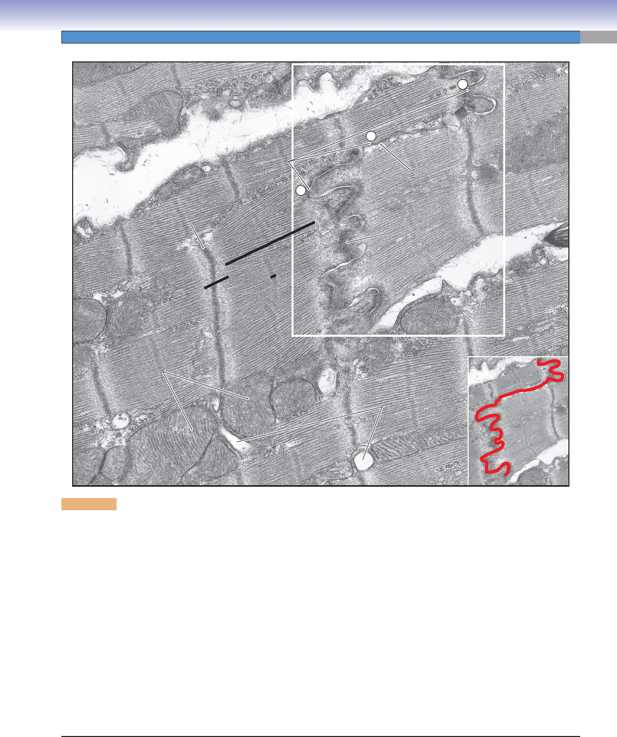
Figure 6-9. Cardiac muscle. EM, 24,800
Cardiac muscle is similar to skeletal muscle in many respects. Both have similar arrangements of actin and myosin fi laments that
interact to produce contraction. The actin fi laments are anchored at Z lines, and myosin fi laments occupy a central position between
two successive Z lines. The structures between two Z lines form a sarcomere. The resulting A band, I band, H band, and Z line are
analogous in the two muscle types. However, there are several notable structural differences. The most obvious is that cardiac myo-
cytes are much shorter than are skeletal muscle fi bers and are joined to each other by complex structures called intercalated disks.
The intercalated disks are specialized regions of the sarcolemma that contain regions of fascia adherens which bind the adjacent cells
together against the stress of contraction, and gap junctions which provide a path for the muscle action potential to travel directly
from one cell to the next. A single intercalated disk typically includes portions that are oriented transversely with respect to the
muscle fi ber (1 and 3) and a portion that is oriented longitudinally (2). The path of this intercalated disk is indicated by the red line
in the inset. Gap junctions are found predominantly in the longitudinal sections. A second major difference is in the T-tubule system.
T tubules (invaginations of the cell membrane) are prominent in cardiac muscle, although there is only one tubule per sarcomere
(located at the Z line) instead of two tubules per sarcomere (located at the A–I junctions) as in skeletal muscle. In addition, the
sarcoplasmic reticulum is not as prominent in cardiac muscle and its function in contraction is not as well understood. Nevertheless,
the release of Ca
++
is critical to contraction, just as in skeletal muscle. The muscle action potential travels along the cell membrane
and T tubules and triggers the fl ow of Ca
++
into the cell from the extracellular space and from the sarcoplasmic reticulum. There
is also a slow leakage of Ca
++
into the muscle fi bers that is responsible for the spontaneous contraction and relaxation rhythm of
isolated cardiac muscle. This natural rhythm is modifi ed by neuronal (autonomic) and hormonal infl uences. Heart rate increases
during physical exercise or stress and decreases during periods of rest and sleep.
G
ap junctions
ap juncti
on
s
Intercalated disk
Intercalated disk
Intercalated disk
3
3
2
2
1
1
Gap junctions
Adherens junction
Adh
ere
n
s junction
Adherens junction
I band
I b
and
I band
H band
H band
H band
A band
A
band
A band
Z line
Z
li
ne
Z line
T-tubules
T-tubules
T-tubules
Mitochondria
Mitochondria
Mitochondria
CUI_Chap06.indd 109 6/2/2010 4:08:52 PM
110
UNIT 2
■
Basic Tissues
Smooth Muscle
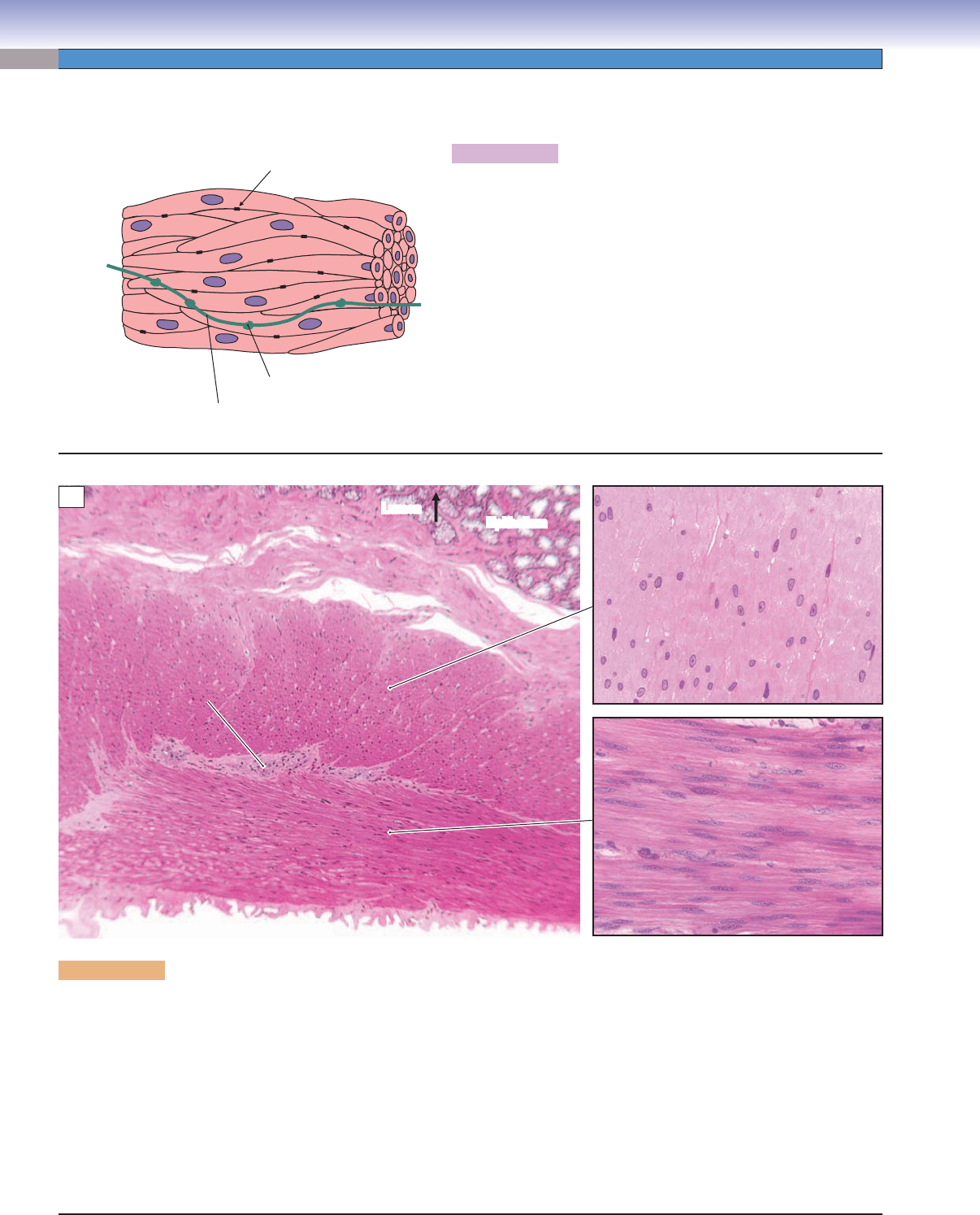
Figure 6-10A. A representation of smooth muscle.
Smooth muscle is similar to skeletal and cardiac muscle in that
contraction is produced by the interaction of actin and myosin fi la-
ments in the presence of Ca
++
. However, there are many differences.
Smooth muscle fi bers are short (15 to 500 μm) and spindle shaped
and have single, centrally placed nuclei. Smooth muscle lacks the stri-
ations observed in skeletal and cardiac muscle because the arrange-
ment of the actin and myosin fi laments is not as orderly. Smooth
muscle is innervated by sympathetic and parasympathetic axons,
but transmitter molecules are released into the intercellular space at
swellings in the axon (varicosities), rather than at specifi c neuromus-
cular junctions (“endplates”) as in skeletal muscle. The sarcolemmas
of some smooth muscles contain gap junctions that permit electrical
excitation to move directly from one fi ber to adjacent fi bers, there-
fore producing a moving wave of contraction.
T. Yang
Varicosity
Gap junction
Autonomic nerve fiber
A
Epithelium
Lumen
Lumen
Epithelium
Circular layer
Myenteric plexus
Connective tissue
Epithelium
Longitudinal layer
B
Figure 6-10B. Smooth muscle, duodenum. H&E, 117; upper inset 485; lower inset 259
In the gastrointestinal tract, smooth muscle is important for keeping food moving at the proper rate to enhance digestion, to permit
the absorption of nutrients, and to prepare waste to be expelled from the body. A low-power section through the duodenum of the
small intestine is shown (see also Fig. 3-7A). The lumen of the intestine, with columnar epithelium specialized for absorption, is at
the top of the picture; beneath it is a layer of connective tissue. Bands of smooth muscles encircle the duodenum. A transverse section
through this circular layer is shown at higher power in the upper inset. The nuclei are scattered randomly through the section. Many
muscle fi bers are cut through a portion of the fi ber that does not contain a nucleus. A second layer of smooth muscle is oriented
along the length of the duodenum and here, it is cut longitudinally. The lower inset shows a longitudinal section at higher power.
Note the long, spindle-shaped nuclei. The smooth muscle of the gut is classifi ed as visceral or unitary smooth muscle and has many
gap junctions. Spontaneous waves of contraction move along the length of the gut, modulated by signals from pacemaker ganglia
or plexuses in the autonomic nervous system. One such plexus, a myenteric plexus, is visible in the low-power photomicrograph
(see also Fig. 7-15B).
CUI_Chap06.indd 110 6/2/2010 4:08:55 PM
CHAPTER 6
■
Muscle
111
CLINICAL CORRELATION
Figure 6-11C.
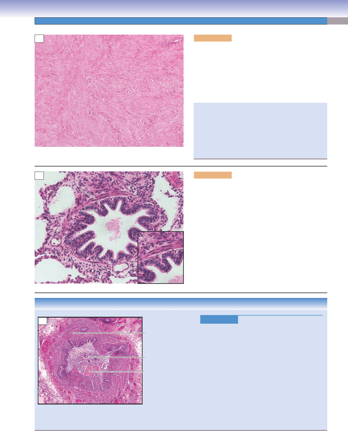
Chronic Asthma. H&E, 27
Asthma is a chronic condition characterized by
wheezing, shortness of breath, chest tightness, and
coughing. Respiratory airways are hypersensitive and
hyperresponsive to a variety of stimuli. Clinical fi nd-
ings include airfl ow obstruction caused by smooth
muscle constriction around airways, airway mucosal
edema, intraluminal mucus accumulation, infl amma-
tory cell infi ltration in the submucosa, and basement
membrane thickening. During acute asthma attacks,
spasms of smooth muscle together with excessive
mucous secretion may close off airways and may be
fatal. Pathologic fi ndings include smooth muscle thick-
ening (hypertrophy and hyperplasia), and remodeling
of nearby small and mid-sized pulmonary blood ves-
sels. Treatment includes using combinations of drugs
and environmental and lifestyle changes.
Thickening (hypertrophy
and hyperplasia)
of smooth muscle layer
Airway plugged by
cell debris
and
mucus
C
Figure 6-11A. Smooth muscle in the wall of the uterus.
H&E, 136
In most locations, fascicles of smooth muscle are oriented in
the same direction. However, in hollow organs in which the
overall size of the organ is reduced by smooth muscle con-
traction, such as the uterus, the fascicles are intertwined and
run in all different directions. In this section, some fascicles
are cut in a longitudinal plane, some in a tangential plane,
and others are cut diagonally.
A
Smooth muscle
Smooth muscle
Bronchiole
Smooth muscle
Ciliated columnar/
Ciliated columnar/
cuboidal epithelium
cuboidal epithelium
Ciliated columnar/
cuboidal epithelium
B
Figure 6-11B. Smooth muscle in a bronchiole. H&E,
136; inset 160
Smooth muscle lines the walls of the bronchioles in the respi-
ratory system (see Figs 11-9 to 11-11). It relaxes to increase
the size of the airway passages under the infl uence of the
sympathetic nervous system and hormones controlled by
the sympathetic nervous system, and it contracts to reduce
the size of the airway passages under the infl uence of the
parasympathetic nervous system. This smooth muscle aids
in expelling air from the lungs during breathing. It is also
important in the cough refl ex, which helps to expel foreign
matter such as dust, smoke, and excess mucus from the lungs.
With age, and in response to irritants such as tobacco smoke,
the contractility of smooth muscle may be reduced, causing
respiratory insuffi ciency. Note the long, thin, spindle-shaped
nuclei of the smooth muscle cells in the inset.
The large forces generated by uterine smooth muscle are
important in expelling the fetus during childbirth and are also
critical for clamping down on blood vessels to stop bleeding
after the placenta is pulled away from its attachment to the
wall of the uterus. Contraction of this smooth muscle can
be enhanced by the administration of oxytocic agents (e.g.,
oxytocin, ergonovine) after delivery to stimulate myometrial
contractions and prevent or treat postpartum hemorrhage.
CUI_Chap06.indd 111 6/2/2010 4:08:58 PM
112
UNIT 2
■
Basic Tissues
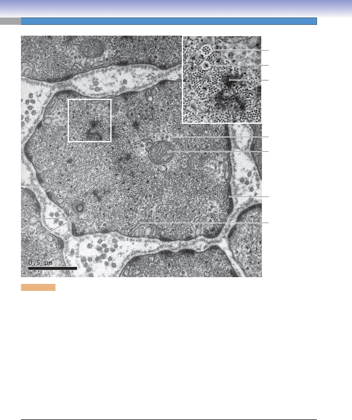
Figure 6-12. Transverse section of smooth muscle of the trachea. EM, 14,750
Although contraction of smooth muscle is produced by a calcium-mediated interaction between actin and myosin fi laments similar
to that described in Figure 6-4C, there are signifi cant differences in the structure of smooth muscle cells and striated (skeletal and
cardiac) muscle cells. Actin and myosin fi laments are clearly visible (see inset), but their arrangement is not as orderly as in skeletal
muscle. Actin fi laments are anchored to the cell walls at dense plaques or at dense bodies in the interior of the cell (see Fig. 6-13B).
The actin fi laments contact myosin fi laments to produce contraction, but the organization is more random and more changeable
than in skeletal or cardiac muscle. Smooth muscle has the property of being able to produce relatively constant contractile force
over a greater range of cell lengths than striated muscle. Skeletal muscle, for example, cannot produce maximum contraction force
when it is fully extended because there is not suffi cient overlap between the actin and myosin fi laments. This property of smooth
muscle is important in organs such as the stomach and uterus where strong contraction may be needed when the organ is distended
and the muscle cells already considerably stretched. Some smooth muscles have the ability to remodel their contractile architecture
in response to different conditions of muscle extension. Intermediate fi laments provide mechanical and structural integrity for many
types of cells, including smooth muscle. They are composed primarily of the proteins vimentin and desmin. Lack of these proteins
impairs the contractility of smooth muscle. Contraction of smooth muscle can be initiated by neural signals (e.g., iris, respiratory
system), mechanical stretch (e.g., gut, urinary tract), electrical signals traveling from one smooth muscle fi ber to another via gap
junctions (e.g., gut, respiratory system), or hormones in the blood stream (e.g., respiratory system, uterus). The calcium necessary
to initiate contraction enters the cell from the extracellular space rather than from the sarcoplasmic reticulum as in striated muscle.
Smooth muscle fi bers have a poorly developed sarcoplasmic reticulum and no T-tubule system at all. Cup-shaped indentations in the
sarcolemma (caveolae) may play a role in sequestering calcium.
Actin filaments
Myosin filament
Dense body
Intermediate filament
Mitochondrion
Dense plaque
Sarcoplasmic
reticulum
Caveolae
Caveolae
Caveolae
CUI_Chap06.indd 112 6/2/2010 4:09:02 PM
CHAPTER 6
■
Muscle
113
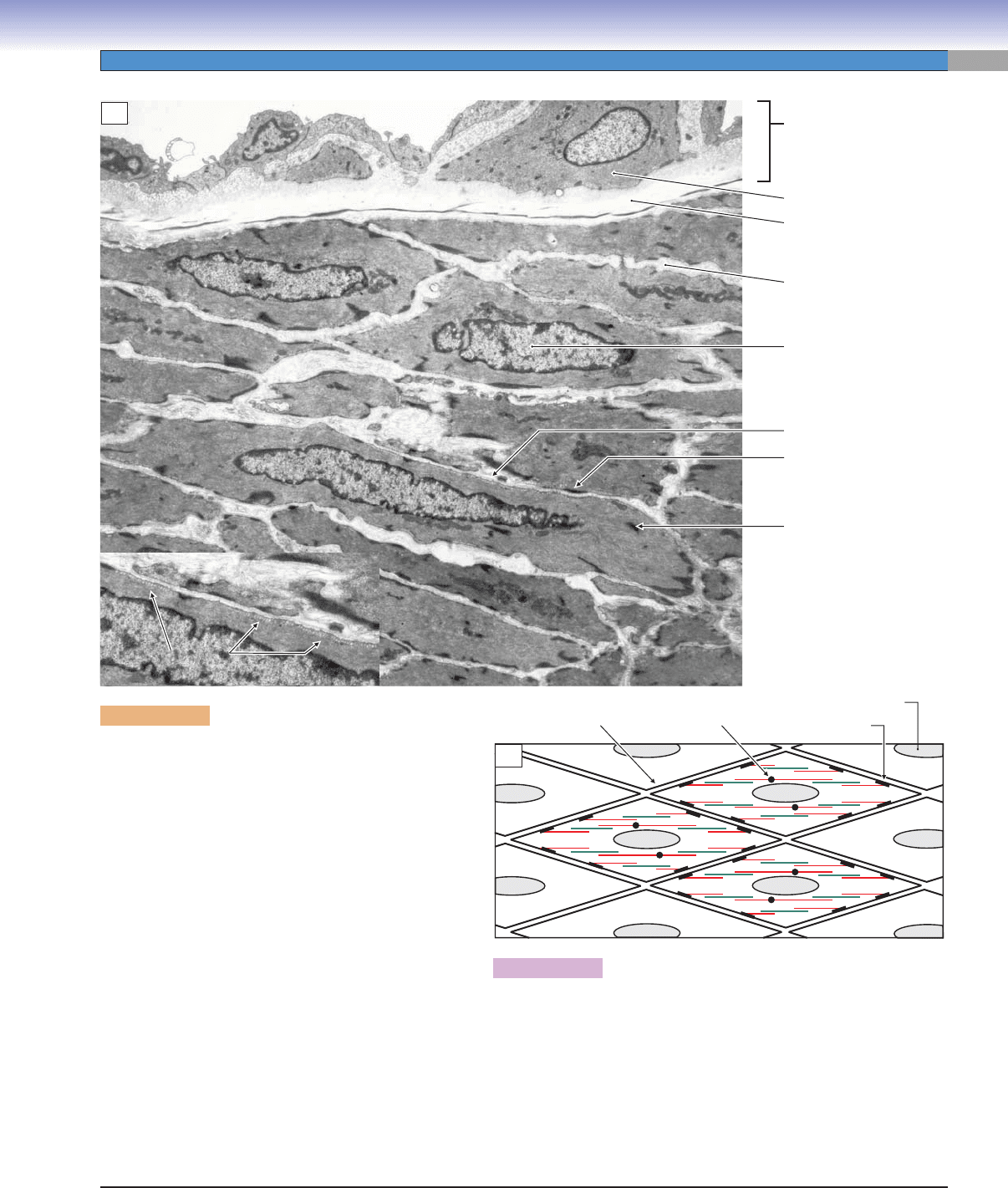
Figure 6-13B. Schematic diagram of the contractile mechanism of
smooth muscle.
Actin fi laments (red) in smooth muscle are anchored in dense plaques
in the cell walls, and the corresponding myosin fi laments (green) are
suspended between two or more actin fi laments. This arrangement is
much less orderly than the arrangement of actin and myosin in skeletal
and cardiac muscles. Furthermore, the organization is, to some extent,
dynamic and can be rearranged in response to changing demands on
the muscle. The cells are attached to each other via junctions with the
collagen and elastin fi bers in the extracellular matrix.
Figure 6-13A. Smooth muscle fi bers in the tunica
media of a medium artery. EM, 4,260; inset 6,530
The smooth muscle fi bers in this illustration are cut
obliquely to their long axis. The thickenings of the cell
membrane, which are termed dense plaques, can be
clearly seen. Actin myofi laments are attached to these
structures and the force of the actin-myosin contrac-
tion is transmitted to the cell wall at these points. Actin
myofi laments are also sometimes anchored to dense
bodies within the cytoplasm. In some types of smooth
muscles (e.g., in the intestine), the myofi laments seem
to be arranged randomly, in a crisscross fashion. In
other types (e.g., in the airway), myofi laments seem to
be arranged in parallel as diagramed in Figure 6-13B.
Smooth muscle cells transmit force from one to another
via the extracellular matrix and, in some cases, via tight
junctions between the cell membranes of adjacent cells.
The extracellular matrix in smooth muscle is composed
of elastin, collagen, and other elements, but in contrast
to other tissues, it is secreted by the myocytes themselves
rather than by fi broblasts. In some tissues, such as in
elastic arteries and in the airway, smooth muscle fi bers
may, with age or disease, gradually lose their ability to
contract and become more and more like fi broblasts.
Lumen of artery
Tunica intima
Endothelial cell
Nucleus
Extracellular matrix
Internal elastic membrane
Dense body
Dense plaque
Caveolae
Caveolae
Caveolae
Caveolae
Dense body
Sarcolemma
Dense plaque
Nucleus
A
B
CUI_Chap06.indd 113 6/2/2010 4:09:05 PM

114
UNIT 2
■
Basic Tissues
Features Skeletal Muscle Cardiac Muscle Smooth Muscle
Striations Yes Yes No
Fibers Long, cylindrical, unbranched Short, branched, anastomosing Short, spindle shaped
Nuclei Multiple, peripheral in cell Single, central in cell Single, central in cell
Cell junctions No Intercalated disks Gap (nexus) junctions
T tubules Well developed Well developed No
Sarcoplasmic
reticulum
Highly developed; has terminal
cisterns
Less well developed; small cisterns Present, but poorly developed
Regeneration Yes, satellite cells No Yes, mitosis
Contraction Initiated by nerve action potential Spontaneous; pacemaker system;
modulated by nervous system and
hormones
Spontaneous; modulated by
nervous system and hormones
Main function Voluntary movement of limbs, digits,
face, tongue, and other muscles
Involuntary rhythmic contractions;
pumps blood to muscles and
organs; modulated by physiological
and emotional factors
Involuntary control of blood
vessel diameter, gut peristalsis,
uterine contractions during
childbirth, airway diameter, and
others
TABLE 6-1 Muscle Characteristics
SYNOPSIS 6-1 Pathological and Histological Terms for Muscle
Anastomose ■ : To join end to end, as in suturing two blood vessels together (Fig. 6-8A).
Autoimmune disease
■ : A condition in which an individual’s immune system mistakes the individual’s own tissue for a for-
eign invader and attacks the tissue, as in myasthenia gravis or multiple sclerosis (Fig. 6-6C).
Caveolae
■ : Small, cup-shaped indentations in the sarcolemma of smooth muscle cells; may be involved in the uptake of
calcium during contraction (Figs. 6-12 and 6-13).
Dystrophin
■ : A large, rod-shaped protein that plays a critical role in connecting the molecular contractile mechanism of
skeletal muscle to the surrounding extracellular matrix so that the force of the actin-myosin contraction can be transferred
to other structures to do useful work. The lack of dystrophin is a key feature of some types of muscular dystrophies
(Fig. 6-3C).
Fibrosis
■ : Abnormal formation of connective tissue, including fi broblasts and connective tissue fi bers, to replace normal
tissues in response to tissue damage caused by disease or injury (Fig. 6-3C).
Hyperplasia
■ : Abnormal proliferation of cells, which may or may not lead to the increase in the size of the affected structure
or organ; may be a precancerous condition (Fig. 6-6C).
Hypertrophy
■ : An increase in the size of a structure produced by an increase in the size of the cells that make up the
structure.
Intrafusal
■ : Structures, particularly muscle fi bers, that are found inside the muscle spindle. The word is derived from the
Latin “fusus” which means “spindle” (Fig. 6-7A).
Necrosis
■ : Pathologic death of cells or tissues as a result of irreversible damage because of disease or injury (Fig. 6-3C).
Synaptic cleft
■ : The small space between a presynaptic axon terminal and the postsynaptic membrane of a muscle cell or a
neuron upon which the axon forms a synapse (Figs. 6-6B,C).
Varicosity
■ : A local swelling in a tubelike structure such as an axon (Fig. 6-10A).
CUI_Chap06.indd 114 6/2/2010 4:09:11 PM
