Cui Dongmei. Atlas of Histology: with functional and clinical correlations. 1st ed
Подождите немного. Документ загружается.


115
Introduction and Key Concepts for the Nervous System
Neurons and Synapses
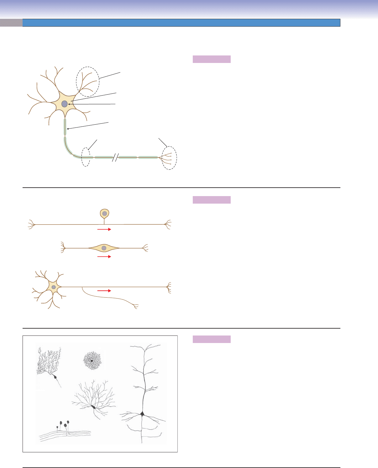
Figure 7-1A The Neuron: The Building Block of the Nervous System
Figure 7-1B,C Types of Neurons
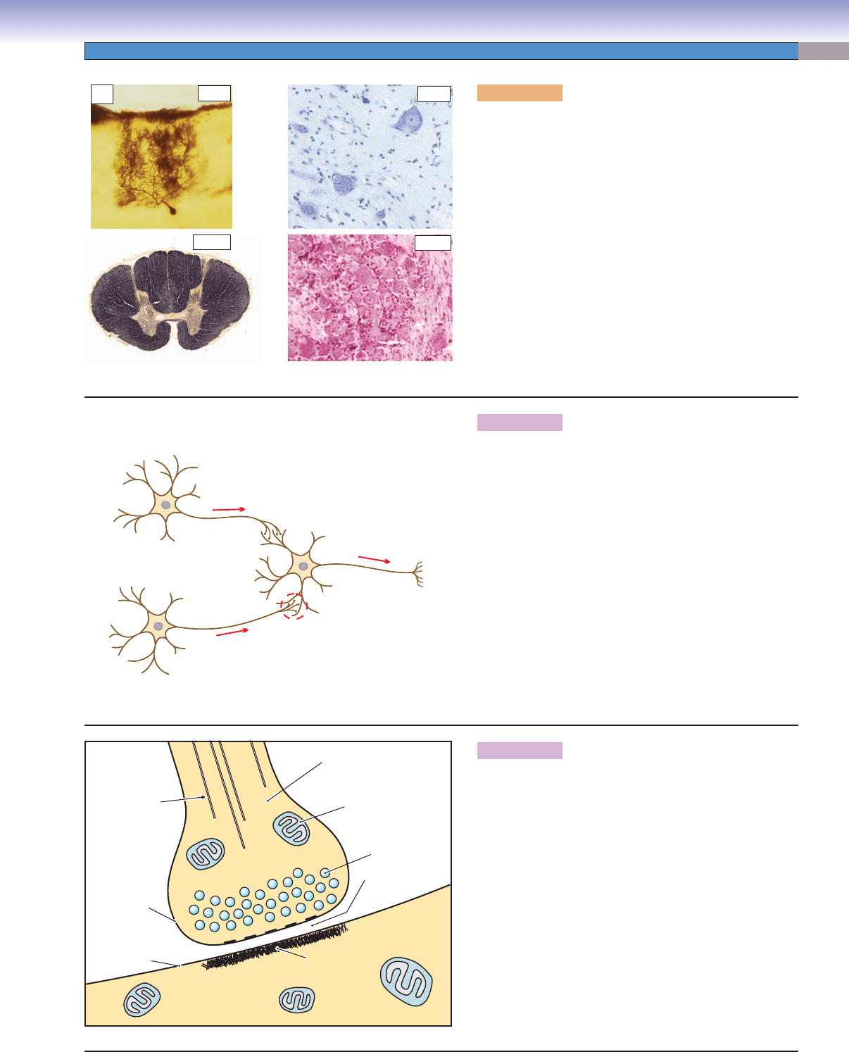
Figure 7-2A Types of Stains for Nervous Tissue
Figure 7-2B Information Transmission in the Nervous System
Figure 7-2C Elements of the Synapse
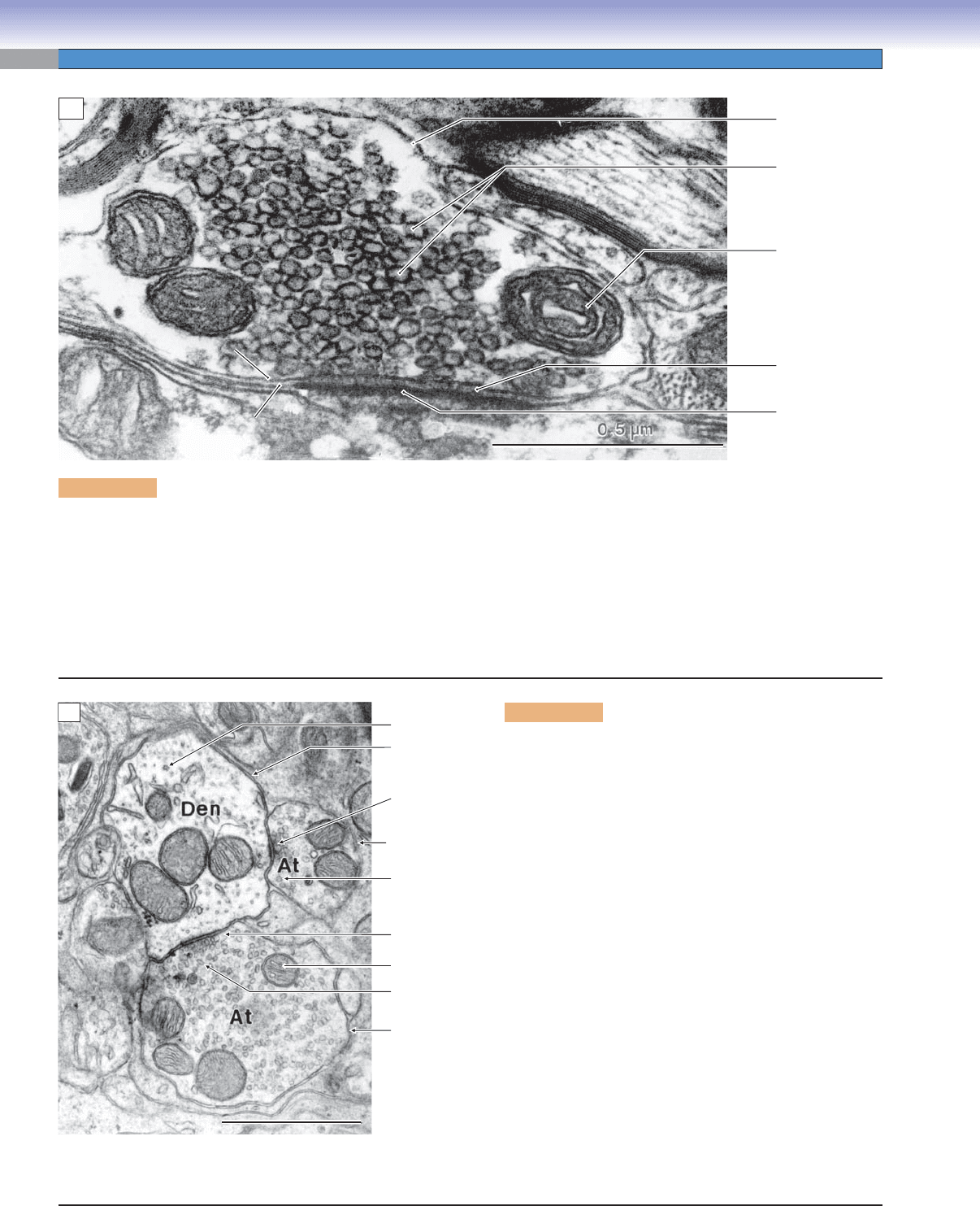
Figure 7-3A,B Structure of the Synapse
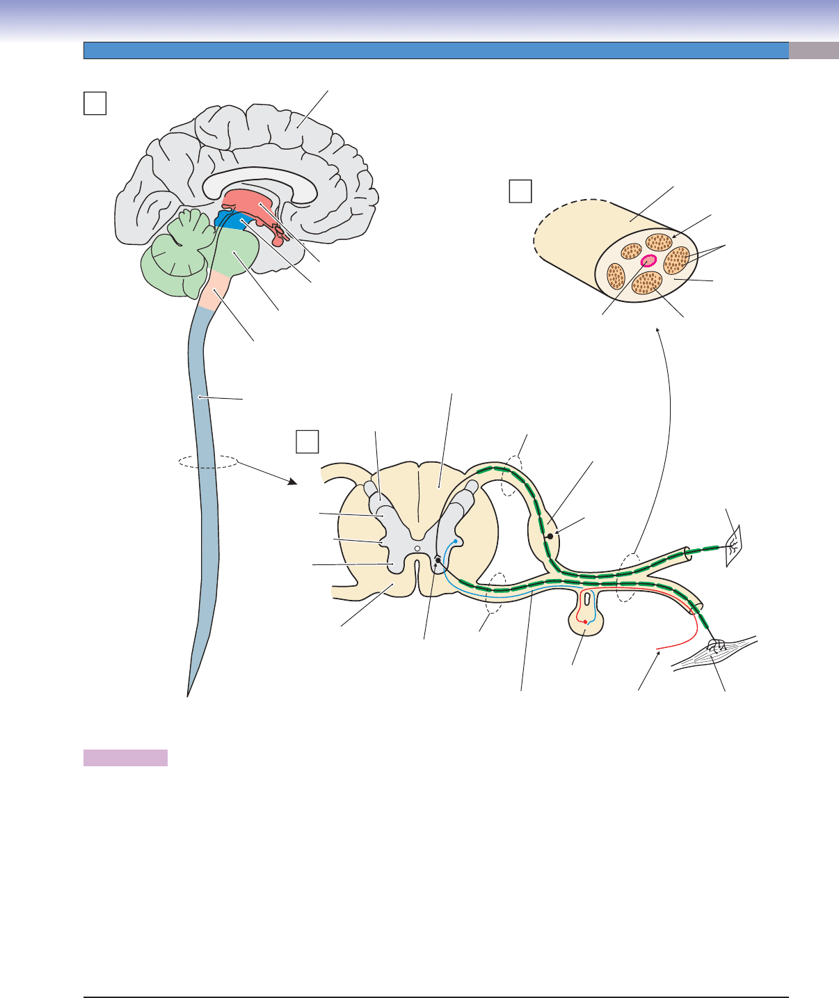
Figure 7-4 Overview of the Central and Peripheral Nervous Systems
Peripheral Nervous System
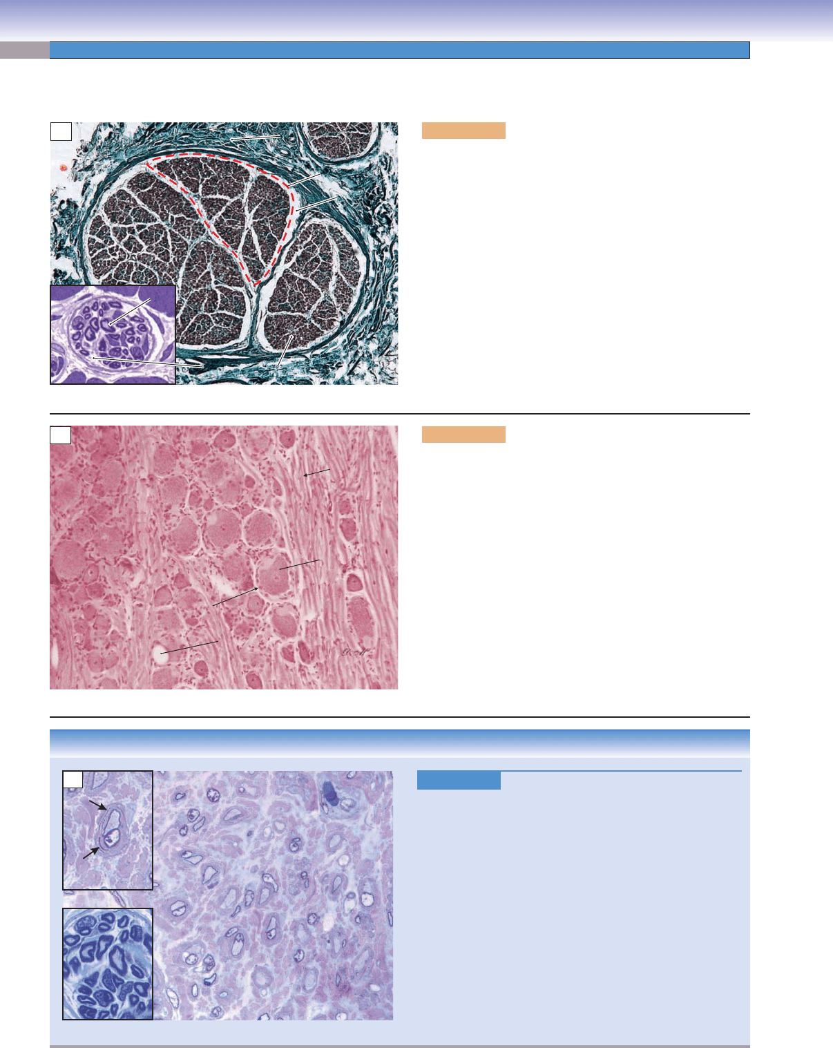
Figure 7-5A Cross Section of a Peripheral Nerve
Figure 7-5B Posterior Root Ganglion
Figure 7-5C Clinical Correlation: Hereditary Sensory Motor Neuropathy, Type III (HSMN III)
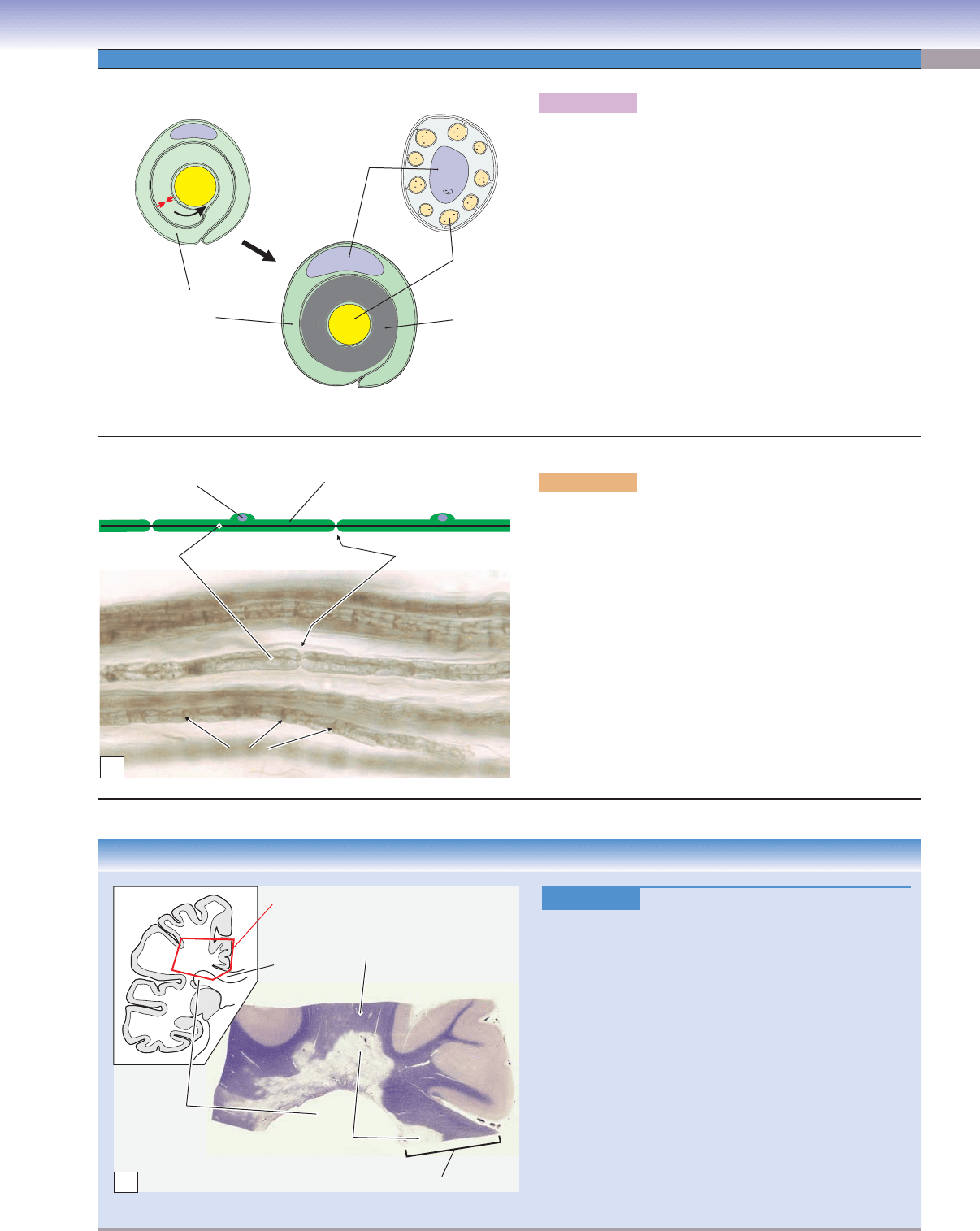
Figure 7-6A Myelinated and Unmyelinated Axons
Figure 7-6B Myelinated Peripheral Nerve Axons (Nodes of Ranvier)
Figure 7-6C Clinical Correlation: Multiple Sclerosis
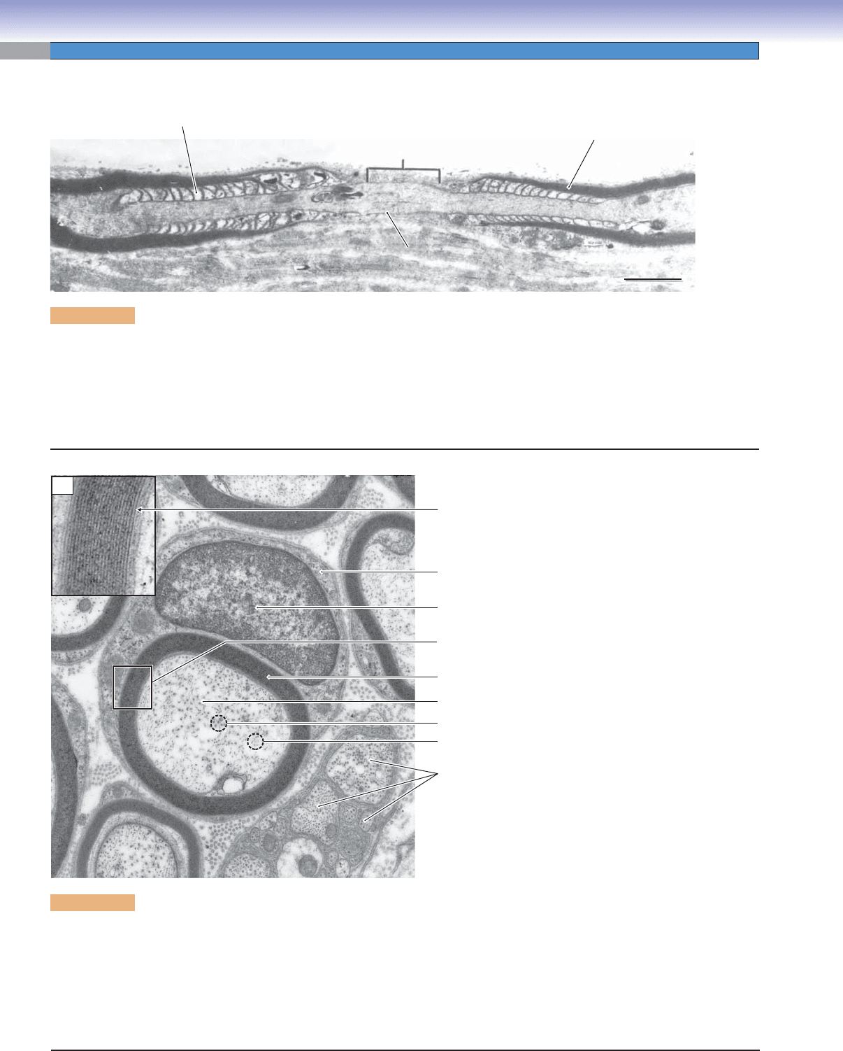
Figure 7-7A Node of Ranvier Between Two Adjacent Schwann Cells
Figure 7-7B Myelinated and Unmyelinated Axons
Figure 7-8A Peripheral Sensory Receptors
Figure 7-8B Meissner Corpuscle
Figure 7-8C Pacinian Corpuscle
Central Nervous System
Figure 7-9A,B Spinal Cord
Figure 7-9C Neurons in the Reticular Formation of the Brainstem
Figure 7-10A,B Cerebral Cortex
Figure 7-10C Clinical Correlation: Alzheimer Disease
Figure 7-11A,B Cerebellar Cortex
Figure 7-11C Clinical Correlation: Encephalocele
Nervous Tissue
7
CUI_Chap07.indd 115 6/2/2010 6:32:00 PM

116
UNIT 2
■
Basic Tissues
Figure 7-12A Dura Mater, Arachnoid, and Pia Mater
Figure 7-12B Spinal Meninges
Figure 7-12C Clinical Correlation: Meningitis
Figure 7-13A Types of Glial Cells
Figure 7-13B Astrocytes
Figure 7-13C Clinical Correlation: Glioblastoma
Autonomic Nervous System
Figure 7-14 Overview of the Autonomic Nervous System
Figure 7-15A Sympathetic Ganglion
Figure 7-15B Myenteric Plexus (Auerbach Plexus)
Figure 7-16 Submucosal Plexus (of Meissner)
Table 7-1 Comparison of Posterior Root and Autonomic Ganglia
Synopsis 7-1 Pathological and Clinical Terms for the Nervous System
Introduction and Key Concepts
for the Nervous System
It is diffi cult to consider the tissue of the nervous system
separately from the nervous system itself. In most organ sys-
tems, the purpose of the tissue is to fi lter, secrete, or transfer
gases or digest and absorb nutrients. The histological structure
of one small region of the liver or kidney or small intestine is
very much like the structure of any other region of that organ,
and the function of one portion of the organ is very much like
the function of any other portion. By contrast, the purpose of
the nervous system is to carry sensory information from the
sensory organs to the brain; to process that sensory informa-
tion in the brain to produce perceptions, memories, decisions,
and plans; and to carry motor information from the brain to
the skeletal muscles in order to exert an infl uence on the indi-
vidual’s surroundings. In truth, all we know of the world that
surrounds us is carried as electrical impulses over our sensory
nerves; the only way we have of interacting with that world is
via electrical impulses carried by motor nerves from our brains
to our muscles.
Neurons and Synapses
The building blocks of the nervous system are cells called
neurons. These cells have a long, thin process, the axon, in
which the cell membrane incorporates specialized protein ion
channels that enable the axon to conduct an electrochemical
signal (action potential) from the cell body to the axon termi-
nals. Axon terminals of one neuron make synaptic contacts
with other neurons, generally on processes called dendrites or
on the cell body itself. When the action potential reaches the
axon terminals, a neurotransmitter is released from synaptic
vesicles into the terminals. The neurotransmitter molecules act
on receptor molecules that are part of ion channels in the den-
drites and soma of the next neuron in a chain. The constant
interplay of excitatory and inhibitory infl uences at the many
billions of synapses in the nervous system forms the basis of our
ability to be aware of our surroundings and to initiate actions
to infl uence our surroundings (Figs. 7-1 to 7-3).
Overview of the Peripheral and
Central Nervous Systems
By defi nition, the brain and spinal cord are classifi ed as the cen-
tral nervous system (CNS), and the nerves and ganglia outside
these structures are classifi ed as the peripheral nervous system
(PNS). Collections of axons that carry action potentials from
one place to another are called nerves in the PNS and tracts
within the CNS. Clusters of neuron cell bodies are called gan-
glia in the PNS and nuclei or cortices in the CNS (Fig. 7-4).
Peripheral Nervous System
Nerves in the PNS carry sensory information from receptors
located in the skin, muscles, and other organs and carry motor
commands from the CNS to muscles and glands. Nerves consist
of clusters of axons surrounded by protective connective tis-
sues (Fig. 7-5A). Nerve axons range in diameter from about
0.5 to 22 μm, with the conduction velocity being higher for
larger axons. In addition, larger axons generally have a dense,
lipid-rich coating, myelin, which further increases conduction
velocity (Fig. 7-6). The cell bodies associated with the sensory
neurons are clustered in a swelling of the posterior spinal root,
the posterior (dorsal) root ganglion.
Central Nervous System
The spinal cord consists of large bundles of myelinated and
unmyelinated axons arranged into ascending (sensory) and
descending (motor) tracts (Fig. 7-9A). The ascending tracts
carry information from peripheral receptors to nuclei in the
brainstem and thalamus and from there to the cerebral cortex.
The descending tracts carry motor information from the cere-
bral cortex and motor centers in the brainstem to interneurons
(relay neurons) in the motor pathways and directly to spinal
motor neurons. These motor neurons innervate muscles directly
to produce movement. The tracts are clustered around a central
region, the spinal gray matter, which contains large numbers
of sensory and motor interneurons, spinal motor neurons, and
preganglionic autonomic visceromotor neurons.
CUI_Chap07.indd 116 6/2/2010 6:32:12 PM

CHAPTER 7
■
Nervous Tissue
117
The higher levels of the CNS include large groups of nuclei
including the thalamus and the basal nuclei as well as sensory
and motor nuclei in the brainstem associated with cranial
nerves. The cerebellum is a large, specialized structure com-
posed of nuclei and cortex, and the cerebral cortex envelops the
surface of the cerebral hemispheres (Figs. 7-10 and 7-11).
MENINGES are brain hemisphere and spinal cord coverings
with three layers of connective tissue membranes that protect the
nervous system, provide mechanical stability, provide a support
framework for arteries and veins, and enclose a space that is
fi lled with cerebrospinal fl uid (CSF), a fl uid that is essential to the
survival and normal function of the CNS. The meninges include
the dura mater, the arachnoid, and the pia mater (Fig. 7-12).
GLIAL CELLS are nonneural cells that provide a variety
of support functions for the neurons that relate to nutrition,
regulation of the extracellular environment including the blood-
brain barrier, immune system, myelin insulation for many
axons, and a host of other support functions (Fig. 7-13).
Autonomic Nervous System
The autonomic nervous system (ANS) is composed of three
divisions: sympathetic, parasympathetic, and enteric. The sym-
pathetic and parasympathetic divisions function under direct
CNS control; the enteric division functions somewhat more
independently. The ANS, together with the endocrine system
and under the general control of certain systems within the
CNS, maintains the homeostasis of the internal environment
of the body. That is, the autonomic and endocrine systems
ensure that the levels of nutrients, electrolytes, oxygen, carbon
dioxide, temperature, pH, osmolarity, and many other related
variables are maintained within optimal physiological limits
(Fig. 7-14).
CUI_Chap07.indd 117 6/2/2010 6:32:12 PM
118
UNIT 2
■
Basic Tissues
Neurons and Synapses
Figure 7-1A. The neuron: The building block of the
nervous system.
Neurons contain the organelles that are common to all cells:
a cell membrane, nucleus, endoplasmic reticulum, and cyto-
plasm. In addition, neurons possess two types of specialized
processes, dendrites and axons. The plasma membranes of den-
drites and of the cell body itself contain specialized receptors
that react to the release of neurotransmitters. These molecules
produce a change in polarization of the membrane. The mem-
brane of axons, by contrast, is specialized to transmit electro-
chemical signals called action potentials. An action potential is
a wave of membrane depolarization that maintains its size as it
travels along the axon. When the action potential reaches the
end of the axon, neurotransmitters are released from the axon
terminals that infl uence the next neuron in line. Some axons
are surrounded by a lipid-rich sheath called myelin, which
facilitates the rapid conduction of action potentials.
J. Lynch
Dendrites
Soma
Nucleus
Myelin
Axon
Axon terminals
A
J. Lynch
Unipolar
Bipolar
Axon
Axon
D
D
D
T
T
T
T
D
Axon
collateral
Multipolar
B
Figure 7-1B. Types of neurons.
Neurons can be classifi ed on the basis of the shapes of their cell
bodies and the general arrangement of their axons and den-
drites. Unipolar neurons have a single process attached to a
round cell body. This process typically divides and forms a long
axon extending from sensory receptors in the various tissues of
the body to synaptic terminals in the CNS. Bipolar neurons have
a process extending from each end of the cell body. This type of
neuron is found primarily in the eye, ear, vestibular end organs,
and olfactory system. Multipolar neurons have many dendrites
extending from the cell body and a single axon (although the
axon may split into two or more collateral axons after it leaves
the cell body). Multipolar neurons are the most numerous in
the nervous system and have many different shapes and sizes.
(D, dendrites; T, axon terminals [“terminal boutons” or “bou-
tons terminaux”] with synaptic endings. Red arrows indicate
the direction of information transmission.)
J. Lynch
C
Purkinje
Unipolar
Stellate
Spinal
motor
Pyramidal
Figure 7-1C. Some representative neurons. Drawings
from Golgi-stained tissue.
Neurons come in a wide variety of shapes and sizes, depend-
ing on their function and location. Purkinje cells are found in
the cerebellar cortex. Pyramidal cells are the most numerous
cells in the cerebral cortex. Stellate cells are also located in the
cerebral cortex. Multipolar motor neurons are found in the
anterior horn of the spinal cord (spinal motor neurons) and in
the motor nuclei of cranial nerves. Other types of multipolar
neurons are found in many central and autonomic nervous
system sites. Unipolar neurons have cell bodies in the poste-
rior root ganglia of the spinal cord. Their peripheral processes
contact sensory receptors in the skin, muscles, and internal
organs; their central processes form synapses on neurons in
nuclei of the CNS. There are many additional types of neurons
in the nervous system, but these represent some of the most
common types and demonstrate the wide variety that exists.
CUI_Chap07.indd 118 6/2/2010 6:32:12 PM
CHAPTER 7
■
Nervous Tissue
119
Figure 7-2A. Types of stains for nervous tissue.
The various features of nervous tissue are more diffi cult to
visualize than are the features of most other histological
tissues. Consequently, several special stains are commonly
used with nervous tissue. Golgi preparations stain all of the
processes of an individual neuron (dendrites, cell body, and
axon) but react with only a very small percentage of the total
number of neurons (Purkinje cell, upper left). Nissl stains
react with rough endoplasmic reticulum and, therefore,
allow the shape and size of cell bodies to be visualized but do
not stain dendrites and axons (spinal motor neurons, upper
right). Myelin stains allow the visualization of myelinated
fi bers but do not react with cell bodies or dendrites (spinal
cord, lower left). Myelinated fi ber tracts are, therefore, dark,
and areas with high concentrations of neuron cell bodies are
light. H&E stains are often used in the diagnosis of patho-
logical conditions and sometimes used to stain normal ner-
vous tissue (posterior root ganglion, lower right).
A
Golgi
Nissl
H&E
Myelin
Figure 7-2B. Information transmission in the nervous
system.
The primary functions of the nervous system are to transfer
information (in the form of action potentials) from one
place to another and to process that information to gener-
ate sensory experience, perceptions, ideas, and motor activ-
ity. Information is carried in the form of action potentials
along axons (red arrows 1 and 2). At the ends of axons,
there are axon terminals, where the electrochemical action
potential causes the release of molecules called neurotrans-
mitters. These molecules act upon receptor complexes in the
dendrites and somas of the next neuron in a series (e.g., 3)
at regions of synapses (red dashed circle). The action of the
neurotransmitters may be either excitatory or inhibitory on
the postsynaptic membrane. When the excitatory infl uences
on a neuron exceed the inhibitory infl uence by a certain
threshold amount, that neuron generates an action poten-
tial that is then transmitted onto yet another neuron.
J. Lynch
1
2
3
Information transmission in the nervous system
B
Figure 7-2C. Elements of the synapse.
A typical chemical synapse consists of a terminal bouton
(a swelling at the end of an axon terminal) that includes
a presynaptic membrane, a specialized postsynaptic mem-
brane, and a space between the two (the synaptic cleft). The
terminal bouton contains many synaptic vesicles that con-
tain neurotransmitter molecules. When an action potential
arrives at the axon terminal, a complex chemical process
is initiated that culminates in the fusion of some vesicles
with the presynaptic membrane and the discharge of their
neurotransmitter molecules via exocytosis into the synaptic
cleft where they can act on receptors in the postsynaptic
membrane. The postsynaptic membrane is thickened in the
immediate vicinity of the synapse as a result of the dense
concentration of receptor protein complexes in that region
(Fig. 7-3A). Both the presynaptic and postsynaptic regions
contain numerous mitochondria, which supply the energy
needed by the synaptic transmission process.
J. Lynch
C
Axon terminal
(terminal bouton)
Mitochondrion
Presynaptic
membrane
Postsynaptic
membrane
Dendrite
Postsynaptic
density
Microtubule
Vesicle
Synaptic cleft
CUI_Chap07.indd 119 6/2/2010 6:32:13 PM
120
UNIT 2
■
Basic Tissues
Figure 7-3A. Structure of the synapse. EM, scale line = 0.5 μm; 104,000
A high magnifi cation electron micrograph of an axon terminal and adjacent postsynaptic membrane is shown. Many synaptic
vesicles and three mitochondria are visible within the axon terminal. When an action potential reaches the axon terminal, some syn-
aptic vesicles fuse with the presynaptic membrane and empty their neurotransmitter molecules into the synaptic cleft. The transmit-
ter molecules bind with receptor complexes in the postsynaptic membrane leading to either depolarization (excitatory infl uence) or
hyperpolarization (inhibitory infl uence) of the postsynaptic membrane. The sum of the excitatory and inhibitory infl uences upon the
postsynaptic neuron determines whether it will fi re an action potential or not. The large difference in the thickness of the presynaptic
and postsynaptic membranes makes this contact an asymmetric synapse. Differences in regions of postsynaptic density in different
synapses are probably a refl ection of different types of receptors in different postsynaptic membranes.
0.5 μm
0.5 μm
Presynaptic
Presynaptic
membrane
membrane
Postsynaptic
Postsynaptic
membrane
membrane
Synaptic vesicles
Mitochondrion
Membrane of
axon terminal
Presynaptic
membrane
Synaptic
cleft
Postsynaptic
density
0.5 μm
Postsynaptic
membrane
A
1.0 μm
1.0 μm
Dendrite
Microtubule
Axon terminal
Synaptic zone
Round
synaptic vesicle
Symmetric
synapse
Flattened
synaptic vesicle
Axon terminal
Mitochondrion
1.0 μm
B
Figure 7-3B. Structure of the synapse. EM, scale line =
1.0 μm; 35,000
Two axon terminals (At) form synaptic contacts with a den-
drite (Den). The dendrite contains many microtubules, which
are more concentrated in dendrites than in axon terminals.
The smaller terminal contains predominantly round vesicles,
which are generally associated with excitatory neurotransmit-
ters, whereas the larger terminal contains many fl attened ves-
icles. Such vesicles are usually associated with inhibitory neu-
rotransmitters. A mixture of round and fl attened vesicles is
termed a pleomorphic distribution. The larger axon terminal
forms a synapse in which the postsynaptic membrane is about
the same thickness as the presynaptic membrane ( symmetric
synapse). This type of synapse is thought to indicate an inhibi-
tory synapse, whereas a synapse in which the postsynaptic
membrane is signifi cantly thicker than the presynaptic mem-
brane (see, e.g., Fig. 7-3A) is an asymmetric synapse and is
thought to be excitatory in its action. Numerous mitochondria
are present in both the dendrite and the axon terminals. Other
types of synapses not illustrated here include axon terminals
that contact neuron cell bodies or the initial segment of axons,
axon terminals that contact other axon terminals, and recip-
rocal synapses at which two adjacent dendrites form synap-
tic contacts with each other. In addition, terminal bundles of
axons sometimes make multiple contacts through boutons en
passage rather than terminal boutons (see, e.g., Fig. 6-10A).
CUI_Chap07.indd 120 6/2/2010 6:32:16 PM
CHAPTER 7
■
Nervous Tissue
121
Figure 7-4. Overview of the central and peripheral nervous systems.
The nervous system is divided into three general divisions: the CNS, the PNS, and the ANS. The CNS consists of the brain and spinal cord
(1). The brain is divided into fi ve regions, labeled above, based on developmental considerations. A typical cross section through the spinal
cord is illustrated in (2). The PNS includes all of the peripheral nerves, which join or exit the spinal cord as 31 sets of spinal roots and 12
sets of cranial nerves. In general, the posterior spinal roots carry sensory information from the body back to the CNS; the anterior roots
carry motor signals from the CNS to the muscles and internal organs. The spinal cord includes large bundles of axons (white matter in 2)
that carry sensory signals to the brain or motor signals to motor neurons located in the gray matter of the spinal cord. The gray matter is
composed of concentrations of neuron cell bodies. These neurons include interneurons in sensory and motor pathways, as well as motor
neurons, which directly innervate muscle fi bers. The color of the white matter derives from the shiny white lipid, myelin, that coats many
of the axons (Figs. 7-6 and 7-7). This coating is sparse in regions of concentrated neuron cell bodies, resulting in the gray color of those
regions. Peripheral nerves (3) are composed of several bundles (fascicles) of axons surrounded by connective tissue (Fig. 7-5A). The ANS
is devoted to the control of internal organs, glands, blood vessels, and associated structures and is diagrammed in Figure 7-14. It includes
sympathetic, parasympathetic, and enteric subdivisions. (In 2, A, anterior; P, posterior; AR, anterior ramus; PR, posterior ramus. Both the
anterior and the posterior rami contain sensory, motor, and autonomic nerve fi bers.)
J. Lynch &T. Yang
Diencephalon
Midbrain
(mesencephalon)
Pons and cerebellum
(metencephalon)
Medulla
(myelencephalon)
Fascicle
Epineurium
Perineurium
Endoneurium
Spinal
nerve
Artery
Axons
Spinal cord
Preganglionic
sympathetic
fiber
Postganglionic
sympathetic
fiber
Skeletal
(striated)
muscle
Posterior root
ganglion
White matter
(myelinated
axons)
Posterior
spinal root
Posterior horn
Intermediate horn
Anterior horn
Anterior
spinal root
Gray matter
(neuron cell
bodies)
White matter
(axons)
Unipolar
sensory
neuron
Multipolar
motor
neuron
Sensory receptor
s
in skin, muscle,
or joints
Sympathetic
chain ganglion
Telencephalon
2
P
A
AR
PR
1
3
P
A
CUI_Chap07.indd 121 6/2/2010 6:32:21 PM
122
UNIT 2
■
Basic Tissues
Peripheral Nervous System
CLINICAL CORRELATION
Figure 7-5C.
Hereditary Sensory Motor Neuropathy,
Type III (HSMN III). Paragon stain of epon section, 680;
inset 1, paragon, 1,150; inset 2, toluidine blue, 907
The profound loss of large myelinated axons, shown here
in a hereditary demyelinating neuropathy, is best appreci-
ated when compared with the density of large myelinated
axons in a normal peripheral nerve (inset 2). Surviving
large axons are remyelinated, encircled by thin sheaths of
compact myelin, which are in turn surrounded by layers
of Schwann cell processes such as the layers of an onion
(onion bulb, inset 1, arrows). Note the Schwann cell nuclei,
marked by sparse central chromatin (open chromatin) and
large nucleoli. HSMN III is both autosomal dominant and
recessive. Affected children are ataxic and have diffi culty
walking. They may have scoliosis, a curvature of the spine.
Sometimes peripheral nerves become so hypertrophic that
they can be palpated beneath the skin.
2
2
1
1
1
2
C
Figure 7-5A. Cross section of a peripheral nerve.
Trichrome, 68; inset toluidine blue 336
Peripheral nerve fi bers carry motor, sensory, and autonomic
nerve fi bers. Peripheral nerves are surrounded by a sheath of
dense, irregular connective tissue, the epineurium. Blood vessels
that run with peripheral nerve trunks typically lie in the epineu-
rium. The axons in a nerve are arranged into clusters called fas-
cicles (red dashed line). Each fascicle is surrounded by a layer of
connective tissue, the perineurium, containing many fl attened
fi broblasts. These cells are connected with each other, forming
a blood-nerve barrier similar to the blood-brain barrier. Within
fascicles, a loose, delicate connective tissue, the endoneurium,
surrounds each axon. The inset shows a small branch of a
motor nerve within a skeletal muscle. Many of the axons in this
branch are surrounded by a dense layer of myelin (M). There
are no separate fascicles within this small nerve branch but only
myelinated axons each surrounded by endoneurium.
M
M
Endoneurium
Endoneurium
Perineurium
Perineurium
Fascicle
Fascicle
Epineurium
Epineurium
Epineurium
Fascicle
M
Perineurium
Endoneurium
A
Figure 7-5B. Posterior root ganglion. H&E, 146
Posterior root ganglia (sensory ganglia) are enlargements in
the posterior peripheral nerve roots of the spinal cord (Fig.
7-4) and contain the cell bodies of unipolar sensory neurons
(Fig. 7-1C) and their axons. The cell bodies are generally
round in shape with centrally located nuclei. There is a wide
range of sizes of neuron cell bodies, with the largest having
axons that are heavily myelinated and carry touch or muscle
stretch information and the smallest having axons that are
lightly myelinated or unmyelinated and that carry pain and
temperature information. Small glialike cells, satellite cells,
surround the neuron cell bodies and regulate the extracellular
ionic environment. Schwann cells provide myelin for the
myelinated axons. The posterior root contains only sensory
neurons, whereas the anterior root contains axons of motor
neurons. In contrast to autonomic ganglia (see below), there
are no synapses in posterior root ganglia.
Satellite cell
Satellite cell
nucleus
nucleus
Blood
Blood
vessel
vessel
Axons
Axons
Unipolar
Unipolar
neuron
neuron
Satellite cell
nucleus
Unipolar
neuron
Axons
Blood
vessel
B
CUI_Chap07.indd 122 6/2/2010 6:32:22 PM
CHAPTER 7
■
Nervous Tissue
123
CLINICAL CORRELATION
Figure 7-6C.
Multiple Sclerosis. Luxol fast blue stain.
Multiple sclerosis (MS) is an autoimmune infl ammatory
demyelinating disease of the CNS, in which the body’
s
immune system destroys the myelin sheath that cov-
ers and protects the nerves. It most often affects young
women between 20 and 40 years of age. Signs and symp-
toms depend on the location of affected nerves and sever-
ity of the damage and may include numbness or weakness
of limbs, visual impairments (double or blurring vision),
unusual sensations in certain body parts, tremor, and
fatigue. A typical case would have multiple episodes with
some resolution between episodes. Genetics and child-
hood infections may play a role in causing the disease.
Pathologically, MS produces multiple plaques of demy-
elination (illustrated) with the loss of oligodendrocytes
and astroglial scarring and possible axonal injury and
loss. Glucocorticoids and immunomodulatory agents are
treatments of fi rst choice.
Region of photomicrograph
Lateral
ventricle
C
Corpus callosum
Normal myelin
Plaques with
degenerated
myelin
Corpus
callosum
Figure 7-6A. Myelinated and unmyelinated axons.
The myelin sheath consists of a tight spiral wrapping of the
lipid-rich cell membrane of a Schwann cell in the PNS or an
oligodendrocyte in the CNS. As the Schwann cell envelops the
axon, the wrapping process proceeds from outside to inside
(black arrow, 1) and the cytoplasm is excluded, bringing the
inner surfaces of the cell membrane together (red arrows, 1).
The closely apposed inner surfaces of the membrane form
the major dense line in the spiraling myelin (Fig. 7-7B, inset).
When the myelination is complete, the axon is surrounded
by many layers of membrane, which function as “insula-
tion,” increasing the speed and effi ciency of nerve conduction
(2). The smallest axons in the PNS and CNS lack the thick
coating of myelin that is present in medium and large axons.
These axons lie in grooves in the cell bodies of supporting
Schwann cells (3 and Fig. 7-7B) and have much slower con-
duction velocities than myelinated axons.
A
Myelinated axon
J. Lynch &T. Yang
1
3
2
Nuclei of
Schwann cells
Schwann cell
cytoplasm
Myelin
Axons
Unmyelinated axons
B
Node
of Ranvier
Schwann cell
Nucleus
Axon
Clefts of Schmidt-Lanterman
Clefts of Schmidt-Lanterman
Clefts of Schmidt-Lanterman
Figure 7-6B. Myelinated peripheral nerve axons (nodes
of Ranvier). Trichrome stain, 272
A preparation of teased myelinated axons is shown here. The
myelin coating is not continuous. Each axon is enveloped by
numerous Schwann cells, each covering a distance of between
a few millimeters and a few tens of millimeters. Between each
pair of Schwann cells is a gap, the node of Ranvier, where
the bare axon membrane is exposed to the extracellular envi-
ronment. It is at these nodes that voltage-gated channels are
concentrated and the membrane becomes active during nerve
conduction. Action potentials jump from one node to the next,
a process which increases both the speed and the metabolic
effi ciency of nerve conduction in large myelinated nerves. The
clefts of Schmidt-Lanterman contain cytoplasm that provides
metabolic support for the membrane of the myelin coating.
CUI_Chap07.indd 123 6/2/2010 6:32:29 PM
124
UNIT 2
■
Basic Tissues
Figure 7-7A. Node of Ranvier between two adjacent Schwann cells. EM, scale bar = 1.0 μm; 14,000
The node of Ranvier is a region between two Schwann cells where the axon membrane lacks a thick coating of insulating myelin.
This region is therefore able to carry out the complicated exchange of sodium and potassium ions across the membrane that is the
basis of the conduction of the action potential. Zones of paranodal cytoplasm are visible in this section. Each layer of the spiral
wrapping of myelin is associated with one of these zones, which provides metabolic support for the attached thin membranous layer
of myelin. Cytoplasm enclosed in the incisures (clefts) of Schmidt-Lanterman plays a similar metabolic support role to the myelin at
various points along the length of the myelin coating.
Exposed axon membrane
Exposed axon membrane
Axon
Axon
Node of Ranvier
Exposed axon membrane
Paranodal cytoplasm
Myelin
Axon
A
Major dense line
Schwann cell
cytoplasm
Schwann cell
nucleus
Microtubules
Myelin sheath
Position of inset
Axon
Neurofilaments
Unmyelinated axons
B
Figure 7-7B. Myelinated and unmyelinated axons. EM, 61,000
A medium-sized myelinated axon together with its associated Schwann cell and myelin sheath is illustrated in the center of this
photomicrograph. The small rectangle shows the position of the inset. The higher power inset shows the individual layers of the
myelin sheath, indicated by major dense lines. Microtubules are important in transporting neurotransmitters and other materials
from the cell body to the axon terminals (anterograde transport) and for transporting other materials (e.g., growth factors) from the
axon terminals back to the cell body (retrograde transport). Both microtubules and neurofi laments are part of the cytoskeleton and
help maintain the structural integrity of the neuron. In the lower right-hand corner of the illustration, several small, unmyelinated
axons are lying in grooves in the body of a single Schwann cell.
CUI_Chap07.indd 124 6/2/2010 6:32:32 PM
