Cui Dongmei. Atlas of Histology: with functional and clinical correlations. 1st ed
Подождите немного. Документ загружается.

CHAPTER 10
■
Lymphoid System
185
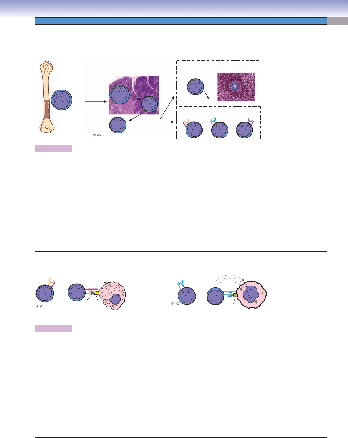
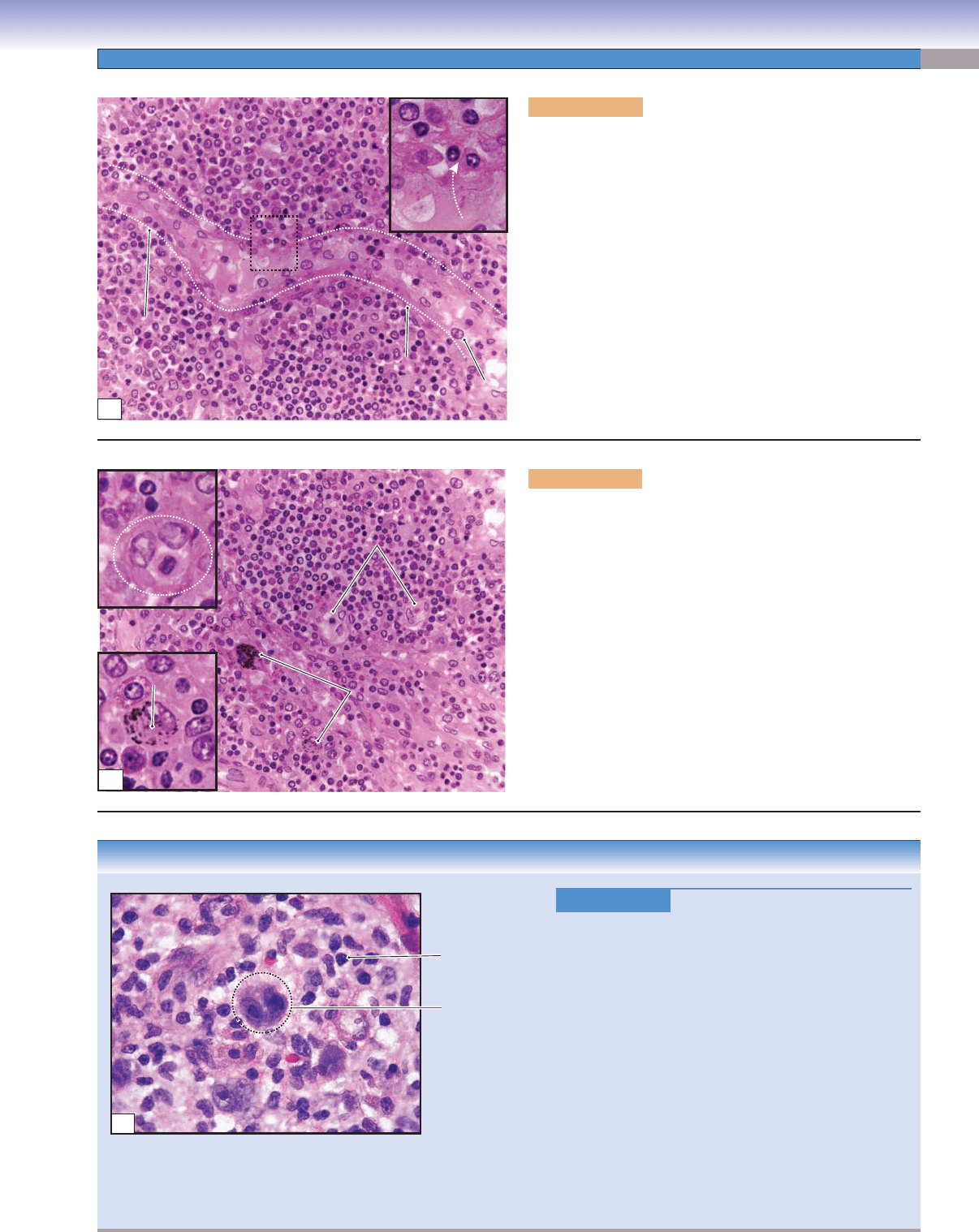
Figure 10-4A. A representation of T-lymphocyte maturation. H&E, 19 (thymus); 200 (spleen)
T cells are derived from pro–T lymphocytes, which migrate from the bone marrow to the thymus where they undergo cell division to generate
a large number of developing lymphocytes (thymocytes). As thymocytes undergo the differentiation process, they begin to express TCR and
other cell-surface proteins. Some of the maturation markers of T cells help them to recognize and interact with MHC molecules. In order to
survive and mature, thymocytes must negotiate both positive and negative selection processes. Positive selection involves promoting survival
of only those thymocytes that are able to interact at an appropriate level with self-MHC molecules, a capacity essential to their ability to
mount effective immune responses. Negative selection involves destruction of thymocytes that have too strong an interaction with self-MHC
molecules; these cells have the potential to contribute to autoimmune disease, and they are removed by macrophages. Positive selection occurs
in the cortex of the thymus and negative selection mainly in the medulla. It has been estimated that only 1% to 2% of thymocytes survive
these selection processes and complete differentiation to become immunocompetent T cells (naive T cells). Naive T cells leave the medulla
of the thymus through the circulation and migrate to the specifi c regions of the secondary lymphoid organs where they may encounter the
foreign antigen that they are programmed to recognize. If antigen stimulation occurs, virgin T cells become active, undergo cell division, and
give rise to clones composed of both memory T cells and effector T cells. Memory T cells can be found in the paracortex of the lymph nodes
and may migrate to infl ammatory sites and give rise to effector T cells. Effector cells include helper T cells, cytotoxic T cells, and regulatory (sup-
pressor) T cells. Each effector cell has either CD4 or CD8 as a surface marker. Effector cells participate in cell-mediated immune responses.
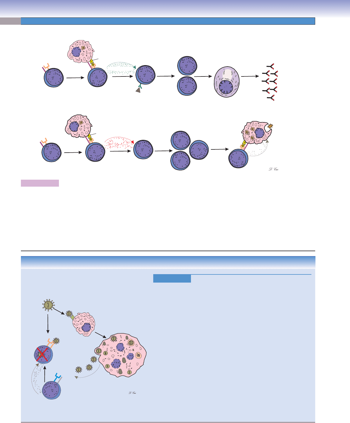
Figure 10-4B. A representation of helper T-cell and cytotoxic T-cell maturation markers.
Each T lymphocyte has in its plasmalemma numerous TCRs, each with the same antigen recognition site. Each T cell also has either CD4 or
CD8 molecules that act as essential coreceptors with the TCR. In the early stages of T-cell development, each thymocyte has both CD4
+
and
CD8
+
markers, and mature T cells have either CD4 or CD8 markers, but not both. CD8
+
cells have the capacity to recognize and react to their
specifi c antigen only if it is presented by another cell in association with MHC class I. All nucleated cells of the body express MHC class I
and present fragments of internally synthesized peptides on their surface MHC class I molecules. If any cell in the body becomes infected
by a virus and synthesizes viral proteins, fragments of these viral proteins are presented as foreign antigens by the cell’s surface MHC class I
molecules. If such a virus-infected cell is encountered by a cytotoxic T cell (CD8
+
cells) that bears TCRs that recognize one of the viral antigens,
the cytotoxic T cell will become activated and destroy the virus-infected cell. CD4
+
cells recognize their specifi c antigen only if it is presented
by another cell in association with MHC class II. MHC class II is expressed by antigen-presenting cells. If an antigen-presenting cell presents
antigen to a CD4
+
(helper T cell) that recognizes the antigen, the helper T cell will become activated to provide signals that promote activation
of other lymphocytes. The illustration on the left shows helper T cells with TCR and surface marker CD4. TCR is an antigen receptor that
is specifi c to the peptide that is attached to the groove of the MHC II molecule on the macrophage. This peptide presents a foreign antigen
to helper T cells. The illustration on the right shows TCR and CD8 markers on the cytotoxic T cells’ surface. TCR of the cytotoxic T cell
responds to antigen presented in association with MHC I molecules of the infected cells. Once a cytotoxic T cell recognizes a nonself antigen,
it releases perforins and enzymes from granules to kill the infected cells as well as some tumor cells, grafted cells, and virus-infected cells.
MHC I
Cytotoxic T cells
CD8
CD8
TCR
Virus-infected cell
MHC II
Peptide
Helper T cells
CD4
CD4
TCR
Antigen-presenting cell
(macrophage)
Perforins
B
Circulation
PALS
PALS
PALS
Pro–T lymphocytes
in bone marrow
Pro–T lymphocytes develop
into lymphoblasts in thymus
Migrate to T–cell regions
(example of PALS in spleen)
Memory T Cell
(CD 4 and CD 8 cells)
Pro–T lymphocytes
Pro–T lymphocytes
CD8
CD4
CD4
TCR
TCR
TCR
Cytotoxic
T cells
Virgin/naive T cells
(medulla)
Activated effector T cells
Helper
T cells
Regulatory
(suppressor) T cells
Thymocytes
Thymocytes
(cortex)
(cortex)
Thymocytes
(cortex)
Pro–T lymphocytes
A
T Lymphocytes
CUI_Chap10.indd 185 6/2/2010 4:11:37 PM
186
UNIT 3
■
Organ Systems
Figure 10-5A. A representation of helper T-lymphocyte activation.
The CD4 surface marker on a helper T cell recognizes MHC II surface proteins on the antigen-presenting cell. The TCR binds with the
peptide-MHC complex on the surface of the macrophage (or other types of antigen-presenting cells); therefore, antigens are presented
to helper T cells. The activating signals (secreted proteins, cytokines) are exchanged between the helper T cells and the macrophages.
There are two main types of helper T cells: (1) Activated helper (T
H
2) cells release a variety of interleukins/cytokines that stimulate
B cells to proliferate and increase the population of plasma cells, thereby increasing production of antibodies. (2) Activated helper
(T
H
1) cells release and bind with IL-2, stimulating proliferation and activation of T
H
1 cells and greatly increasing their own numbers.
Activated T
H
1 cells provide signals that promote proliferation of cytotoxic T cells (CD8
+
cells) and activation of macrophages. In turn,
activated macrophages kill bacteria by a variety of mechanisms (see Fig. 8-6) and stimulate additional infl ammatory processes.
M
M
M
MHC II
MHC II
Antigen
(1) Helper (T 2) cells
H
(2) Helper (T 1) cells
H
B cells
T 1 cells
H
T 1 cells
H
T 1 cells
H
Plasma cell Antibodies
CD4
CD4
TCR
TCR
IL-2
IL-4, IL-5, IL-6
IFN-„
Antigen-presenting
cell (macrophage)
Antigen-presenting
cell (macrophage)
Activated macrophage
(kill bacteria)
A
CLINICAL CORRELATION
Figure 10-5B.
Human Immunodefi ciency V
irus Infection.
Infection by the retrovirus HIV leads to acquired immunodefi ciency
syndrome (AIDS). Infection may be transferred from an infected indi-
vidual through exposure to body fl uids including blood, semen, and
breast milk. It is associated with a progressive decline in CD4
+
T cell
numbers. The stage of infection can be determined by measuring the
patient’s CD4
+
T cell number and the level of HIV in the blood. HIV
primarily infects CD4
+
helper T cells, macrophages, and dendritic cells
(antigen-presenting cells). The low level of CD4
+
T cells in the blood
of HIV-infected patients may be because of (1) the HIV virus killing
infected CD4
+
T cells directly, (2) increased rates of apoptosis in infected
CD4
+
T cells, or (3) CD8
+
cytotoxic lymphocytes recognizing and kill-
ing CD4
+
T cells after the virus has infected them. The HIV virus enters
macrophages (CD4
+
T cells as well), replicates in the host cells, and the
new viruses are released from the host cells. Greatly reduced numbers
of CD4
+
T cells result in the loss of cell-mediated immunity. Without
stimulation from CD4
+
T helper cells, humoral immunity function is
compromised. AIDS patients are vulnerable to opportunistic infections;
common diseases include Pneumocystis jiroveci pneumonia, toxoplas-
mosis, and thrush. Histologically, lymph nodes in the early stage of HIV
infection reveal large, irregular lymphatic nodules and an increased
number of macrophages in the germinal centers (Fig. 10-9C).
Perforins
CD4
TCR
Helper
T cells
Infected
macrophage
Macrophage
HIV
CD8
TCR
Cytotoxic T cells
B
CUI_Chap10.indd 186 6/2/2010 4:11:41 PM
CHAPTER 10
■
Lymphoid System
187
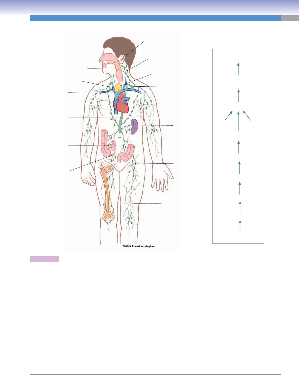
Figure 10-6. Overview of the lymphoid organs.
Locations of the principal lymphoid organs and vessels are shown on the left; the route of lymph drainage is shown on the right.
Structures of the Lymphoid Tissues and Organs
Thoracic
duct
Subclavian
vein
Right lymphatic duct
Peyer
patches
(ileum)
Appendix
Bone marrow
Palatine
tonsil
Lymph node
(inguinal node)
Lymph node
(popliteal node)
Lymph node
(axillary node)
Thoracic duct
Lymphatic vessel
Thymus
Lymph node
(cervical node)
Spleen
Pharyngeal
tonsil
Lymph
Lymphatic capillaries
Lymphatic vessels
Lymph nodes
Lymphoid
organs
Lymphatic
tissue
Lymphatic vessels
Afferent lymphatic vessels
Efferent lymphatic vessels
Right lymphatic duct
Subclavian veins
Thoracic duct (left)
I. Mucosa-associated lymphoid tissue
A. Gut-associated lymphoid tissue: Lymphatic tissue in the
mucosa of the digestive tract, such as Peyer patches in
ileum and nodules in the appendix.
B. Bronchus-associated lymphoid tissue: Lymphatic tissue
in the mucosa of the respiratory tract, such as lymphatic
tissue in bronchi, bronchioles.
C. Tonsils
1. Palatine tonsils
2. Pharyngeal tonsils
3. Lingual tonsils
II. Lymphoid organs
A. Bone marrow (see Chapter 9, “Circulatory System”)
B. Thymus
1. Cortex
2. Medulla
C. Lymph nodes
1. Afferent lymphatic vessels
2. Efferent lymphatic vessels
3. Cortex
4. Paracortex
5. Medulla
D. Spleen
1. White pulp
2. Red pulp
CUI_Chap10.indd 187 6/2/2010 4:11:43 PM
188
UNIT 3
■
Organ Systems
Figure 10-7. Orientation of detailed lymphoid organ illustrations.
Structures of Lymphoid Organs with Figure Numbers
Fig. 10-14A,B,C
Fig. 10-15A,B
Fig. 10-16
Fig. 10-8B
Fig. 10-8A
Fig. 10-10
Fig.10-11 A,B,C,D
Fig. 10-12 A,B
Fig. 10-13A,B,C
Fig. 10-9A
Pharyngeal tonsil
Figure 10-8A
Palatine tonsil
Figure 10-8B
Appendix
Figure 10-9A
Lymph node
Figure 10-9B
Figure 10-9C
Figure 10-10
Figure 10-11A
Figure 10-11B
Figure 10-11C
Figure 10-11D
Figure 10-12A
Figure 10-12B
Figure 10-12C
Thymus
Figure 10-13A
Figure 10-13B
Figure 10-13C
Spleen
Figure 10-14A
Figure 10-14B
Figure 10-14C
Figure 10-15A
Figure 10-15B
Figure 10-16
CUI_Chap10.indd 188 6/2/2010 4:11:45 PM
CHAPTER 10
■
Lymphoid System
189
Mucosa-Associated Lymphoid Tissue
Name Location Epithelial
Covering
Crypts Capsule Lymphatic
Nodules (Follicles)
Palatine tonsils (2) Posterolateral walls
of the oral cavity
Stratifi ed squamous
epithelium
(nonkeratinized)
Yes, deep and
branched crypts
divide tonsil into
lobules
Thick, incomplete
connective tissue
capsule; par-
tially covered by
epithelium
Each lobule contains
numerous lymphatic
nodules, most having
a germinal center
Pharyngeal
(adenoid) tonsil (1)
Posterior roof of the
nasopharynx
Pseudostratifi ed
ciliated columnar
epithelium
No, only epithelial
invagination
Thin, incomplete
connective capsule;
partially covered
by epithelium
Mostly diffuse
lymphoid tissues
and some lymphatic
nodules
Lingual tonsils (2) Posterior fl oor of the
mouth (surface of
the posterior third of
the tongue)
Stratifi ed squamous
epithelium
(nonkeratinized)
Yes, wide
nonbranched crypt;
duct of mucous
gland opens into
the crypt
No capsule;
partially covered
by epithelium
Rows of lymphatic
nodules supported
by connective tissue
septa
TABLE 10-1 Tonsils
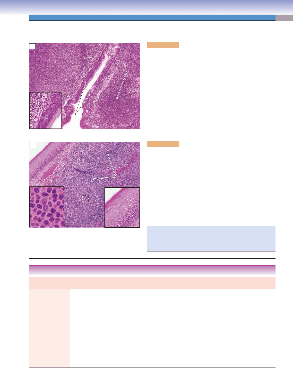
Figure 10-8A. Pharyngeal tonsil, MALT. H&E, 76; inset 184
MALT refers to diffuse lymphatic tissues or aggregate lymphatic nod-
ules in the mucosa of the digestive, respiratory, and genitourinary
tracts. Comparable tissue is GALT in the gut and BALT in the respi-
ratory system. Tonsils are composed of aggregate lymphatic nodules
and belong to MALT. Tonsils include pharyngeal, palatine, and lingual
tonsils. The pharyngeal tonsil is located in the roof of the nasophar-
ynx (Fig. 10-6). It has epithelial invaginations, but no crypts, and is
covered by pseudostratifi ed columnar epithelium. The pharyngeal ton-
sil traps bacteria and viruses and is one of the lymphoid organs that
provides an environment for lymphocytes to meet antigens. It mostly
consists of secondary nodules and a few primary nodules. A secondary
nodule is composed of a germinal center and mantle zone. Activated
B cells are found mainly in the germinal centers of secondary nodules
and inactivated B cells primarily in primary nodules.
Pseudostratified
Pseudostratified
columnar
columnar
epithelium
epithelium
Pseudostratified
columnar
epithelium
Pseudostratified
columnar
epithelium
Mantle
Mantle
zone
zone
Primary
Primary
nodule
nodule
Germinal
Germinal
center
center
Pseudostratified
Pseudostratified
columnar
columnar
epithelium
epithelium
Mantle
zone
Primary
nodule
Germinal
center
Pseudostratified
columnar
epithelium
Pseudostratified
columnar
epithelium
A
Germinal
Germinal
center
center
Large lymphocytes
Large lymphocytes
in germinal center
in germinal center
Mantle
Mantle
zone
zone
Mantle
zone
Stratified squamous
Stratified squamous
epithelium
epithelium
Germinal
center
Large lymphocytes
in germinal center
S
S
tr
tr
a
a
tifie
tifie
d
d
s
s
q
q
u
u
a
a
m
m
o
o
u
u
s
s
e
e
p
p
ith
ith
e
e
liu
liu
m
m
Stratified squamous
epithelium
Stratified squamous
ep
ithelium
B
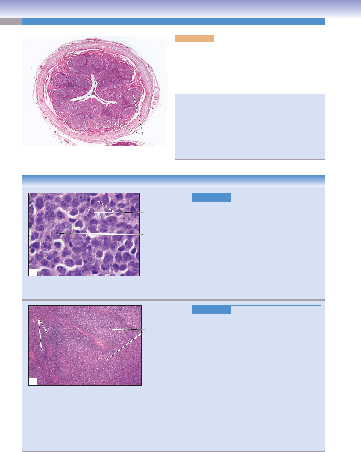
Figure 10-8B. Palatine tonsil, MALT. H&E, 83; inset 750
(left); 197 (right)
Palatine tonsils are paired and are located in the posterior and lateral
portions of the oral cavity. They have 10 to 20 crypts and the portion
facing the oral cavity is covered by stratifi ed squamous epithelium.
The nodules usually lie as a row beneath the epithelium and sur-
round each crypt. They safeguard the entrance of the respiratory and
digestive tracts against microbe invasion. They also function in the
recirculation of lymphocytes and provide sites for the lymphocyte
to interact with antigens. The germinal center of a nodule contains
large-sized B cells and antigen-presenting cells where B cells encoun-
ter antigens and continue to proliferate and develop into plasma
cells. The mantle zone of the nodule contains mostly small inactive
B cells. The peripheral region of the nodule contains mostly T cells.
Palatine tonsils are common sites for infection, such as acute tonsil-
litis, recurrent tonsillitis, or tonsillar hypertrophy due to lymphoid
hyperplasia. Tonsillectomy may be a choice in some children with
recurrent tonsillitis.
CUI_Chap10.indd 189 6/2/2010 4:11:47 PM
190
UNIT 3
■
Organ Systems
Figure 10-9A. Appendix, MALT. H&E, 18
The appendix and Peyer patches in the ileum of the digestive system
are GALT. The appendix is a small, blind tube that extends from
the cecum in the lower right quadrant of the abdomen. It contains
large numbers of lymphatic nodules in its lamina propria. Most
of the nodules are secondary nodules with germinal centers. The
secondary nodules often penetrate into the submucosa.
Lymphatic
nodules
A
CLINICAL CORRELATIONS
Figure 10-9B.
Diffuse Large B-Cell Lymphoma. H&E,
1,000
Diffuse large B-cell lymphoma (DLBCL) is the most
common type of non-Hodgkin lymphoma (25% of all
lymphomas), characterized by a fast-growing and often
symptomatic mass at a nodal or extranodal site. The
most common extranodal site is the gastrointestinal
tract, but other sites include skin, soft tissue, Waldeyer
ring, lung, spleen, and kidneys. Patients may experi-
ence fever, weight loss, and drenching night sweats.
Histologically, tumor cells are large with large nuclei,
open chromatin, and prominent nucleoli. The tumor
grows in a diffuse pattern. Treatment for DLBCL
includes intensive combination chemotherapy with
possible radiotherapy to the involved tumor site.
Figure 10-9C.
Lymph Node, HIV Infection. H&E, 40
HIV infection is associated with a progressive decline
in helper T lymphocytes, resulting in immunosuppres-
sion (Fig. 10-5B). Patients with acute HIV infection
may experience fever
, lymphadenopathy, pharyngitis,
rash, and myalgia. The chronic phase of HIV infection
may last from months to years with patients exhibiting
few symptoms. During the fi nal crisis phase, patients
are at an increased risk of opportunistic infections and
neoplasms. Lymph nodes in the early stage of HIV
infection show marked follicular lymphoid hyperplasia
with enlarged, irregularly shaped follicles (lymphatic
nodules) and increased numbers of macrophages in
the germinal center. The enlarged lymph nodes may be
found fi rst in the upper body, then around the lungs,
and fi nally around the bowel. Patients with compro-
mised immunity are highly likely to be infected by
bacteria and other microbes. Anti-HIV drugs include
four major classes: Reverse transcriptase inhibitors,
protease inhibitors, entry and fusion inhibitors, and
integrase inhibitors.
Lymphoma cells
with large nucleolus
Large irregular
lymphoma cells
B
Enlarged and
irregular-shaped
lymphatic nodules
Lymphatic
Lymphatic
nodules
nodules
Lymphatic
nodules
C
Appendicitis is a common disease, which may be triggered by
bacterial and viral infections resulting in hyperplasia of lymphatic
nodules and obstruction of the lumen of the appendix. Patients
may experience abdominal pain, which most likely will be local-
ized at the McBurney point (one third of the distance between the
anterior superior iliac spine and the umbilicus on the right side) as
the disease progresses. Fever, nausea, and vomiting are the com-
mon symptoms. Emergency appendectomy is the fi rst treatment
choice for most cases.
CUI_Chap10.indd 190 6/2/2010 4:11:50 PM
CHAPTER 10
■
Lymphoid System
191
SYNOPSIS 10-2 Lymphoid Organs
In ■ primary lymphatic organs, lymphocytes differentiate and mature; B cells’ primary lymphoid organ is bone marrow;
T cells’ primary lymphoid organ is the thymus.
In
■ secondary lymphatic organs, lymphocytes encounter and respond to foreign antigens; secondary lymphoid organs
include MALT, lymph nodes, and spleen.
Lymphatic nodules
■ are spherical structures that contain accumulated lymphocytes. They include primary nodules and
secondary nodules.
Primary nodules
■ contain mostly small (inactivated) B cells and do not have a germinal center.
Secondary nodules
■ contain mostly large (activated) B cells and have a germinal center (light area in the center).
Lymphatic nodules
■ contain mostly B cells.
The
■ thymus, paracortex of the lymph node, and PALS in the spleen contain mostly T cells.
Afferent
lymphatic vessels
Artery
Medullary
sinuses
Efferent
lymphatic vessel
Postcapillary venules
(HEV in paracortex)
Venules
Small vein (medulla)
Vein
Subcapsular
sinuses
Small artery
(b
in medulla)
ranches of artery
Peritrabecular
sinuses
Arterioles
(paracortex and cortex)
capillaries
(nodules of cortex)
Vascular channels (blood):
Lymphatic channels (lymph):
Secondary
nodule
Afferent
lymphatic vessel
Subcapsular sinus
Primary
nodule
Medullary
cords
Medullary
sinuses
Efferent
lymphatic vessel
Trabecula
Trabecula
Germinal center
Germinal center
of secondary
of secondary
nodule in cortex
nodule in cortex
HEV in
paracortex
Trabecula
Peritrabecular
sinus
Germinal center
of secondary
nodule in cortex
Medulla
Paracortex
Artery
Vein
Cortex
Cortex
Cortex
Capsule
P
P
a
a
r
r
a
a
c
c
o
o
r
r
te
te
x
x
Medulla
Medulla
Paracortex
Para
cortex
Paracortex
Medulla
Paracortex
Figure 10-10. Overview of the lymph node.
This is a representation of a lymph node. It is covered by a capsule consisting of a layer of connective tissue, which extends into the
substance of the node to form trabeculae. The lymph node is divided into three regions: cortex, paracortex, and medulla. (1) The
cortex is composed of a row of lymphatic nodules; the majority are secondary nodules with germinal centers. Occasionally, primary
nodules (without germinal centers) may be found in the cortex region. (2) The paracortex lies between the cortex and medulla; most
T cells reside in this region. HEVs are located in paracortex and are the sites where circulating lymphocytes enter the node. (3) The
medulla is composed of medullary cords and medullary sinuses. Lymph enters the lymph node through afferent lymphatic vessels;
courses through the subcapsular, peritrabecular, and medullary sinuses; and exits the lymph node through the efferent lymphatic
vessel (follow the dotted magenta line). The artery and vein enter and exit by passing through the hilum of the lymph node.
Lymph Nodes
Comparison of Lymph and Blood Flow
CUI_Chap10.indd 191 6/2/2010 4:11:53 PM
192
UNIT 3
■
Organ Systems
Figure 10-11A. Lymph node. H&E, 43
Lymph nodes are bean shaped and are the only lymphoid
organs that have afferent lymphatic vessels (Fig. 10-9). This
is a cross section of a lymph node. (1) The cortex is the
peripheral region of the lymph node and consists of a row of
nodules. (2) The medulla stains lighter and is located at the
center area; it is composed of medullary sinuses and medul-
lary cords (Fig. 10-11B,D). (3) The paracortex lies between
the cortex and the medulla. Lymph nodes are the major sites
to fi lter incoming lymph and are the sites for lymphocytes to
meet antigens.
Figure 10-11B. Lymph node. H&E, 100
The subcapsular sinus carries lymph from afferent lymphatic
vessels into the node by passing through peritrabecular sinuses
to the medullary sinuses. The nodules in the cortex consist of a
germinal center (loosely packed large B cells) and a mantle zone
(containing tightly packed small B cells). T cells mainly reside
in the paracortex region where they interact with antigen-
presenting cells; lymphocytes enter the lymph node through
HEVs in the paracortex region (Fig. 10-12A,B).
Figure 10-11C. Germinal center, lymph node. H&E, 658
The germinal center is composed of activated B cells in various
stages of maturation. Cell size and nuclear shape are varied.
The large immature cells with round nucleus and dispersed
euchromatin are lymphoblasts and plasmablasts. They dif-
ferentiate into memory B cells and plasma cells. The germinal
center also contains follicular dendritic (antigen-presenting)
cells, which help pass antigens to B cells. They are diffi cult to
recognize in H&E stain.
Figure 10-11D. Medullary sinuses and cords, lymph node.
H&E, 658
A medullary sinus surrounded by a medullary cord is shown
here. Medullary sinuses carry lymph to where antigens are
removed by macrophages from slow-fl owing lymph. The
medullary cords contain B cells, plasma cells, dendritic cells,
and macrophages held within a network of reticular fi bers.
Medullary cord
Medullary cord
Medullary
Medullary
cord
cord
Medullary cord
Lumen of the
Lumen of the
medullary sinus
medullary sinus
Lumen of the
medullary sinus
Medullary
cord
Macrophages
Macrophages
Macrophages
Medullary cord
Medullary cord
Medullary cord
Lymphocytes in the
Lymphocytes in the
medullary sinus
medullary sinus
Lymphocytes in the
medullary sinus
D
Lymphatic
nodules
M
M
e
e
d
d
u
u
lla
lla
Medulla
Paracortex
Paracortex
Paracortex
Cortex
Cortex
Cortex
A
Subcapsular sinus
Subcapsular sinus
Subcapsular sinus
Peritrabecular
Peritrabecular
sinus
sinus
Peritrabecular
sinus
Medullary
Medullary
cords
cords
Medullary
cords
Germinal
Germinal
center
center
Germinal
center
Medullary
Medullary
sinuses
sinuses
Medullary
sinuses
Medullary
Medullary
sinuses
sinuses
Medullary
sinuses
Paracortex
Paracortex
Paracortex
Mantle
Mantle
zone
zone
Mantle
zone
B
Lymphoblast
Lymphoblast
Lymphoblast
Small
Small
lymphocytes
lymphocytes
Small
lymphocytes
Lymphoblast
Lymphoblast
Lymphoblast
C
CUI_Chap10.indd 192 6/2/2010 4:11:56 PM
CHAPTER 10
■
Lymphoid System
193
CLINICAL CORRELATION
Figure 10-12C.
Hodgkin Lymphoma. H&E, 824
Hodgkin lymphoma, also known as Hodgkin disease,
is one of the two major categories of malignant lym-
phoid cancers, characterized by painless enlargement of
lymph nodes, spleen, and liver
. Patients often experi-
ence fever, night sweats, unexpected weight loss, and
fatigue. The cancer cells are transformed from normal
lymphoid cells, which reside predominantly in lym-
phoid tissues. Characteristic Reed-Sternberg cells, of
B cell origin, can be found in affected lymphoid tis-
sues. These cells are large (20–50 μm) and contain
abundant, amphophilic, and fi nely granular/homoge-
neous cytoplasm with two mirror-image nuclei (“owl’s
eyes”), each with an eosinophilic nucleolus and a thick
nuclear membrane. Radiotherapy and chemotherapy
are both effective in treatment of Hodgkin lymphoma.
The 5-year survival rate is approximately 90% when
the disease is detected and treated early.
C
Reed-Sternberg
cell
Lymphocyte
Figure 10-12A. High endothelial venules (HEVs), paracor-
tex of lymph node. H&E, 272; inset 720
Arteries that serve a lymph node enter the hilum and give rise
to branches that pass through the medulla and reach the cortex
where they form a network of capillaries in the nodule (fol-
licle) region. Postcapillary venules (in the paracortex region)
carry blood from the capillary bed back to the venule system
and out of the lymph node at the hilum (Fig. 10-10). HEVs
are specialized postcapillary veins, which are lined by cuboidal
cells instead of squamous endothelial cells. The apical surfaces
of these cuboidal cells contain rich glycoproteins that attract
lectinlike receptors (L selectin) on the surface of the lympho-
cytes, which helps lymphocytes stop and attach to the HEVs.
Lymphocytes pass through HEVs by way of diapedesis and
enter the lymph node from blood circulation. The inset shows
a lymphocyte escaping from a HEV into the lymphatic tissue.
Cubodial
Cubodial
cell
cell
Lymphatic tissue
Lymphatic tissue
Lymphatic tissue
Lymphatic tissue
High
High
endothelial
endothelial
venule
venule
High
High
endothelial
endothelial
venule
venule
Lymphatic tissue
Lymphatic tissue
High
endothelial
venule
Cuboidal
cell
High
endothelial
venule
A
Active
Active
macrophages
macrophages
High
High
endothelial
endothelial
venules
venules
Active
macrophages
High
endothelial
venules
High endothelial
High endothelial
venule
venule
High endothelial
venule
Active
Active
macrophage
macrophage
Active
macrophage
B
Figure 10-12B. High endothelial venules, paracortex of
lymph node. H&E, 281; insets 725
HEVs can be found in all of the secondary lymphoid organs
except the spleen. They are the major sites for both naive B and
T lymphocytes that have migrated from circulation into the lym-
phatic tissue. After they enter the lymph node, B cells migrate to
the cortex region where they differentiate in the germinal center.
Most T cells remain in the paracortex region where they inter-
act with antigen-presenting cells (macrophages). Once T cells
acquire antigens, they release cytokine (IL-4, IL-5, and IL-6),
which stimulates B cells’ division and maturation to become
memory B cells and plasma cells with the consequent produc-
tion of antibodies (Fig. 10-5A). Endothelial cells of HEVs are
cuboidal cells and have large round or oval nuclei with pale
chromatin. The insets show a lymphocyte in the cross section
of a HEV (upper); and an active macrophage in the paracortex
region (lower).
CUI_Chap10.indd 193 6/2/2010 4:12:03 PM
194
UNIT 3
■
Organ Systems
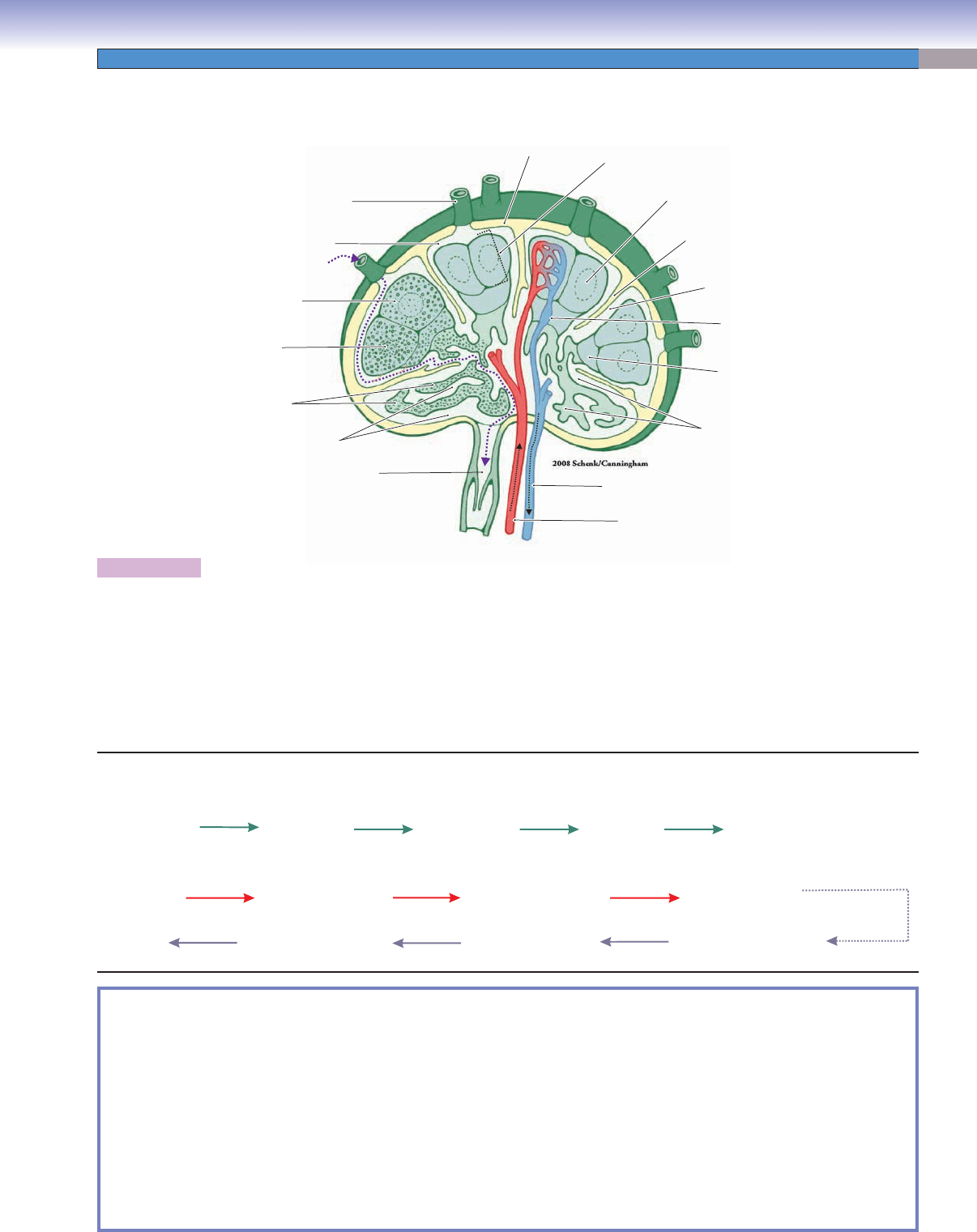
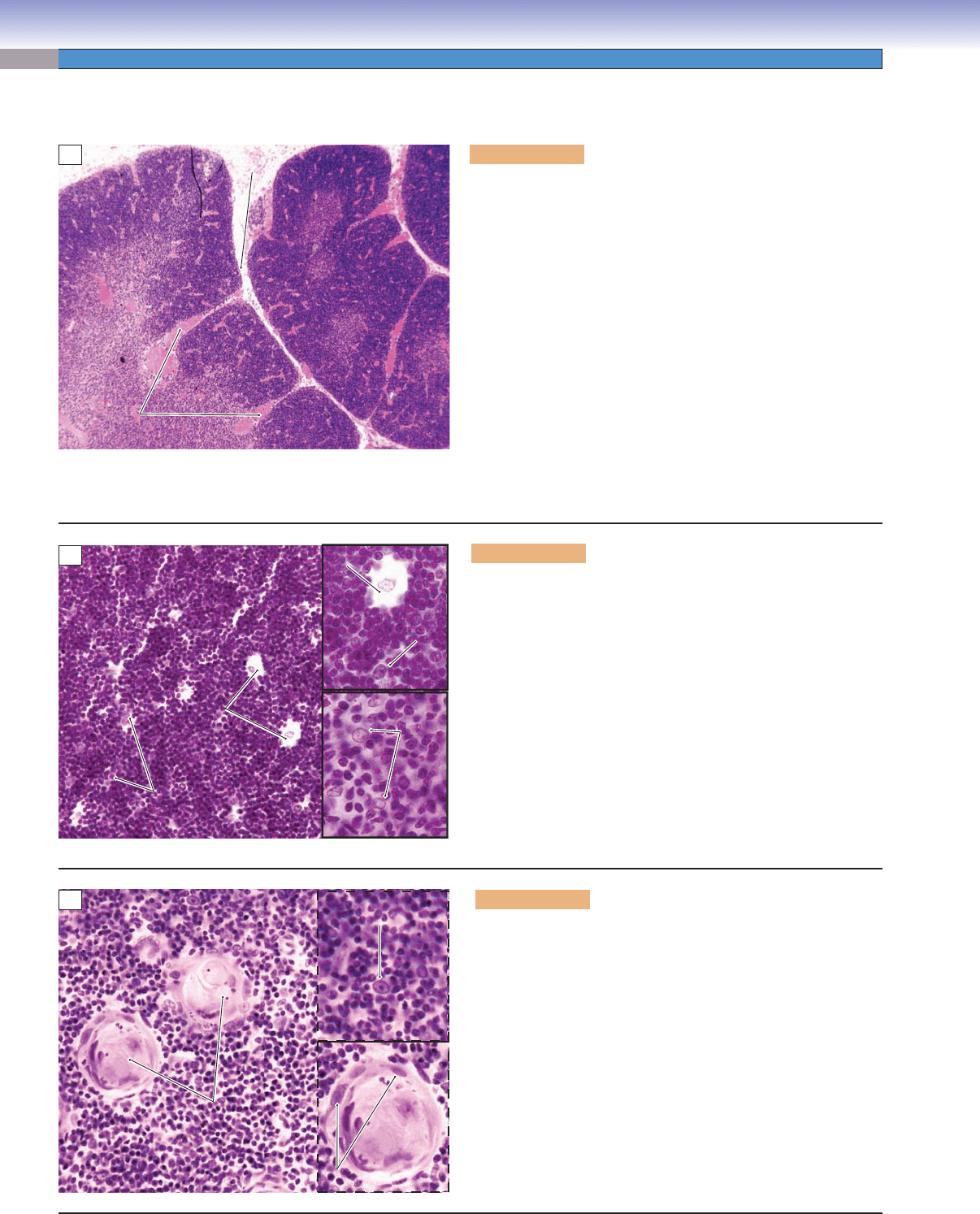
Figure 10-13A. Thymus. H&E, 46
The thymus is a primary lymphoid organ for T cells where T-cell
maturation takes place. The thymus is large in children and gradu-
ally atrophies to be replaced by fat after puberty. The thymus is
located in the superior mediastinum and is divided into smaller
units called lobules by connective tissue septae, which extend
inward from the surface of the organ. The thymus does not have
lymphatic nodules; it is organized into cortex (peripheral) and
medulla (center). There are no afferent lymphatic vessels; its effer-
ent lymphatic vessels arise from the corticomedullary junction and
medulla and leave the thymus in company with the blood vessels.
Thymocytes (developing T cells) are concentrated in the cortex
region, and as they undergo differentiation, they move down to
the medulla. The blood vessels pass through the interlobar septa
and enter the thymus at the junction of the cortex and medulla.
Thymic capillaries are continuous capillaries with thick basement
membranes. They are surrounded by epithelial reticular cells and
form an effective thymic-blood barrier, which prevents foreign
antigens from entering the thymus.
Septum
Septum
Medulla
Medulla
Cortex
Cortex
Medulla
Septum
Cortex
Medulla
Medulla
C
C
o
o
rte
rt
e
x
x
Medulla
Cortex
M
M
e
e
d
d
u
u
lla
ll
a
Cortex
Cortex
C
C
o
o
rte
rt
e
x
x
Medulla
Cortex
Cortex
Septum
Septum
Septum
A
B
Epithelial
Epithelial
reticular
reticular
cells
cells
Epithelial reticular cells
Epithelial reticular cells
Epithelial reticular cells
Epithelial reticular cells
Macrophage
Macrophage
Macrophage
Macrophage
Epithelial reticular cells
Epithelial
reticular
cells
Epithelial reticular cells
Macrophage
Macrophage
Figure 10-13B.
Thymus, cortex. H&E, 278; insets 510
The cortex region contains thymocytes, macrophages, dendritic
cells, and epithelial reticular cells. The macrophages and dendritic
cells are antigen-presenting cells; they present self-antigens to
thymocytes. Only 1% to 2% of thymocytes survive and continue to
develop. Epithelial reticular cells are derived from endoderm (lym-
phocytes are derived from mesoderm). They are interconnected with
each other to form a framework to hold T lymphocytes together.
They have large, ovoid nuclei and long processes and make contact
with each other by desmosomes. They contain secretory granules
and produce thymosin, serum thymic factor, and thymopoietin hor-
mone. These hormones play an important role in T-cell maturation.
The epithelial reticular cells can be classifi ed into six types based on
their functions and locations. Types I to III are located in the cortex
region, and type IV in the corticomedullary junction. Types V and
VI are located in the medulla of the thymus.
C
Epithelial reticular cells
Epithelial reticular cells
Epithelial reticular cells
Epithelial reticular cells
Epithelial reticular cells
Epithelial reticular cells
Hassell
Hassell
corpuscle
corpuscle
Hassall
corpuscle
Figure 10-13C. Thymus, medulla. H&E, 624; insets 843
The medulla region contains naive (virgin) T cells, macrophages,
and types V and VI epithelial reticular cells. The naive T cells
are immunocompetent cells. They mature from thymocytes in the
cortex and migrate from the medulla to secondary organs where
they become effective or memory T cells if they meet with specifi c
foreign antigen. The medulla of the thymus is also the place where
T cells are selectively removed by macrophages. Both types V and
VI epithelial reticular cells are located in the medulla. The type
VI epithelial reticular cells show various degrees of keratinization
and are arranged into concentric layers forming a spherical struc-
ture called a Hassall corpuscle. Although the function of Hassall
corpuscles is not fully understood, their numbers are increased
in older individuals. Hassall corpuscles can be used as one of the
unique features to distinguish the thymus from other lymphatic
organs during the histological slide examination.
Thymus
CUI_Chap10.indd 194 6/2/2010 4:12:08 PM