Cui Dongmei. Atlas of Histology: with functional and clinical correlations. 1st ed
Подождите немного. Документ загружается.

CHAPTER 11
■
Respiratory System
205
Conducting Portion: Upper Respiratory Airway
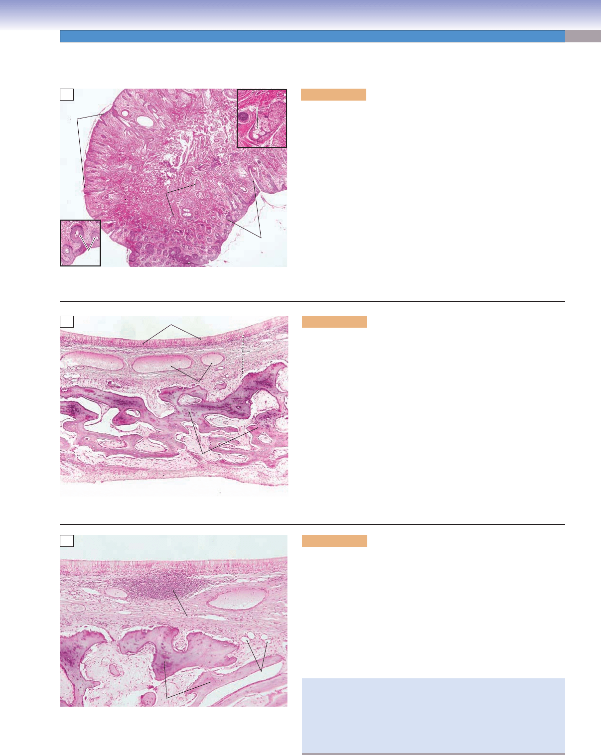
Figure 11-3A. Nasal vestibule, nose. H&E, 12; insets 42 (left);
34 (right)
The nasal cavity contains pairs of chambers separated by the nasal
septum; the air passing through these chambers is moistened and
warmed before it enters the lungs. There are three types of epithelia
lining the nasal cavity in different regions: (1) the vestibule region is
lined by stratifi ed squamous epithelium; (2) the nasal mucosa region is
lined by respiratory epithelium, which occupies most of the area in the
nasal cavity, such as the conchae and nasal cavity wall; (3) the olfac-
tory mucosae are covered by a specialized olfactory epithelium and
are concerned with the sense of smell (Figs. 11-1 and 11-4). The exter-
nal surface of the nasal vestibule is covered by the skin and its internal
surface is covered by stratifi ed squamous epithelium with numbers of
vibrissae (stiff hairs) that entrap dust particles and prevent them from
entering the lungs. The vibrissae are greater in number at the anterior
end and gradually decrease at the posterior end of the vestibule. Seba-
ceous glands are found around the roots of the vibrissal follicles.
A
Sebaceous
Sebaceous
gland
gland
Sebaceous
gland
Sebaceous
Sebaceous
gland
gland
Sebaceous
gland
Vibrissal
follicles
Vibrissal
Vibrissal
follicles
follicles
Vibrissal
follicles
Skin
B
Lamina
propria
Respiratory epithelium
Bone
Bone
Bone
Venous
Venous
plexuses
plexuses
Venous
plexuses
Figure 11-3B. Nasal mucosa, nose. H&E, 42
Nasal mucosa lines most of the nasal cavity. It is made up of respira-
tory epithelium (a layer of ciliated pseudostratifi ed columnar epithe-
lium) and a layer of connective tissue beneath the lamina propria. The
nasal mucosa is attached to the bone for skeletal support. Respiratory
epithelium is composed of ciliated cells, goblet cells, and basal cells
as well as rarer cell types such as endocrine cells (Fig. 11-7; see also
Figs. 3-9 and 3-10). This type of epithelium lines most regions of the
respiratory system. There are many blood vessels (venous plexuses) in
the lamina propria of the nasal mucosa; these small veins provide a
rich blood fl ow, which warms the air passing through the nasal cavity
before air enters the lungs.
The large venous plexuses within the lamina propria of the nasal
conchae are called swell bodies. Small arteries empty blood directly
into the venous plexuses within the conchae; this causes the lam-
ina propria to swell, reducing airfl ow through the nasal cavity and
increasing air contact with the nasal mucosa.
C
Small vein
Capillaries
MALT
Bone
Figure 11-3C. Nasal mucosa, nose. H&E, 70
Lymph nodules or diffuse lymphocytes are often found in the lamina
propria of the nasal mucosa, bronchi, and bronchioles (Fig. 11-10B).
They are called mucosa-associated lymphatic tissue (MALT) and are
usually located in the connective tissue where they can infi ltrate the
epithelium (see inset). The lymphoid tissues immunologically support
the wet epithelial membranes of the body’s mucosae and can be found
in mucosae of other organs, such as the appendix and the ileum of the
digestive tract (see Chapter 10, “Lymphoid System,” Fig. 10-9A and
Chapter 15, “Digestive Tract,” Fig. 15-15). Mucous and mixed muco-
serous glands may be found in the lamina propria in some specimens.
In response to upper respiratory airway infection or allergic reac-
tion, the nasal mucosa may become swollen (especially the inferior
concha) and infl amed, blocking air passage through the nasal cav-
ity. This condition is called rhinitis. Symptoms may include a stuffy
or runny nose; common treatments are antihistamine and decon-
gestant pills and sprays, etc.
CUI_Chap11.indd 205 6/16/2010 7:34:43 PM
206
UNIT 3
■
Organ Systems
Ducts of
Ducts of
Bowman glands
Bowman glands
Connective
Connective
tissue
tissue
Basal cells
Basal cells
Blood vessel
Blood vessel
Nuclei of
Nuclei of
olfactory cells
olfactory cells
Olfactory
Olfactory
epithelium
epithelium
Lamina
Lamina
propria
propria
Cilia
Cilia
Basal cells
Blood vessel
Connective
tissue
Ducts of
Bowman glands
Olfactory fila
Olfactory fila
(axons with nuclei of
(axons with nuclei of
unmyelinated Schwann cells)
unmyelinated Schwann cells)
Olfactory fila
(axons with nuclei of
unmyelinated Schwann cells)
Nuclei of
olfactory cells
Olfactory
epithelium
Lamina
propria
Cilia
Nuclei of
Nuclei of
supporting cells
supporting cells
Nuclei of
supporting cells
Bowman
Bowman
glands
glands
Bowman
glands
B
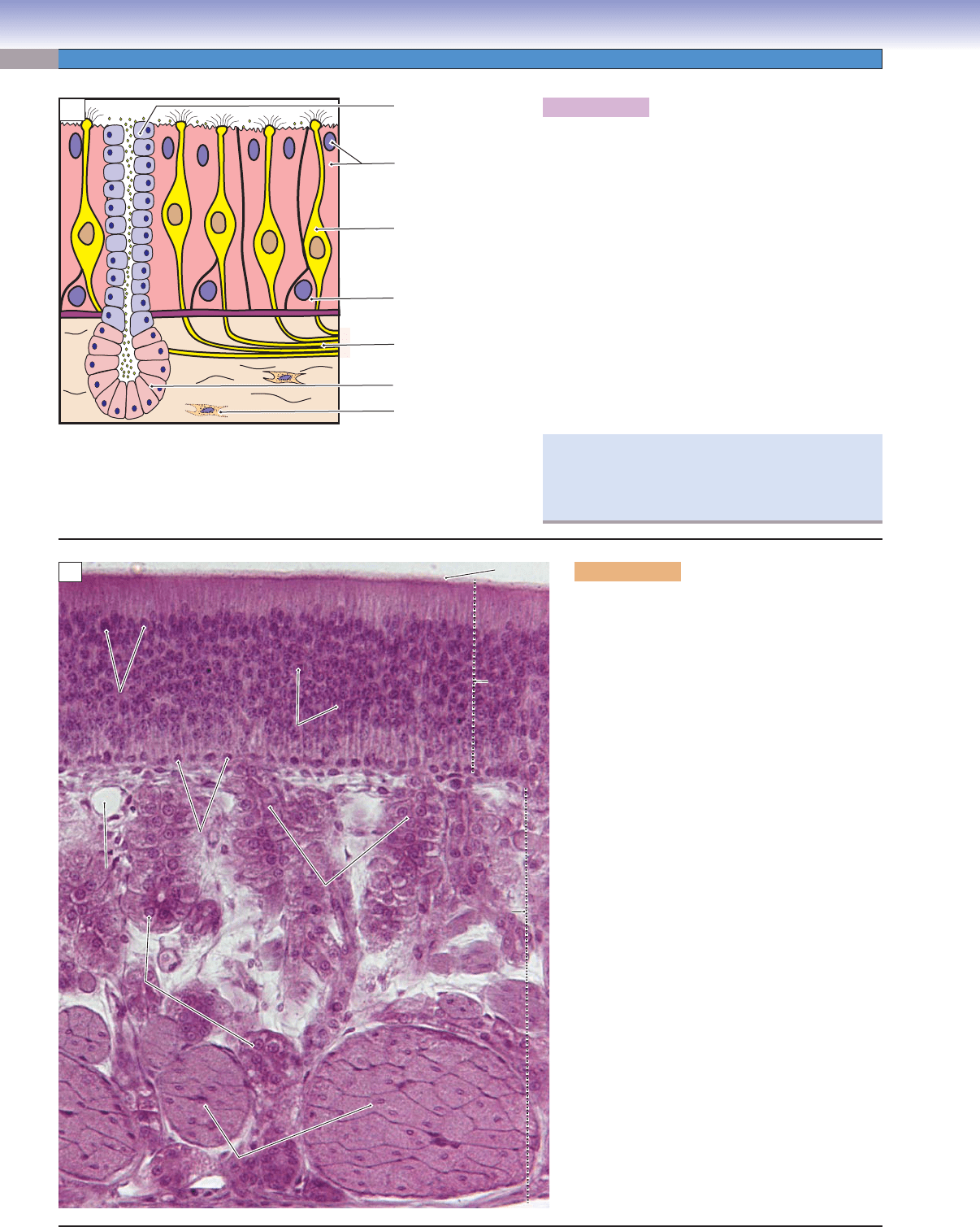
Figure 11-4B. Olfactory mucosa, nose. H&E,
326
Olfactory mucosa is composed of specialized
epithelium (olfactory epithelium) and lamina
propria with Bowman glands, nerve axons
(olfactory fi la), and blood vessels. The olfac-
tory epithelium looks like other pseudostrati-
fi ed columnar epithelium but contains different
types of cells: olfactory cells (receptor neurons),
supporting (sustentacular) cells, and basal cells.
Olfactory receptor neurons are bipolar cells with
long, nonmotile cilia, which function as recep-
tors for odorants. Supporting cells are colum-
nar shaped; their nuclei are dark and ovoid and
are positioned in the apical region of the cells.
Microvilli and a terminal web of the supporting
cells may be seen at the electron microscopy level.
The function of the supporting cells is to provide
mechanical support; they may also be involved
in binding or inactivation of odorant molecules.
Basal cells are short cells with round nuclei. They
lie in a single layer at the basal region of the epi-
thelium, serve as stem cells, and are capable of
regenerating into the other types of cells in the
epithelium. The lamina propria of the olfactory
mucosa contains Bowman glands, olfactory fi la
(collective unmyelinated axons), and blood ves-
sels. Bowman glands are serous glands, which
release a watery secretion onto the surface of the
epithelium. These watery secretions (containing
water-soluble proteins) serve to bathe the surface
of the olfactory epithelium and help trap and dis-
solve odorant molecules. The olfactory fi la are
axons of the olfactory receptor neurons.
Figure 11-4A. A representation of olfactory
mucosa.
The olfactory mucosa is located in the roof of the
nasal cavity (Fig. 11-1); it is composed of olfactory
cells (olfactory receptor neurons), supporting cells,
and basal cells in the epithelium, and of olfactory
fi la (unmyelinated axons) and Bowman glands in the
lamina propria. Bowman glands release their product
onto the surface of the epithelium via ducts. The main
function of the olfactory mucosa is to detect odor.
Odorant molecules come into contact with the surface
of the olfactory epithelium in the nasal cavity and bind
to receptors on the cilia of the olfactory cells. Olfac-
tory cells transmit signals through the olfactory fi la to
the olfactory bulb and to the olfactory centers of the
central nervous system. Olfactory neurons are able to
proliferate after being damaged.
T. Yang &D. Cui
Olfactory cell
(olfactory receptor
neuron)
Duct of
Bowman gland
Basal cell
Olfactory fila
(unmyelinated axons)
Bowman gland
Fibroblast in
connective tissue
Supporting cell
A
Clinically, loss of smell is called anosmia, and
decreased sensitivity to odorants is called hyposmia.
These symptoms are often associated with upper air-
way infections.
CUI_Chap11.indd 206 6/16/2010 7:34:46 PM
CHAPTER 11
■
Respiratory System
207
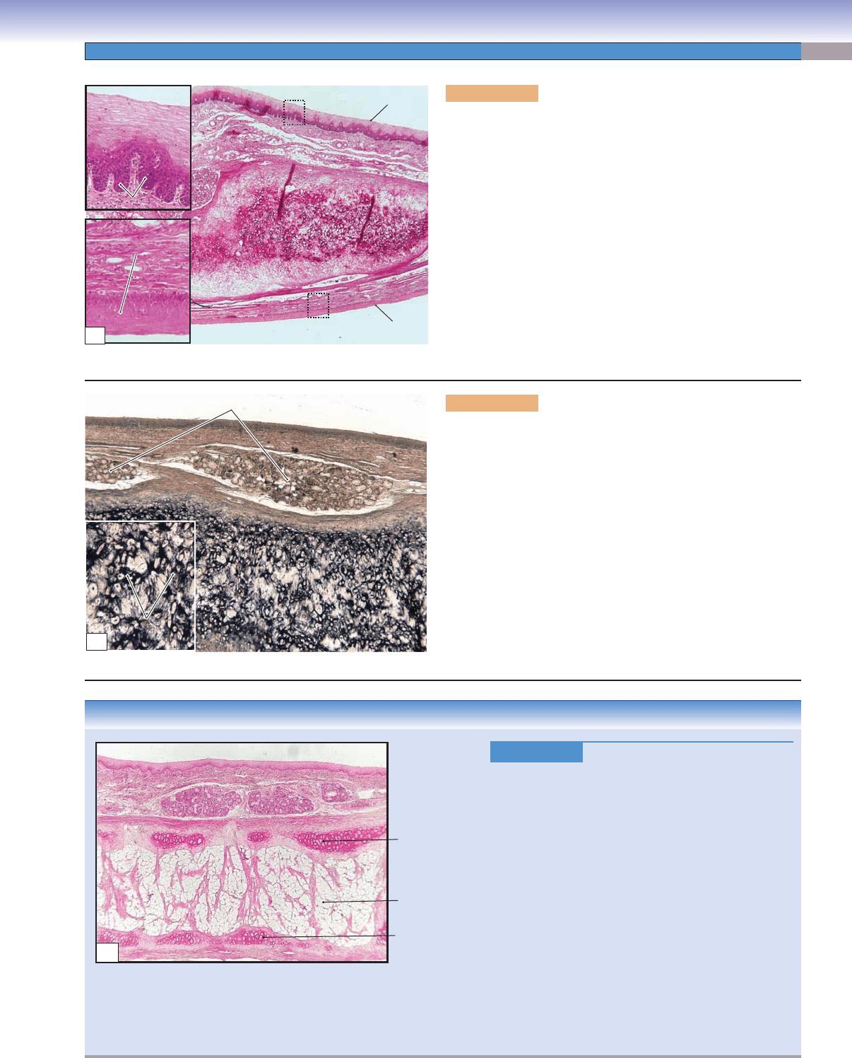
Figure 11-5A. Epiglottis, larynx. H&E, 18; inset 99
The larynx is the short passage that connects the pharynx with
the trachea; its main function is to produce sound and prevent
food or liquid from entering the trachea. Laryngeal structures
include the epiglottis, vocal cords, and nine pieces of cartilage
located in its wall (including the thyroid cartilage—“Adam’s
apple”). The epiglottis is a fl attened, leaf-shaped structure with
elastic cartilage support. Classically, the epiglottis is covered by
two types of epithelia: stratifi ed squamous on the lingual surface
facing the oropharynx and respiratory epithelium on the laryn-
geal surface facing the larynx. However, this normal condition
is rarely found, because of squamous metaplasia (see Fig. 3-9C),
resulting from aging and irritation (even in young individuals).
The stratifi ed squamous epithelium on the lingual surface has the
distinguishing feature of rete ridges on its basal region, whereas
the stratifi ed squamous epithelium (squamous metaplasia) on the
laryngeal surface is fl attened on its basal region.
CLINICAL CORRELATION
Figure 11-5C.
Age Impact on the Epiglottis. H&E,
21
The integrity of the epiglottis is affected by age and
other factors. Structurally, variations include not only
the change from pseudostratifi ed columnar epithelium
to stratifi ed squamous epithelium (squamous meta-
plasia [Fig. 11-5A]), but also reduction in cartilage
size due to replacement of the central portion of the
cartilage by a large amount of adipose tissue. Loss of
elastic cartilage is associated with loss of elastic fi bers
in the epiglottis. These changes result in a reduction of
the elasticity and stiffness of the epiglottis. Epiglottis
abnormalities, such as the hypoplastic, bifi d epiglottis
associated with cleft palate in children, can cause epi-
sodic choking during food or fl uid intake. Recurrent
foreign substances entering the respiratory tract can
cause chronic infl ammation of the respiratory tract.
Nasogastric tube feeding and surgical repair of the
deformed epiglottis may be necessary in severe cases.
Reduced size
of cartilage
Remaining
cartilage
Adipose tissue
C
Rete ridge
Rete ridge
Stratified squamous
Stratified squamous
epithelium
epithelium
Stratified squamous
Stratified squamous
epithelium
epithelium
Elastic cartilage
Elastic cartilage
Lingual surface
Elastic cartilage
Laryngeal surface
Rete ridge
Stratified squamous
epithelium
Stratified squamous
epithelium
A
Elastic fibers
Elastic fibers
Elastic cartilage
Elastic cartilage
Elastic cartilage
Elastic cartilage
Mixed (mucous and serous) gland
Elastic cartilage
Elastic cartilage
Elastic fibers
B
Figure 11-5B. Epiglottis, larynx. Elastic fi ber stain, 35; inset
105
This sample of epiglottis tissue was stained with an elastic fi ber
stain. The elastic fi bers are visible in black. Chondrocytes are
embedded with the elastic fi bers in the cartilage matrix. Elastic
cartilage has different properties than hyaline cartilage; it forms
the framework and provides a fi rm and elastic support for the
epiglottis. There are some mixed glands (most are mucous) in the
lamina propria. These glands produce mucin and a watery fl uid
on the surface of the epiglottis. The inferior portion of the epiglot-
tis is attached to the rim of the thyroid cartilage and hyoid bone.
The superior portion is free to move up when making sounds,
and to move down to close the airway while food and fl uid are
passing through the pharynx. The main functions of the epiglot-
tis are to prevent food and fl uid from entering the trachea and to
cooperate with the vocal cords to produce sound.
CUI_Chap11.indd 207 6/16/2010 7:34:49 PM
208
UNIT 3
■
Organ Systems
Conducting Portion: Lower Respiratory Airway
Figure 11-6A. Trachea. H&E, 7
The trachea is a fl exible tube that connects the larynx to the
primary bronchi. It is about 10 to 12 cm long, 2 to 2.5 cm in
diameter, and is located immediately anterior to the esopha-
gus. It is composed of mucosa, submucosa, hyaline cartilage,
and adventitia. (1) The mucosa covers the inner surface of
the trachea and contains respiratory epithelium and the lam-
ina propria. (2) The submucosa contains connective tissue,
which is denser than the lamina propria. (3) Hyaline cartilage
has a unique C-shape (some animals, e.g., the rat, may have
O-shaped cartilage), and there are about 16 to 20 rings in the
trachea. (4) The adventitia is composed of connective tissue,
which covers the outer surface of the cartilage and connects
the trachea to the adjacent structures. There are some elastic
connective tissues and smooth muscles (trachealis muscle) in
the opening between the two ends of the cartilage; this stabi-
lizes the opening.
Cartilage ring
Mucosa
Submucosa
Adventitia
Trachealis muscle
A
L
L
a
a
m
m
in
in
a
a
p
p
ro
ro
p
p
ria
ria
S
S
u
u
b
b
m
m
u
u
c
c
o
o
s
s
a
a
Adventitia
Adventitia
Respiratory
epithelium
Respiratory
epithelium
Perichondrium
Tracheal cartilage
(hyaline cartilage)
Glands in
submucosa
Submucosa
Lamina propria
L
L
a
a
m
m
in
in
a
a
p
p
ro
ro
p
p
ria
ria
Lamina
propria
Adventitia
B
Figure 11-6B. Trachea. H&E, 35; inset 146
The luminal surface of the trachea is covered by ciliated pseu-
dostratifi ed columnar epithelium, also called respiratory epi-
thelium (Fig 11-7A,B). The epithelium plus the lamina propria
constitute the mucosa. The lamina propria is a layer of loose
connective tissue beneath the epithelium. The submucosa is
a layer of dense connective tissue located between the lam-
ina propria and cartilage; it contains many trachealis glands
(seromucous glands). Mucin and watery secretions from tra-
cheal glands are delivered through their ducts to the surface
of the epithelium (Fig. 11-6C). The C-shaped hyaline cartilage
rings (tracheal cartilages) provide support for the trachea.
They are covered by perichondrium and many chondrocytes
are embedded in their matrix (see Fig. 5-2B).
Trachealis muscle
Trachealis muscle
(smooth muscle)
(smooth muscle)
Trachealis
Trachealis
glands
glands
Duct of
Duct of
glands
glands
Trachealis muscle
Trachealis muscle
(smooth muscle)
(smooth muscle)
Tracheal cartilage
(hyaline cartilage)
Trachealis muscle
(smooth muscle)
Trachealis
glands
Duct of
glands
Trachealis muscle
(smooth muscle)
C
Figure 11-6C. Trachealis muscle, trachea. H&E, 35;
inset 100
The trachealis muscle is a smooth muscle located between the
open ends of the C-shaped cartilage rings. Trachealis muscle
fi bers attach directly to the perichondrium of the cartilage
together with connective tissue, which stabilizes the cartilage’s
open ends. The contraction and expansion of smooth muscle
help to adjust the airfl ow through the trachea.
If a foreign object enters the airway, smooth muscle in the
trachea and bronchi contracts, narrowing the lumina, help-
ing to induce coughing. (Cough refl ex: cooperation among
the epiglottis, vocal cords, trachea, bronchi, lungs, respira-
tory muscles, and the autonomic nervous system). In asthma,
the smaller airways (bronchi and bronchioles) are narrowed
because of excessive contraction of the smooth muscle of the
lower airway in response to histamine released in an allergic
reaction (see Fig. 6-11C).
CUI_Chap11.indd 208 6/16/2010 7:34:53 PM
CHAPTER 11
■
Respiratory System
209
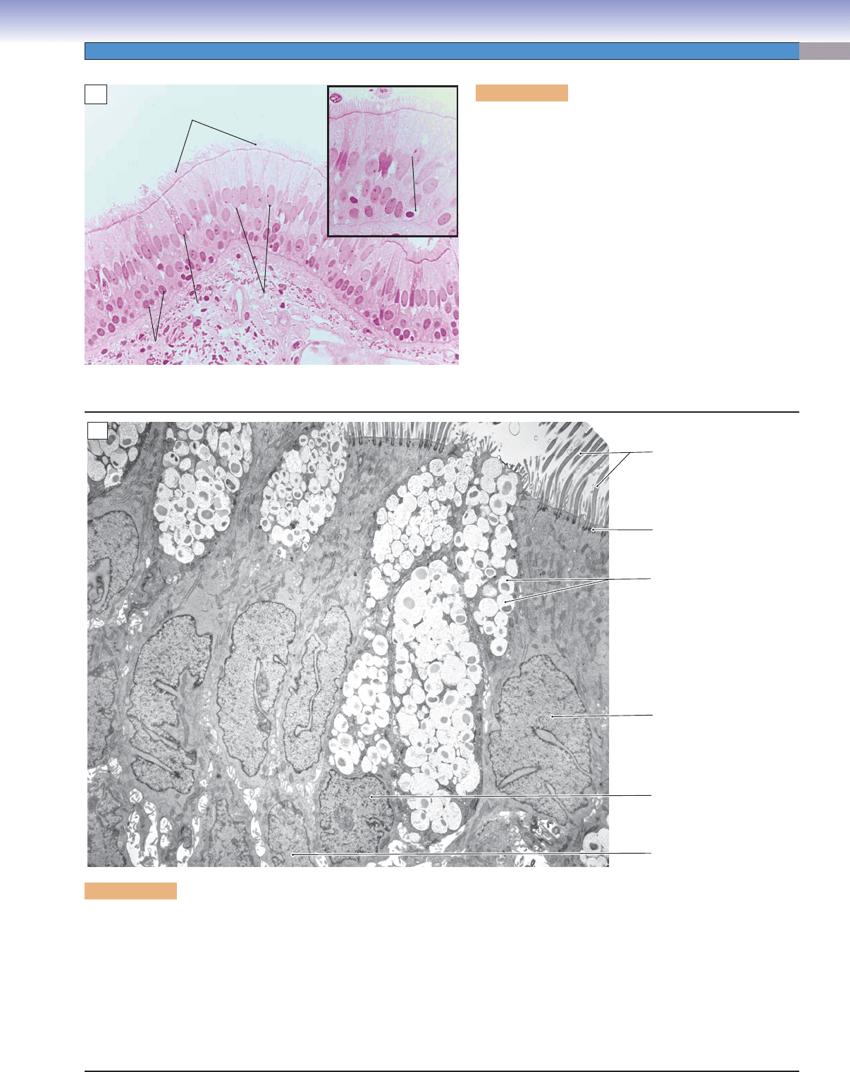
Figure 11-7A. Respiratory epithelium, trachea. H&E,
284; inset 403
The respiratory epithelium is a ciliated pseudostratifi ed
columnar epithelium that lines the inner surface of the respi-
ratory tract. It is composed of several types of cells: ciliated
columnar cells, goblet cells, basal cells, and neuroendocrine
cells (DNES). Ciliated columnar cells are tall and have long,
actively motile cilia, which help to move mucus and trapped
dust toward the mouth. Goblet cells are goblet-shaped cells
without cilia; they secrete mucus onto the surface of the epi-
thelium. The mucus captures dust particles when air passes
through the trachea. Basal cells are short cells capable of
differentiating into other cell types in the epithelium. DNES
cells have a round, dark nucleus with clear cytoplasm and
contain granules at the basal region of the cytoplasm fac-
ing the basement membrane. These cells secrete serotonin
and peptide hormones that act as local mediators. This may
affect nerve endings as well as regulate mucous secretion
and ciliary beating of nearby cells.
Ciliated
columnar cell
Neuroendocrine
cells
Goblet
cell
Basal cell
Cilia
A
Figure 11-7B. Respiratory epithelium. TEM, 4,200
The epithelium that lines the nasal cavities, trachea, bronchi, and larger bronchioles has a characteristic composition of cell types
arranged as pseudostratifi ed columnar epithelium. Ciliated cells and goblet cells are the most prominent cell types, and they function
together to generate a mechanism called the mucociliary escalator, which functions to entrap airborne debris in mucus and transport
it along the surface toward the oral cavity. The numerous cilia projecting from the apical surface of the ciliated cells (most abundant
cell type) beat in a coordinated fashion to move material toward the oropharynx. Parts of basal cells are visible at the bottom of the
fi eld; these serve as stem cells for replacement of the other cell types. Not visible here are two other cell types that occur in lower
numbers in respiratory epithelium. Brush cells are columnar cells with microvilli at the apical surface. These cells are contacted by
nerve endings, indicating a sensory function. DNES are the fi fth cell type; they are short basal cells with small cytoplasmic granules
that contain signaling molecules. (For microstructure of cilia, see Fig. 3-12A.)
Cilia
Basal body
Mucin granules
Nucleus of
ciliated cell
Nucleus of
goblet cell
Nucleus of
basal cell
B
CUI_Chap11.indd 209 6/16/2010 7:34:57 PM
210
UNIT 3
■
Organ Systems
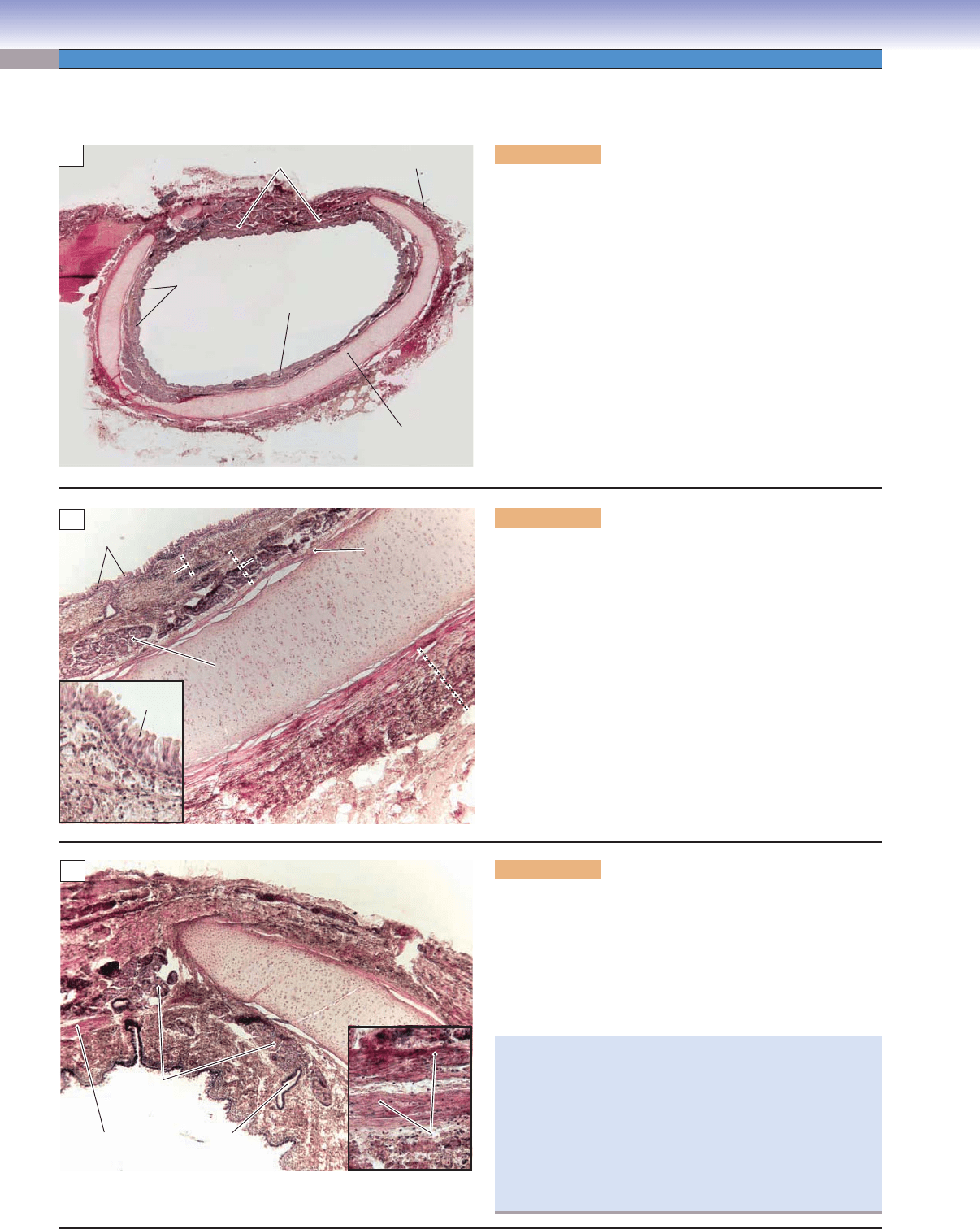
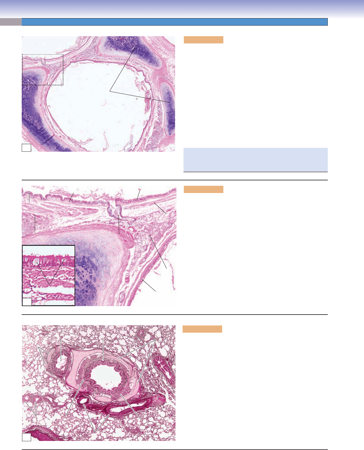
Figure 11-8A. Bronchus, secondary bronchus. H&E, 11
The trachea bifurcates to give rise to two main bronchi
( primary bronchi), which are also called extrapulmonary
bronchi because they have not yet entered the lungs. Primary
bronchi give rise to secondary bronchi and continue to divide
into tertiary bronchi. The extrapulmonary bronchi have a
similar structure to the trachea; the cartilage is still C-shaped
and lined with ciliated pseudostratifi ed columnar epithelium.
Bronchi that enter the lung tissue are called intrapulmonary
bronchi; they include the secondary bronchi and tertiary (seg-
mental) bronchi. Here is an example of a secondary bronchus,
which has bifurcated from a primary bronchus at the hilus just
above the entry to the lung. Large plates of hyaline cartilage,
no longer C-shaped, provide support for the secondary bron-
chi. Bronchi are also covered by respiratory epithelium.
Hyaline cartilage
plates
Fig. 11-8B
Lumen of bronchus
Lumen of
bronchus
A
Respiratory epithelium
Respiratory
epithelium
Respiratory
epithelium
Smooth muscle
Goblet cells
Mucosa
Mucosa
Mucosa
Submucosa
Duct of
the glands
Hyaline
Hyaline
cartilage
cartilage
Hyaline
cartilage
Smooth
muscle
Bronchial
glands
Lamina propria
B
Figure 11-8B. Bronchus, secondary bronchus. H&E, 37;
inset 198
Secondary bronchi are also called lobar bronchi. The right
lung has three lobar bronchi, and the left lung has two lobar
bronchi. The epithelial lining of secondary bronchi is similar
to that of the trachea and primary bronchi. Goblet cells can
be seen in this fi gure interspersed in the ciliated pseudostrati-
fi ed columnar epithelium. There is a band of smooth muscle
that is arranged in a spiral fashion between the mucosa and
submucosa that surround the lumen of the bronchi. This
smooth muscle is controlled by the sympathetic and parasym-
pathetic nervous systems. Sympathetic fi bers cause relaxation
of the smooth muscle; parasympathetic fi bers cause smooth
muscle to contract, reducing the diameter of the lumen of the
bronchi. Bronchial glands (seromucous glands) located in the
submucosa, and the ducts of these glands, are visible in this
specimen.
Figure 11-8C. Bronchus, tertiary bronchus. H&E, 17
Secondary bronchi divide into tertiary bronchi, also known
as segmental bronchi. These decrease in size as they branch
distally within the lung. Two tertiary bronchi are shown here.
The luminal surfaces of the tertiary bronchi are covered with
respiratory epithelium; smooth muscle and submucosal glands
are also present. Elastic fi bers are prominent in the lamina pro-
pria and are stained red in this example. The cartilage plates
of the tertiary bronchi are smaller than the plates in the sec-
ondary bronchi. As the tertiary bronchi continues to branch,
their diameters gradually decrease; as the cartilage plates
become smaller and fewer, the bronchial glands and goblet
cells decrease in number as well.
The right primary bronchus is wider and shorter and more
vertical than the left one; foreign body aspiration happens
more often to the right lung than to the left lung.
Mixed
Mixed
glands
glands
Mixed
Mixed
glands
glands
Cartilage
Cartilage
plates
plates
Cartilage
Cartilage
plates
plates
Cartilage
plates
Cartilage
Cartilage
plates
plates
Cartilage
plates
Lumen of a
Lumen of a
small bronchus
small bronchus
Lumen of a
small bronchus
Smooth
Smooth
muscle
muscle
Smooth
muscle
Bronchioles
Bronchioles
Bronchioles
Blood vessels
Blood vessels
Blood vessels
Mixed
Mixed
glands
glands
Mixed
glands
Lumen of a
Lumen of a
bronchus
bronchus
Lumen of a
bronchus
Elastic fibers
Elastic fibers
Elastic fibers
C
CUI_Chap11.indd 210 6/16/2010 7:34:59 PM
CHAPTER 11
■
Respiratory System
211
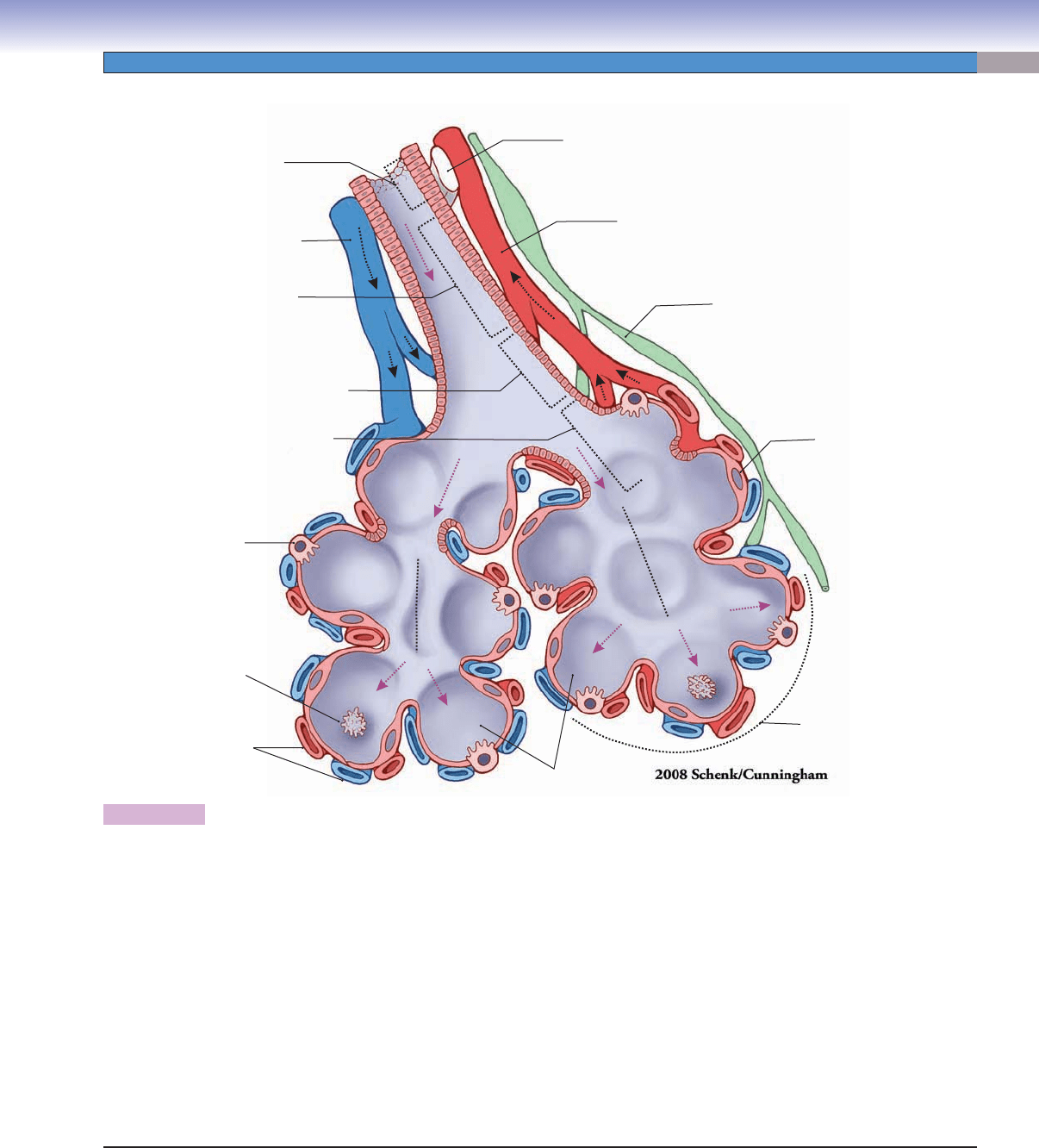
Figure 11-9. Overview of the bronchioles and alveoli.
This is a representation of the bronchioles and alveoli; the length of the different types of bronchioles is not drawn to scale. Segmental
bronchi bifurcate into bronchioles, which give rise to many branches as they move distally within the lung (Fig. 11-1). Bronchioles
have no cartilage and continue to divide into smaller bronchioles. Terminal bronchioles are the fi nal parts of the conducting airway.
They extend into alveolar sacs to give rise to respiratory bronchioles, which connect to the alveolar ducts. Respiratory bronchioles
are small in diameter, are lined by cuboidal cells, and contain increased numbers of alveoli. Respiratory bronchioles mark the transi-
tion from the conducting portion to the respiratory portion in which gas exchange occurs. An alveolar duct is a hallway that con-
nects the respiratory bronchiole to an alveolar sac. Alveolar ducts are lined by squamous alveolar epithelium and knobs of cuboidal
epithelium lying on the smooth muscle cells. An alveolar sac is the blind end of an alveolar duct and includes a common opening for
two or more alveoli. Alveoli have very thin walls lined by alveolar epithelium that contains type I and II pneumocytes (alveolar cells).
The basement membrane of the type I pneumocytes and endothelial cells of the capillaries are fused together to form the air-blood
barrier (Fig 11-12). Type I pneumocytes are squamous cells that line the alveoli (Fig. 11-13). Type II pneumocytes are pulmonary
surfactant–producing cells that are important for reducing the surface tension of the alveoli (Fig. 11-14). Alveolar macrophages, also
called dust cells, lying free on the alveolar wall, are shown here and can also be found in the septa of the alveoli (Fig. 11-15). Dust
cells move around on the alveolar surface like vacuum cleaners to clear dust particles and other debris on the surface of the alveoli
and also help remove excess surfactant.
Alveoli
Bronchioles
(multiple branches)
Pulmonary
vein (to the left atrium)
Cartilage plate
Lymphatic vessel
Type I pneumocyte
Type II
pneumocyte
Alveolar
capillaries
Alveolar macrophage
(dust cell)
Pulmonary artery
(from the right ventricle)
Terminal bronchiole
(multiple branches)
Respiratory bronchiole
Alveolar sac
Alveolar duct
Alveolar duct
Smallest segmental
bronchus
(multiple branches)
CUI_Chap11.indd 211 6/16/2010 7:35:03 PM
212
UNIT 3
■
Organ Systems
CLINICAL CORRELATION
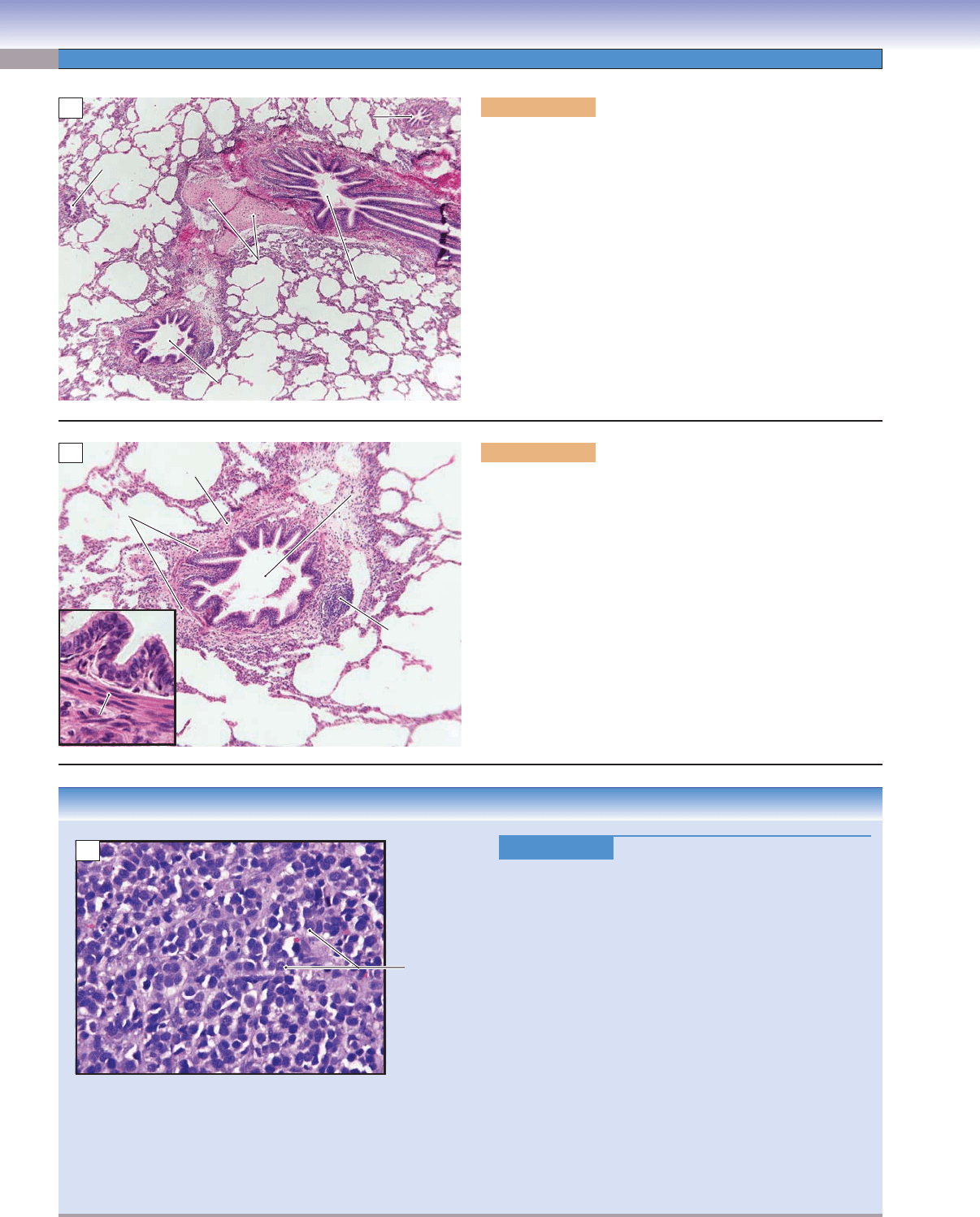
Figure 11-10C.
Small Cell Neuroendocrine Carcinoma.
H&E, 213
Small cell neuroendocrine carcinoma is a highly malignant
lung tumor characterized by its origin from the epithelium of
the central airways, rapid growth, infi ltration, gradual obstruc-
tion of the airways, and early metastases. It is associated
with genetic mutations, air pollution, and cigarette smoking.
Patients will likely present with large hilar lymph nodes with
prominent mediastinal adenopathy in computed tomography
or other radioimagings. Symptoms include weight loss, cough,
chest pain, and dyspnea. The liver, adrenals, bones, bone
marrow, and brain are the common sites of metastasis. The
tumor cells are round, small, and spindle shaped with spare
cytoplasm, ill-defi ned cell borders, prominent nuclear mold-
ing, and fi nely dispersed chromatin without distinct nucleoli.
The tumor cells are about twice the size of lymphocytes and
have characteristic “blue” cell features. Small cell carcinoma
is initially very sensitive to chemotherapy and radiotherapy,
but loses its sensitivity within months. Treatment also includes
surgery if the cancer is discovered at an early stage.
Figure 11-10A. A small tertiary bronchus and bronchioles,
lung. H&E, 25
A small tertiary bronchus and several different sizes of
bronchioles are shown here. Small tertiary bronchi have much
smaller diameters than large tertiary bronchi (Fig. 11-8C). Its
hyaline cartilage is reduced to a few plates and the epithelial
lining has decreased numbers of goblet cells and glands in the
submucosal layer. These submucosal glands gradually disappear
as the airways become smaller. Small tertiary bronchi give rise
to smaller airways called bronchioles, which, because of the ran-
dom branching pattern of the airway, appear at various places
in the section. The glands and the cartilage plates of the bron-
chioles have completely disappeared at this level. Bronchioles
continue to branch and decrease in size and give rise to terminal
bronchioles (Fig. 11-11B,C).
Small tertiary
Small tertiary
bronchus
bronchus
Bronchiole
Bronchiole
Bronchiole
Bronchiole
Bronchiole
Bronchiole
Cartilage plates
Cartilage plates
Small tertiary
bronchus
Bronchiole
Bronchiole
Bronchiole
Cartilage plates
A
B
Bronchiole
Bronchiole
Lymph
Lymph
nodule
nodule
Smooth
Smooth
muscle
muscle
Smooth
Smooth
muscle
muscle
Bronchiole
Lymph
nodule
Adventitial layer
of the bronchiole
Smooth
muscle
Smooth
muscle
Figure 11-10B. Bronchioles, lung. H&E, 71; inset 612
The bronchioles are lined by ciliated columnar or cuboidal
epithelium with decreased numbers of goblet cells and increased
numbers of Clara cells. Goblets cells occasionally can be found
in larger bronchioles. Clara cells are present in small bronchi-
oles, and their numbers are greatly increased in terminal bron-
chioles (Fig.11-11A). There are many elastic fi bers in the lamina
propria, which are not easy to see here with H&E stain. A layer
of smooth muscle on the bronchiole wall is shown in the inset.
The connective tissue (adventitial) layer is attached to the sur-
rounding alveoli. Lymph nodules or diffuse lymphocytes are
occasionally found in the connective tissue layer. The epithelium
lining the bronchioles changes from columnar to cuboidal cells.
Each bronchiole gives rise to several terminal bronchioles as it
branches distally in the lung.
C
Tumor cells
CUI_Chap11.indd 212 6/16/2010 7:35:09 PM
CHAPTER 11
■
Respiratory System
213
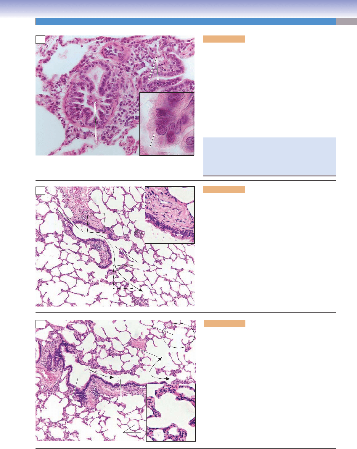
Figure 11-11A. Clara cells, terminal bronchioles.
H&E, 284; inset 1,181
Clara cells are secretory cells that are scattered among
ciliated cells and often project into the lumen of the bron-
chioles. They are dome-shaped cells without cilia and con-
tain apical granules (visible only with a special stain); they
are more abundant in terminal bronchioles. The substances
(including glycosaminoglycans and secretory proteins) pro-
duced by Clara cells help to form the lining of the bron-
chiole. Clara cells play a role in immunomodulatory and
anti-infl ammatory activities, thereby helping to protect the
bronchiolar epithelium. They also function as progenitor
cells that can differentiate into other epithelial cell types,
especially in epithelial repair after airway injury.
Clara cells
Clara cells
Clara cells
Clara cells
Clara cells
Clara cells
Alveolus
Alveolus
Alveolus
Alveolus
Alveolus
Alveolus
Lumen of a
Lumen of a
terminal bronchiole
terminal bronchiole
Lumen of a
terminal bronchiole
A
Fig.11-11C
Fig.11-11C
inset
inset
S
S
m
m
o
o
o
o
th
t
h
m
m
u
u
s
s
c
c
le
le
Smooth muscle
Respiratory
Respiratory
bronchiole
bronchiole
Respiratory
bronchiole
Terminal
Terminal
bronchiole
bronchiole
Terminal
bronchiole
Fig.11-11C
inset
B
Figure 11-11B. Terminal bronchiole, lung. H&E, 70;
inset 179
The terminal bronchioles are the last segment (most distal)
of the conducting portion of the respiratory system. They
are lined by simple cuboidal cells consisting mainly of
Clara cells, with some ciliated cells and a few basal cells.
Gradually, as the bronchioles proceed distally in the lung,
the epithelium changes from columnar to cuboidal cells.
The terminal bronchioles contain large amounts of smooth
muscle in the airway wall. This smooth muscle is con-
trolled by the sympathetic and parasympathetic nervous
systems. At this point, cartilage and submucosal glands
are absent from all bronchioles.
Alveolar sac
Alveolar sac
Alveoli
Alveoli
Terminal
Terminal
bronchiole
bronchiole
Respiratory
Respiratory
bronchiole
bronchiole
Alveolar
Alveolar
duct
duct
Alveolar
Alveolar
duct
duct
Terminal
bronchiole
Respiratory
bronchiole
Alveolar sac
Alveoli
Alveolar
duct
Alveolar
duct
C
Figure 11-11C. Respiratory bronchioles, lung. H&E,
71; inset 179
The terminal bronchioles give rise to respiratory
bronchioles. Respiratory bronchioles are the fi rst airways
that function in gas exchange. They are lined by cuboidal
cells and have gradually increasing numbers of alveoli.
Respiratory bronchioles connect to alveolar ducts. Alveo-
lar ducts are lined by squamous alveolar cells (type I pneu-
mocytes) and knobs of cuboidal epithelium lying on the
smooth muscle cells. Each alveolar duct functions struc-
turally as a corridor, which connects to several alveoli
(Fig. 11-9). Each alveolar sac is composed of two or more
alveoli that share a common opening. The arrows indicate
the direction of the airfl ow, from the terminal bronchiole
to the respiratory bronchiole, then to the alveolar duct
and, eventually, into the alveolar sac.
Research has shown that in heavy smokers, Clara cells
are greatly decreased and goblet (mucus-producing) cells
are greatly increased in the epithelium of the bronchi-
oles. These changes are caused by directly inhaled irri-
tants and chronic exposure to harmful substances.
CUI_Chap11.indd 213 6/16/2010 7:35:13 PM
214
UNIT 3
■
Organ Systems
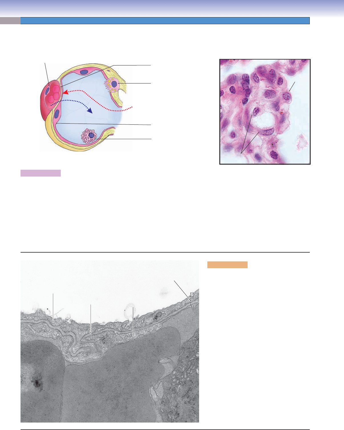
Figure 11-12A. Alveolus and gas exchange. H&E, 1,077
An alveolus is the terminal unit of the respiratory system. It functions as a primary site for gas exchange. Its wall is composed of type
I and II pneumocytes. Type I pneumocytes are primary cells, which form the structure of alveolar walls and are in contact with the
capillary walls. Type II pneumocytes are septal cells, which are located in the connective tissue of the septal junction. They produce a
surfactant, which reduces surface tension of the alveolus. Gas exchange occurs between the alveolus and capillary wall in a structure
called the blood-air barrier. The blood-air barrier is composed of type I pneumocytes, the endothelial cells of the capillary, and the
fused basement laminae of these cells (Fig. 11-12B). The exchange of O
2
and CO
2
occurs by passive diffusion across the thin blood-
air barrier. The difference in O
2
and CO
2
tensions across the membrane determines the driving pressure for diffusion of the gases.
In normal conditions, at sea level, air has a high O
2
and a low CO
2,
concentration whereas blood in the pulmonary capillaries has a
low O
2
and a high CO
2
concentration. The net driving pressure will force CO
2
out of the blood into the alveolar space and O
2
into
the blood from the alveoli.
Figure 11-12B. Blood-air barrier. TEM,
27,000
Three elements separate the air in alveoli from
the blood in the underlying capillaries: (1) Air
fi rst passes through the highly fl attened type I
pneumocyte with its coating of surfactant. The
cytoplasm can be even thinner (25 nm) than
the segment highlighted in this view. (2) The
middle element is the fused basal laminae of
the type I cell and the underlying endothelial
cell. (3) The endothelial cell lining the alveolar
capillary, like the type I cell, has an extremely
thin cytoplasm, so that the total thickness of
the blood-air barrier can be as little as 0.1 μm.
T. Yang
Type II pneumocyte
Fused basement
laminae
Type I pneumocyte
Alveolar macrophage
(dust cell)
Alveolar septum
Alveolar septum
Alveolar septum
Capillary
Type II
Type II
pneumocyte
pneumocyte
Type II
pneumocyte
Endothelial cell
Endothelial cell
Endothelial cell
Endothelial cell
O
2
CO
2
A
B
Cytoplasm of capillary
Cytoplasm of capillary
endothelial cells
endothelial cells
2. Fused basal
laminae
3. Cytoplasm of capillary
endothelial cell
Air space
Cytoplasm of
Cytoplasm of
type I pneumocyte
type I pneumocyte
1. Cytoplasm of
type I pneumocyte
0.10
0.10
m
m
μ
μ
0.10 mμ
Erythrocyte in
Erythrocyte in
lumen of the capillary
lumen of the capillary
Erythrocyte in
lumen of the capillary
Respiratory Portion: Alveolar Ducts and Alveoli
CUI_Chap11.indd 214 6/16/2010 7:35:16 PM