Caballero B. (ed.) Encyclopaedia of Food Science, Food Technology and Nutrition. Ten-Volume Set
Подождите немного. Документ загружается.


transport fluid containing high levels of dissolved
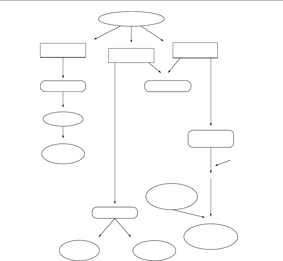
solids, starch or sugar compounds.
0023 The ideal geometry of a fluid disinfection system
for maximum utilization of the UV energy is a single
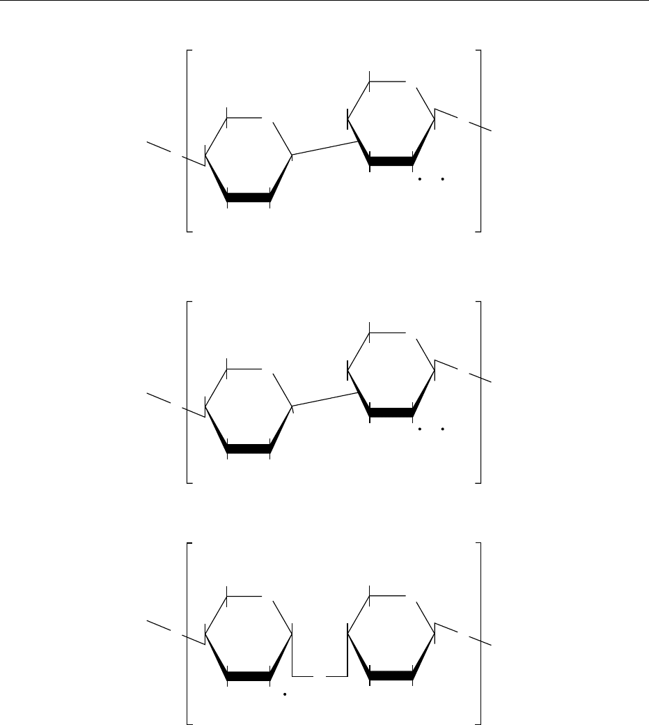
lamp running along the axis of a cylindrical treatment
chamber.
Practical Applications of UV
0024 Microbiological contamination can occur at any
stage of a production process from the incoming
process water to the surfaces of packaging. In many
cases, UV disinfection can offer a safe and effective
method of providing microbe-free process water,
fluids and air. It is effective against food pathogens,
including viruses and fungal spores (see Table 2).
0025 Unlike chemical biocides, UV does not introduce
toxins or residues into process water and does not
alter the chemical composition, taste, odor, or pH of
the fluid being disinfected. This feature is especially
important in food and drink processing plants, where
the chemical dosing of incoming process water can
cause off-flavors and alter the chemical properties of
the product.
0026 Ultraviolet treatment systems can be used as the
primary disinfection system or as a back-up for
other water purification methods, such as activated
carbon filtration, reverse osmosis or pasteurization.
As UV has no residual effect, the best position for a
treatment system is immediately prior to the point of
use. This ensures that incoming microbiological
contaminants are deactivated and there is minimum
chance of posttreatment contamination. Many users
install UV systems after filter beds and storage tank
outlet valves, to reduce the likelihood of contamin-
ation from these sources.
0027 Ultraviolet systems can also be installed as an
alternative to, or in conjunction with, air filtration
systems. The UV source will deactivate any micro-
organisms present and prevent colonization of air-
filtration systems.
Clean-in-place (CIP) Rinse Systems
0028It is essential that CIP final rinse water, which is used
to flush out foreign matter and disinfecting solutions,
is microbiologically safe. Fully automated UV dis-
infection systems can be integrated with CIP rinse
cycles to insure that final rinse water does not
reintroduce microbiological contaminants. Although
most town supplies are free from coliforms, they are
rarely sterile. Resistant to the effects of acid, deter-
gents, steam and chemical sterilants, UV systems are
an effective method of providing disinfected water for
final rinse systems.
Sugar Syrups
0029Specially modified UV treatment chambers are rou-
tinely used in the brewing and soft drinks industry to
disinfect concentrated sugar solutions and syrup. Al-
though high-Brix syrups will not support microbial
growth, any dormant spores present may become
active after the syrup has been diluted. Treating the
syrup and dilution water with UV prior to use will
insure that any dormant microorganisms are deacti-
vated.
0030Systems for syrup treatment are designed with a
chamber of relatively small diameter. This helps to
insure that sufficient UV light penetrates the liquid
and deactivates spores, which generally require higher
doses of UV than other microorganisms for complete
deactivation. In food and dairy processing plants, the
same equipment can be used to disinfect injection
brine and cheese whey.
Air Disinfection
0031For machine blanketing, and in sanitary and food
preparation areas, the exclusion of airborne contam-
inants, such as bacterial spores, yeasts and viruses, can
be a major problem. Ultraviolet disinfection systems
are now available for treating airflows entering a
sterile environment. Air-disinfection systems are fitted
into ductwork, and any microorganisms present are
deactivated as they are exposed to the UV source. (See
Plant Design: Designing for Hygienic Operation.)
0032Ultraviolet systems can also be used to disinfect
displacement air for pressurizing tanks, or pipelines
holding perishable fluids or free-flowing solids. Stor-
age tanks are particularly susceptible to bacterial col-
onization and contamination by airborne spores. To
prevent this, immersible UV treatment systems have
been designed to fit in the tank head airspace and
disinfect the air present.
tbl0002 Table 2 Experimentally determined UV dose, at 254 nm,
required for a 90% kill rate (D
10
) of various organisms
Organism D
10
(mJ cm
2
)
Bacteria
Escherichia coli 5.4
Bacillus subtilis 7.1
Clostridium botulinum 12.0
Micrococcus candidus 6.05
Pseudomonas aeruginosa 5.5
Mold spores
Aspergillus niger 100
Cladosporium herbarum 30–70
Mucor mucedo 50–70
Penicillium roqueforti 13
Yeasts
Saccharomycescerevisiae 6.0
5888 ULTRAVIOLET LIGHT

Packaging Applications
0033 Another aspect of contamination control that can
benefit from UV technology is the disinfection of fin-
ished products and their packaging. A typical system
incorporates a UV light source, shielded on three sides
and mounted over a conveyor or production line. As
foodstuffs or packaging pass under the light, any
microbes present on the surface are deactivated.
0034 After treatment by UV, the risk of microbial con-
tamination in filling and packaging lines is reduced,
and the shelf-life of treated products is extended. This
surface sterilization equipment can also be used to
disinfect bottle crowns, foil lids and plastic films.
(See Packaging: Packaging of Solids.)
Recent Advances in UV
0035 Modern UV treatment systems are microprocessor-
controlled with automatic UV intensity and lamp
status monitors. This facilitates the interfacing of
UV systems with other process control systems to
coordinate their operation.
Integral Wiping Systems
0036 The development of integral wiping systems has in-
creased the applications of UV to include treatment of
turbid fluids containing processing residues or high
solids concentrations. The wiping system is fitted to
the outside of the quartz sleeve. At preset intervals
during operation, a mechanical wiper moves along
the lamp surface, displacing any processing residues.
These would quickly foul conventional UV systems,
inhibiting the transmission of UV light and reducing
germicidal effectiveness. Wiped systems can con-
stantly reduce contamination to acceptable levels in
poultry, egg and meat processing, in the transport and
cooling of vegetables, and in the disinfection of
effluent flows. (See Effluents from Food Processing:
Disposal of Waste Water.)
Lamp Output
0037 High-output, single-lamp chambers emit the high
energy levels required to penetrate the turbid waters
that can be found in processing plants. This includes
virtually opaque fluids, such as transport water for
flour- and pasta-based products, and industrial efflu-
ent or recirculation water. Currently, the minimum
single lamp power available is 8 W (low-pressure),
increasing to a maximum of 5 kW (medium-pressure).
Limitations of UV Technology
System Sizing
0038 In theory, UV systems can be built to treat any flow
rate, air volume, or surface area. As UV demand
increases in terms of size, degree of contamination
and quality of water, the number and power output
of the UV lamps are increased proportionally to
maintain UV dose above the required minimum.
0039Obviously, practical constraints, such as the size of
the UV system, pressure drop through the system,
and power consumption, vary with each installation.
Single-chamber UV systems treating up to 250 m
3
h
1
are often more than adequate for process-water ap-
plications. Duty stand-by units, programmed to come
on-line when flow exceeds preset levels, can also be
installed as a back-up measure.
0040Ultraviolet systems have been successfully installed
to treat water flows over 1 10
5
m
3
per day. At these
flow rates, medium-pressure systems have the advan-
tage of fewer lamps, compared with low-pressure
systems, and therefore have lower maintenance
requirements.
Residual Disinfection
0041The only potential drawback with UV water disinfec-
tion, compared with chemical treatment, is that UV
does not leave a residual disinfectant effect. In food
and drink manufacturing, this is a positive advantage
as the presence of chlorine could adversely affect
product quality. In industrial applications, the lack
of residual is rarely a problem, provided that down-
stream pipework is maintained in a hygienic condi-
tion, and no ‘dead legs’ or leaks are allowed where
recolonization could occur.
0042Ultraviolet systems are recommended for installa-
tion as close to the point-of-use as possible to reduce
the likelihood of recontamination. It is mainly in the
drinking-water treatment industry, where there may
be miles of distribution pipework following disinfec-
tion, that a small chlorine residual is often added to
the water after UV disinfection. A combination of UV
disinfection and electrochlorination is most common,
both systems requiring little maintenance and able to
operate almost unsupervised.
0043With air and surface disinfection, there is a remote
risk that an operator may be exposed to the UV light
source. Prolonged exposure to high-intensity UV
light an damage the eyes, but this kind of accident is
very rare and can be avoided by carefully designed
shielding around the UV source. In addition, access
to the UV source can be interlocked with the power
supply, ensuring that access can be obtained only
when the power supply has been turned off.
Conclusion
0044The unique ability of UV light to deactivate micro-
organisms in water and air, and on surfaces, without
creating by-products or residual effects, enables
ULTRAVIOLET LIGHT 5889

UV disinfection to be used for a wide variety of appli-
cations. Whether installed as the main disinfection
system, as a back-up to alternative methods, or in
specific clean-room environments where high stand-
ards of sterility must be maintained, UV treatment
systems can ensure that levels of microbial contamin-
ants are effectively controlled.
See also: Effluents from Food Processing: Disposal of
Waste Water; Packaging: Packaging of Solids; Plant
Design: Designing for Hygienic Operation
Further Reading
Anonymous (1988) High UV output disinfects high solids
process water. Prepared Foods 157: 114.
Mans J (1987) Disinfect water with ultraviolet light.
Prepared Foods 156: 72–74.
Maunder DT (1977) Possible use of ultraviolet sterilisation
of containers for aseptic packaging. Food Technology
31: 36–370.
Phillips R (1983) Sources and Applications of Ultraviolet
Radiation. London: Academic Press.
United States Food and Drug Administration See Food and Drug Administration
URONIC ACIDS
F G Huffman, Florida International University, Miami,
FL, USA
Copyright 2003, Elsevier Science Ltd. All Rights Reserved.
Chemistry
0001 Uronic acids are produced by the oxidation of the
alcohol group of monosaccharides. These compounds
are named by substituting -ose with uronic acid. The
structures of the most common uronic acids are
shown in Figure 1. d-Mannuronic acid is the 2-
epimer of d-glucuronic acid, and l-iduronic acid is
the 5-epimer of d-glucuronic acid.
0002 The uronic acids are important constituents of cer-
tain natural heteropolysaccharides. They play a sig-
nificant role in the detoxification of substances such
as drugs. Glucuronic acids are found in human urine
bound with glycosidic linkages to hydroxylated com-
pounds such as menthol, borneol, and estrogens.
Owing to the increased solubility of hydroxylated
compounds (when conjugated with glucuronic acid),
they are readily disposed by the body. Glucuronic
acid also forms a conjugate with bilirubin, a bile
pigment.
0003 The most important uronic acid in humans is the
d-glucuronic acid. It, or its epimer, is a constituent
of many glycosaminoglycans (GAG) as well as glu-
curonide derivatives of drugs and hormones. d-
Glucuronic acid is also the precursor of l-ascorbic
acid in animals. The schematic utilization of d-
glucuronate in microorganisms, animals, and plants
is shown in Figure 2. Glucuronic acid is synthesized
from glucose in the uronic pathway, an alternative
oxidative pathway for glucose without the produc-
tion of adenosine triphosphate (ATP). In the uronic
pathway, glucose 6-phosphate is converted to glu-
cose 1-phosphate which subsequently reacts with
uridine triphosphate to form uridine diphosphate glu-
cose (UDPGlc). This compound is then oxidized at
the six-carbon position in a two-step process by the
COOH
H
H
H
HO
H
H
OH
OH
H
OH
H
OH
H
OH
OH
O
COOH
H
H
HO
HO
H
OH
H
O
H
H
COOH
H
HO
H
OH
OH
O
COOH
HO
H
H
H
H
OH
OH
O
(a) (b)
(c) (d)
fig0001Figure 1 Structures of uronic acids: (a) D-glucuronic acid
(Glu U);(b)
D-mannuronic acid (Man U); (c) D-galaturonic acid
(Gal U); (d)
L-iduronic acid (L-guluronic acid; Gul U).
5890 URONIC ACIDS

nicotinamide adenine dinucleotide (NAD)-dependent
enzyme (UDPGlc dehydrogenase) to form UDP-
glucuronate.
0004 UDP glucuronate is the form of glucuronic acid
which can be incorporated into proteoglycans or
conjugated with steroid hormones, certain drugs, or
bilirubin. In bilirubin, two molecules of glucuronic
acid are attached with ester linkages to the two pro-
pionic acid groups to form an acylglucuronide. This
conjugation process increases the water solubility
of bilirubin, thus allowing its secretion into the
bile, ultimately to be excreted via the gastrointestinal
tract.
0005 A second product of the uronic pathway is l-
ascorbic acid, which is produced in mammals, with
the exception of humans, primates, and the guinea-
pig. In a series of reactions glucuronate is reduced to
l-gulonate, which is subsequently converted to l-
ascorbic acid. The glucuronic acid pathway is most
active in the liver, kidneys, and intestines. The pri-
mary function of the pathway is to produce UDP-
glucuronic acid, which is needed for detoxification
of various compounds by elimination of glucuronides
in the urine or bile. Many carcinogens, and drugs,
including antipyretics, hypnotics, and antimalarial
drugs, may be eliminated by this mechanism. There
may be certain organs which may have an active
uronic pathway specific for a drug. Glucuronic acid
synthesis may be stimulated when the consumption
of substances that are excreted as glucuronides is
increased, or when steroids and barbiturates, which
induce the microsomal P-450 system, are consumed.
D-Glucuronate
In
microorganism
In
animals
In
plants
D-Fructuronate D-Glucaric acid
Pyruvate
Triose
phosphate
UDP-glucuronic
acid
Xylulose
L-Gulonate
L-Ascorbic
acid
Pectin,
hemicellulose,
etc.
Glucuronate
1-phosphate
UTP
fig0002 Figure 2 The fate of glucuronate in plants, animals and microorganisms. UTP, uridine triphosphate; UDP, uridine diphosphate.
URONIC ACIDS 5891

Uronic Acids in Animal Tissue
0006 Uronic acids are an integral part of GAGs, formerly
known as the mucopolysaccharides. They have
structural importance in vertebrate animals. Some
important examples of GAGs are hyaluronic acid,
the chondroitin sulfates of connective tissue, the der-
matan sulfates of skin, and heparin (Table 1 and
Figure 3).
Hyaluronic acid
0007 Hyaluronic acid functions in the body as an agent
which increases the viscosity of body fluids and acts
as a lubricant. It is present in the joints and in the
vitreous humor of the eye. Hyaluronic acid is
composed of equal molecules of d-glucuronic acid
and 2-acetamido-2-deoxy-d-glucose, which alternate
in the heteropolysaccharide. The linkages from the
amino sugar to the acid are b-1,4; those from
the acid to the amino sugar are b-1,3. Hyaluronic
acid is also present in skin, aorta, heart valve fibro-
blast, and the umbilical cord.
Chondroitin Sulfates
0008 Chondroitin sulfates are the major constituents of
hyaline cartilage. They are located in cartilage and
also at the calcification site of the bone. The disac-
charide unit of the chondroitin sulfates contains glu-
curonic acid and N-acetylgalactosamine. Glucuronic
acids are connected by b-1,3 linkages. The two major
chondroitin sulfates, A and C, differ from one
another in the position of the sulfates. Chondroitin
sulfate A is sulfated at position 4, and chon-
droitin sulfate C is sulfated at position 6. Thus the
names chondroitin 4-sulfate and chondroitin 6-sul-
fate are used for these two polysaccharides.
0009 The number of repeating uronic acids in the chon-
droitin chain varies depending on the source. The
number may even differ within the same tissue. The
ratio of chondroitin 6-sulfate to chondroitin 4-sulfate
units increases progressively with age. In humans, a
plateau is reached around the age of 50.
Dermatan Sulfate
0010 Dermatan sulfate contains l-iduronic acid as the
major uronic acid. Glucuronic acid is also present in
smaller amounts. Dermatan sulfate is found in the
cornea and the sclera of the eye, which helps to main-
tain corneal transparency and the shape of the eye.
Many other tissues in animals and humans also con-
tain dermatan, such as blood vessel walls, heart valve,
and the umbilical cord. Dermatan sulfates may occur
at the C4 or C6 positions of l-iduronic acid. They
are found to increase normally with age, except in
diseased conditions.
Heparin
0011Heparin is mainly stored intracellularly in the gran-
ules of the mast cells. It may be released in response to
a specific stimulus. The main uronic acid of heparin
is the l-iduronic acid, although d-glucuronic acid is
also present. Heparin is a well-known anticoagulant
that binds to factors IX and XI but, most importantly,
it interacts with plasma antithrombin III.
Heparan
0012Heparan sulfate differs from heparin in many ways.
The dominant uronic acid in heparan sulfate is d-
glucuronic acid. The degree of sulfation is reduced
in heparan sulfate because there are fewer l-iduronic
acids. Heparan sulfates are located in the extracellu-
lar medium of mast cells. They differ in their physio-
logical functions in that they may become receptors
and participate in cell growth and communication.
Alterations in Glycosaminoglycans Metabolism
0013The preceding discussion shows that GAG has spe-
cific functions in vertebrate animals. Recent research
in this field suggests that certain disease conditions
may alter the functions, synthesis, and composition of
GAGs. For example, increased chondroitin sulfate
and uronic acids and decreased dermatan were ob-
served in the breast biopsies of women with carcin-
oma, when compared with controls. Some tumor cells
have less heparan sulfate, which may reduce their
adhesiveness. Hyaluronic acid may facilitate tumor
cell migration through the extracellular matrix and
tumor cells are capable of synthesizing increased
amounts of GAG. Samples of degenerated cartilage
from human patellas had increased dermatan sulfate
and hyaluronate and decreased chondroitin 6-sulfate
and uronic acid content. Dermatan sulfate binds to
low-density lipoproteins (LDL) in plasma and is be-
lieved to play a significant role in plaque development
in arteriosclerosis. Significantly increased urinary ex-
cretion of glycosamines was reported in subjects with
hypothyroidism. The levels of heparan sulfate and
chondroitin sulfate were higher in subjects with
hypothyroidism than the controls, indicating alter-
ation in the metabolism of connective tissue in
tbl0001 Table 1 Composition of glycosaminoglycans
Glycosaminoglycan Uronic acid composition
Hyaluronic acid b-glucuronic acid
Chondroitin 4-sulfate b-glucuronic acid
Heparin Sulfated iduronic acid
Dermatan
L-iduronic acid
5892 URONIC ACIDS

hypothyroidism. Specific reactions responsible for
these changes need to be studied.
0014 In different types of arthritis proteins of GAG may
act as autoantigens, causing additional symptoms of
the disease. Recent research indicates that anti-DNA
antibodies cross-reacting with GAG are present in
patients with lupus erythematosus. Increased produc-
tion of hyaluronic acid was observed with increased
severity of the disease in patients with autoimmune
thyroid disease. It appears that GAG not only plays a
role in the pathogenesis of autoimmune diseases but
also may be used as an activity marker of the disease.
0015Increases in hyaluronic acid and keratan and
decreases in chondroitin sulfate may be contrib-
uting factors in the development of osteoarthritis. A
recent surge in the use of chondroitin sulfate and
COO
−
O
O
H
H
H
HO
O
H
OH H
H
H HN CO CH
3
OH
O
O
H
H
H
n
n
n
H
H
1
4
COO
−
O
O
OH H
HOH
HH
H
1
1
4
COSO
3
COO
−
OO
O
O
O
OH H
H NH SO
3
OSO
3
H
H
H
H
HH
OH H
H
14
1
3
3
−
SO
3
O
HOCH
2
O
H
OH
H
O
HOCH
2
H HN CO CH
3
β-Glucuronic acid N-Acetylgalactosamine sulfate
β-Glucuronic acid N-Acetylglucosamine
Sulfated
g
lucosamine Sulfated iduronic acid
Chondroitin 4-sulfate
Hyaluronic acid
Heparin
−
−
−
fig0003 Figure 3 Structures of glycosaminoglycans.
URONIC ACIDS 5893

glucosamine by sufferers of osteoarthritis is currently
being investigated.
0016 Heparan and dermatan sulfates are found to inhibit
calcium oxalate crystallization and thus prevent
kidney stones. Recently a uronic acid-rich protein
has been isolated from the urine of normal and
stone-forming individuals. This protein appears to be
a most efficient inhibitor of calcium oxalate nephro-
lithiasis.
Uronic Acids in Plant Tissue
0017 In plants, uronic acids are associated with the fiber
component of the plant cell. The fibers which have
significance in human and animal life are called diet-
ary fibers. Not all dietary fibers contain uronic acids.
0018 Dietary fibers have three major components:
1.
0019 Structural polysaccharides and derivatives, such as
cellulose and cellulose derivatives (microcrystal-
line cellulose, semisynthetic gums, and noncellulo-
sic polysaccharides in land and sea plants).
2.
0020 Structural nonpolysaccharides, such as lignin.
3.
0021 Nonstructural polysaccharides, such as mucilages,
seed gums, plant exudates, and microbial gums.
The uronic acids are found in the noncellulosic, struc-
tural polysaccharides, and derivatives – hemicellu-
lose, pectic substances (land plants), and alginate
(sea plant) – and in the nonstructural polysaccharides
– mucilages, seed gums, plant exudes, and microbial
gums (xanthan). These substances are also referred to
as soluble dietary fiber. The most common uronic
acids in the plants are d-galacturonic and d-glucuro-
nic acids (Table 2). The uronic acid content and
physiochemical properties of dietary fibers are as
follows.
Hemicellulose
0022 Hemicellulose is a branched polymer of pentose and
hexose sugars, found in the plant cell wall. The uronic
acid composition is mainly d-glucuronic acid and 4-
O-methyl-d-glucuronic acid. There are two distinct
hemicelluloses in plants: the acidic and the neutral.
Acidic hemicelluloses contain a larger number of uro-
nic acids than neutral hemicelluloses. Hemicelluloses
are partially fermented by the microorganisms of the
colon, producing some volatile fatty acids. Hemicel-
luloses are insoluble in water but soluble in alkaline
solutions. They, along with other insoluble dietary
fibers, decrease the intestinal transit time; hemicellu-
loses also increase fecal weight and slow down starch
hydrolysis. Acidic hemicelluloses may bind to cations.
These characteristics of hemicellulose may be respon-
sible for its physiological effects.
Pectins
0023Pectins are a complex mixture of polysaccharides
containing d-galacturonic acid as the main constitu-
ent. They are found in the primary cell walls and
intercellular layers in land plants. Citrus fruits,
apples, and pears contain large amounts of pectin.
In the intestinal tract they are almost completely fer-
mented by the microflora. Pectins are water-soluble
and form gels under specific conditions. They are
hydrophilic and have ion-binding and water-holding
capacities. Pectins delay gastric emptying and in-
crease bile acid excretion.
Gums and Mucilages
0024Gums and mucilages are water-soluble, viscous, and
highly fermentable by the microorganisms of the
intestinal tract. The uronic acids that dominate gums
are d-glucuronic and d-galacturonic acids. The most
important gums used in the food industry are gum
arabic, gum karaya, tragacanth, carob, and guar,
which are obtained as exudates from trees or shrubs.
Xanthan gum, which is synthetically produced, con-
tains d-glucuronic acid. Because of their gel-forming,
water-holding ability, gums delay gastric emptying.
Alginate
0025Alginate is a noncell-wall component of seaweed
which contains d-mannuronic and iduronic acids. It
forms gels and is highly fermentable by intestinal
microorganisms. Alginates are used in food process-
ing and they enter into the human food chain as food
additives.
Physiological Effects of Dietary Fiber
0026The physical and chemical properties of dietary fiber
create physiological effects in animals. Because
dietary fiber cannot be hydrolyzed by the intestinal
enzymes it is unavailable for absorption and therefore
continues to exert effects in the gastrointestinal tract.
Soluble dietary fibers have been shown to reduce
plasma levels of glucose and cholesterol. These
physiological effects of soluble dietary fibers are at-
tributed to their ability to form gels and delay gastric
emptying. Viscous fibers bind to bile acids and
tbl0002 Table 2 Uronic acid composition of fiber
Fiber Uronic acid composition
Hemicellulose
D-glucuronic acid and/or
4-methyl
D-glucuronic acid
Pectin
D-galacturonic acid
Gums, mucilages
D-glucuronic acid and/or D-galacturonic acid
Alginate
D-mannuronic acid and/or iduronic acid
5894 URONIC ACIDS

prevent their reabsorption, causing body cholesterol
to be excreted and thus reducing plasma cholesterol.
Second, fiber interferes with digestive enzymes by
sequestering lipids, carbohydrates, and proteins, pre-
venting their absorption. Fiber may also interfere
with micelle formation, mixing of intestinal contents,
and inhibition of cholesterol synthesis.
0027 The viscous fibers are highly fermentable by the
intestinal microorganisms, producing volatile fatty
acids, which, by being absorbed into the portal
blood, suppress hepatic cholesterol synthesis by in-
hibiting human menopausal gonadotropin coenzyme
reductase activity. Some diabetics and hypercholester-
olemics have experienced alleviation of their condi-
tion by increasing intakes of dietary fiber.
0028 Isolated dietary fiber components have been used
to elicit a specific response. However, It has become
clear that fiber exerts its primary effect as a compon-
ent of whole foods rather than as an isolated entity.
0029 The low incidence of colon cancer in populations
consuming high levels of fibrous foods has prompted
scientists to study the effects of fiber on preventing
colon cancer. The roles of specific dietary fiber are
unclear, but diets high in fiber and low in fat are
increasingly recommended.
0030 Research indicates that the effect of fiber is not
always favorable. Reduced bioavailability of certain
vitamins and minerals has been reported. Among the
vitamins studied, availability of vitamins B
12
,B
6
,A,
and E was reduced as a result of high fiber intake.
Among the minerals, the absorption of sodium, po-
tassium, magnesium, calcium, zinc, and iron was re-
duced by dietary fiber, especially by the fiber fraction
containing uronic acids. Research to determine the
effect of fiber on nutrient bioavailability continues
with vigor.
Methods of Analysis for Uronic Acids
0031 Since uronic acids are found as integral parts of
animal and plant tissues, they must be separated
from their native materials.
0032 GAGs which contain mammalian uronic acids may
be separated by density gradient centrifugation and
chromatography (ion exchange or gel). Hydrolysis by
acid or by specific enzymes is used. Uronic acids may
be measured either by colorimetry or by decarboxyla-
tion techniques.
0033 There are two primary methods for quantification
of dietary fiber. The gravimetric method measures
total fiber content and uses enzymes or detergents to
solubilize the nonfiber components, such as starch
and protein. Defatting using organic solvents is
usually performed prior to sample analysis. Acid
and base solutions are used to separate acid- or
base-soluble fractions.
0034The fractionation method, developed by Southgate
in 1969, has gone through many modifications over
the years. This method allows measurements of total
dietary fiber as well as the fiber fractions. It is neces-
sary to use the fractionation method for fiber analysis
to be able to free uronic acids for quantification.
Most fiber analyses have three steps:
1.
0035Preparation of an extractive-free residue (alcohol-
insoluble residue)
2.
0036Removal of starch and protein from the residue
(enzyme or detergent hydrolysis)
3.
0037Analysis of destarched, deproteinated residue for
neutral sugars and uronic acids
In recent years highly specific enzyme preparations
have become available, improving recovery of various
fiber fractions. The detergent method of fiber analysis
is simple and fast, but the soluble fiber component,
which contains the uronic acids, is lost during extrac-
tion. The method of choice for the quantitative analy-
sis of uronic acids appears to be the enzymatic
method followed by either colorimetry or decarbox-
ylation.
Quantification of the Uronic Acids
0038Uronic acids may be determined in the hydrolysate
of food samples following enzyme and/or acid
hydrolysis. Dilutions of the hydrolysate to give
25–100 mgml
1
may be necessary and can be achieved
with a mixture of sodium chloride and boric acid.
Diluted samples are heat-treated in the presence of
concentrated sulfuric acid. After cooling to room
temperature, a 3,5-dimethylphenol solution is added
and 10–15 min later the absorbance is read at
400 and 459 nm. Appropriate glucuronic acid stand-
ards are used to develop a standard curve. Differences
in absorbance readings of the sample are plotted on
the standard curve and sample concentrations are
read.
0039The colorimetric methods using carbazole reagent
may also be used in determining uronic acids. The
sample hydrolysate is placed in a test-tube containing
cold acid borate. The tubes are placed in a boiling
water bath, followed by cooling to room tempera-
ture. Carbazole reagent is added and tubes are again
placed in the boiling water bath. After cooling to
room temperature, the intensity of the color is
measured by reading absorbance at 530 nm. Sample
concentrations may be calculated using values from
standard curve. Recently a colorimetric method using
1,9-dimethylmethylene blue was used to determine
GAG in partial urine from pediatric patients. Results
indicated the efficiency and sensitivity of this method
and it is recommended for widespread use. Decarbox-
ylation of uronic acids with hydroiodic acid seems
URONIC ACIDS 5895

to improve the accuracy of measurements over
colorimetry procedures.
0040 The gas–liquid chromatography (GLC) method has
improved the accuracy and specificity of uronic acid
determinations. Although free, low-molecular uronic
acids are readily analyzed by GLC, it is impossible to
measure the specific uronic acids by this method.
0041 Recently a microtiter plate assay for the determin-
ation of uronic acids in biological samples has been
validated. Modification of a commonly used proced-
ure promises to have less risk in handling strong hot
acid and increases accuracy of measurement.
See also: Ascorbic Acid: Properties and Determination;
Carbohydrates: Classification and Properties; Digestion,
Absorption, and Metabolism; Metabolism of Sugars;
Cellulose; Dietary Fiber: Properties and Sources;
Determination; Physiological Effects; Glucose: Function
and Metabolism; Gums: Food Uses; Pectin: Properties
and Determination; Single-cell Protein: Algae
Further Reading
Atkins EOT (1985) Polysaccharides: Topics in Structure
and Morphology. Deerfield Beach, FL: VCH Publishers
Boons GJ (ed.) (1998) Carbohydrate Chemistry. New York:
Blackie Academic & Professional.
David S (1997) The Molecular and Supramolecular Chem-
istry of Carbohydrates: A Chemical Introduction to the
Glycosciences. New York: Oxford University Press.
Dreher ML (1987) Handbook of Dietary Fiber: An Applied
Approach. New York: Marcel Dekker.
Hecht SM (ed.) (1999) Bioorganic Chemistry: Carbo-
hydrates. New York: Oxford University Press.
Horowitz I and Pigman W (eds) (1978) The Glycoconju-
gates. II – Mammalian Glycoproteins, Glycolipids,
Proteoglycans. New York: Academic Press.
Horwitz W (ed.) (2000) Official Methods of Analysis
of AOAC International. Gaithersburg: AOAC Inter-
national.
Leeds R and Avenell A (eds) (1985) Dietary Fiber Perspec-
tives: Reviews and Bibliography, vol. 1. London: John
Libbey.
Leeds AR and Burley VJ (eds) (1990) Dietary Fiber Perspec-
tives: Reviews and Bibliography, vol. 2. London: John
Libbey.
Prosky L and Harland B (1985) Dietary fibre method-
ology. In: Trowell H, Burkitt D and Heaton K (eds)
Dietary Depleted Foods and Disease. London: Aca-
demic Press.
Robyt JF (1998) Essentials of Carbohydrate Chemistry.
New York: Springer-Verlag.
Southgate DAT, Waldron K, Johnson IT and Fenwick GR
(eds) (1990) Dietary Fibre: Chemical and Biological
Aspects. RSC Special Publications no. 83. Cambridge:
Royal Society of Chemists.
Spiller GA (ed.) (1993) Handbook of Dietary Fiber in
Human Nutrition. Boca Raton, Florida: CRC Press.
Stipanuk MH (2000) Biochemical and Physiological
Aspects of Human Nutrition. Philadelphia, PA: W.B.
Saunders.
5896 URONIC ACIDS

V
VEAL
R G Cassens, University of Wisconsin-Madison,
West Madison, WI, USA
Copyright 2003, Elsevier Science Ltd. All Rights Reserved.
Introduction
0001 Veal is the meat from a young calf. It is tender, pale in
color, and has a high moisture and low fat content. It
is a delicate, almost bland, meat. Consumption of
veal comprises a small proportion of total meat con-
sumption, and demand for veal is often concentrated
in geographic areas or within certain ethnic popula-
tions. Veal calves may be slaughtered soon after birth
or they may be specially fed for several weeks.
Methods of Production
0002 The traditional production of veal was a segment of
the dairy industry. The calves were normally fed only
their mother’s milk and slaughtered at a few days of
age in an effort to recover some value. The meat from
such immature animals was pale, delicate and tender.
It is pointed out in Larousse Gastronomique that the
ancient method of feeding the calf exclusively on
the mother’s milk results ‘in a very pale pink meat
smelling of milk, with satiny white fat having no tinge
of red.’
0003 At present, there are three methods of veal produc-
tion:
1.
0004 The calf may be fed only on milk and slaughtered
at 3 weeks or less of age. This produces so-called
bob veal. Another alternative is to feed the calves
up to about 20 weeks of age.
2.
0005 Of greatest importance now is the production of
special-fed veal, a process in which the calf is fed
on milk replacers.
3.
0006 When some pasture, roughage, or grain is incorp-
orated into the diet, the result is so-called pink
veal.
0007 In the USA, the current types of production and
importance of the veal industry to a dairying state
such as Wisconsin can be assessed from the findings
of Piwoni and Kliebenstein. They reported that the
approximately 1.8 million dairy cows in Wisconsin
produce about 800 000 bull calves annually. About
20% are sold as feedlot or herd replacements. About
30% are sold and slaughtered as bob veal – milk-fed
calves of 3 weeks or less age. The production of bob
veal continues to decline. The remaining 50% are
used in the production of special-fed, fancy, or prime
veal. These calves are raised in confinement, receive
only milk replacer, and are slaughtered at 14–17
weeks of age when they weight 145–170 kg.
0008The special feeding programs have evolved into a
highly intensified industry. The producers follow a
detailed plan which specifies all aspects such as
purchase of replacement calves, housing, feeding,
management, health, and marketing. These produc-
tion systems have been criticized as being factory
farms in which the animals are mistreated. Such is
not the case. In fact, most of these production units
are indeed family operations because of the close and
frequent supervision required to raise the veal calves
successfully. Adequate nutritional status and comfort
of the animal are prerequisites to successful produc-
tion.
0009Wholesalers characterize veal as follows. Baby veal
(bob veal) is produced from calves of only a few days’
age, but they may range up to a month. They are
usually male calves from the dairy industry, and the
carcasses range from 10 to 25 kg in weight. Vealers
range in age from 1 to 3 months, and the carcasses
weigh from 35 to 70 kg. They are also raised primar-
ily on milk. Another category comes from calves of
3–8 months of age with carcasses weighing 55–
135 kg. They have been grown primarily on feeds
other than milk, and their meat is maturing to resem-
ble beef more than veal. Nature veal is produced from
calves approximately 16 weeks old, and the carcass
weight varies from 80 to 110 kg. They are fed a
controlled and scientifically designed diet, and they
have limited activity, thereby minimizing muscular
development. This controlled regimen of diet and
activity produces a pinkish-white color of meat, and
