Bhushan B. Handbook of Micro/Nano Tribology, Second Edition
Подождите немного. Документ загружается.


© 1999 by CRC Press LLC
and Europe with the following industry/university and government laboratory splits: 50/50, 70/30, and
30/70, respectively. According to some estimates, over 3000 users of SPMs exist with $400 million in
support. It is clear that research and industrial applications of SPMs are rapidly expanding.
STMs, AFMs, and their modifications can be used at extreme magnifications ranging from 10
3
to 10
9
×
in x-, y-, and z-directions for imaging macro- to atomic dimensions with high-resolution information
and for spectroscopy. These instruments can be used in any sample environment such as ambient air
(Binnig et al., 1986a), various gases (Burnham et al., 1990), liquid (Marti et al., 1987; Drake et al., 1989;
Binggeli et al., 1993), vacuum (Binnig et al., 1982; Meyer and Amer, 1988), low temperatures (Coombs
and Pethica, 1986; Kirk et al., 1988; Giessibl et al., 1991; Albrecht et al., 1992; Hug et al., 1993), and high
temperatures. Imaging in liquid allows the study of live biological samples, and it also eliminates water
capillary forces present in ambient air present at the tip–sample interface. Low-temperature (liquid
helium temperatures) imaging is useful for the study of biological and organic materials and the study
of low-temperature phenomena such as superconductivity or charge density waves. Low-temperature
operation is also advantageous for high-sensitivity force mapping due to the reduction in thermal
vibration. These instruments are used for proximity measurements of magnetic, electrical, chemical,
optical, thermal, spectroscopy, friction, and wear properties. Their industrial applications include micro-
circuitry and semiconductor industry, information storage systems, molecular biology, molecular chem-
istry, medical devices, and materials science.
1.3.1 Scanning Tunneling Microscope
The principle of electron tunneling was proposed by Giaever (1960). He envisioned that if a potential
difference is applied to two metals separated by a thin insulating film, a current will flow because of the
ability of electrons to penetrate a potential barrier. To be able to measure a tunneling current, the two
metals must be spaced no more than 10 nm apart. Binnig et al. (1982) introduced vacuum tunneling
combined with lateral scanning. The vacuum provides the ideal barrier for tunneling. The lateral scanning
allows one to image surfaces with exquisite resolution, lateral less than 1 nm and vertical less than 0.1 nm,
sufficient to define the position of single atoms. The very high vertical resolution of STM is obtained
because the tunnel current varies exponentially with the distance between the two electrodes, that is, the
metal tip and the scanned surface. Typically, tunneling current decreases by a factor of 2 as the separation
is increased by 0.2 nm. Very high lateral resolution depends upon the sharp tips. Binnig et al. (1982)
overcame two key obstacles for damping external vibrations and for moving the tunneling probe in close
proximity to the sample; their instrument is called the STM. Today’s STMs can be used in the ambient
environment for atomic-scale imaging of surfaces. Excellent reviews on this subject are presented by Pohl
(1986), Hansma and Tersoff (1987), Sarid and Elings (1991), Durig et al. (1992), Frommer (1992),
Guntherodt and Wiesendanger (1992), Wiesendanger and Guntherodt (1992), Bonnell (1993), Marti and
Amrein (1993), Stroscio and Kaiser (1993), and Anselmetti et al. (1995) and the following dedicated
issues of the Journal of Vacuum Science Technolology (B9, 1991, pp. 401–1211) and Ultramicroscopy (Vol.
42–44, 1992).
TABLE 1.1 Comparison of Various Conventional Microscopes with SPMs
Optical Confocal SEM/TEM SPM
Magnification 10
3
10
4
10
7
10
9
Instrument price, U.S. $ 10k 30k 250k 100k
Technology age 200 yrs 10 yrs 30 yrs 9 yrs
Applications Ubiquitous New and unfolding Science and technology Cutting-
edge
Market 1993 $800M $80M $400M $100M
Growth rate 10% 30% 10% 70%
Data provided by Topometrix.
© 1999 by CRC Press LLC
The principle of STM is straightforward. A sharp metal tip (one electrode of the tunnel junction) is
brought close enough (0.3 to 1 nm) to the surface to be investigated (second electrode) that, at a
convenient operating voltage (10 mV to 1 V), the tunneling current varies from 0.2 to 10 nA, which is
measurable. The tip is scanned over a surface at a distance of 0.3 to 1 nm, while the tunneling current
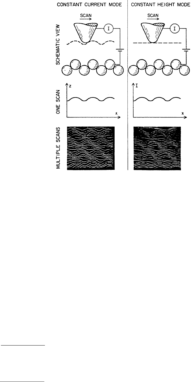
between it and the surface is sensed. The STM can be operated in either the constant-current mode or
the constant-height mode, Figure 1.3. The left-hand column of Figure 1.3 shows the basic constant
current mode of operation. A feedback network changes the height of the tip z to keep the current
constant. The displacement of the tip given by the voltage applied to the piezoelectric drives then yields
a topographic picture of the surface. Alternatively, in the constant-height mode, a metal tip can be scanned
across a surface at nearly constant height and constant voltage while the current is monitored, as shown
in the right-hand column of Figure 1.3. In this case, the feedback network responds only rapidly enough
to keep the average current constant (Hansma and Tersoff, 1987). A current mode is generally used for
atomic-scale images. This mode is not practical for rough surfaces. A three-dimensional picture [z(x, y)]
of a surface consists of multiple scans [z(x)] displayed laterally from each other in the y direction. It
should be noted that if different atomic species are present in a sample, the different atomic species
within a sample may produce different tunneling currents for a given bias voltage. Thus, the height data
may not be a direct representation of the topography of the surface of the sample.
1
FIGURE 1.3 Scanning tunneling microscope can be operated in either the constant-current or the constant-height
mode. The images are of graphite in air. (From Hansma, P. K. and Tersoff, J. (1987), J. Appl. Phys., 61, R1–R23. With
permission.)
1
In fact, Marchon et al. (1989) STM imaged sputtered diamond-like carbon films in barrier-height mode by
modulating the tip-to-surface distance, with lock-in detection of the tunneling current. The local barrier-height
measurements give information on the local values of the work function, thus providing chemical information, in
addition to the topographic map.

© 1999 by CRC Press LLC
1.3.1.1 Binnig et al.’s Design
Figure 1.4 shows a schematic of one of Binnig and Rohrer’s designs for operation in an ultrahigh vacuum
(Binnig et al., 1982; Binnig and Rohrer, 1983). The metal tip was fixed to rectangular piezodrives P
x
, P
y
,
and P
z
made out of commercial piezoceramic material for scanning. The sample is mounted on either a
superconducting magnetic levitation or two-stage spring system to achieve the stability of a gap width
of about 0.02 nm. The tunnel current J
T
is a sensitive function of the gap width d; that is, J
T
αV
T
exp(–Aφ
1/2
d), where V
T
is the bias voltage, φ is the average barrier height (work function) and A ~ 1 if
φ is measured in eV and d in Å. With a work function of a few eV, J
T
changes by an order of magnitude
for every angstrom change of h. If the current is kept constant to within, for example, 2%, then the gap
h remains constant to within 1 pm. For operation in the constant-current mode, the control unit (CU)
applies a voltage V
z
to the piezo P
z
such that J
T
remains constant when scanning the tip with P
y
and P
x
over the surface. At the constant-work functions φ, V
z
(V
x
, V
y
) yields the roughness of the surface z(x, y)
directly, as illustrated at a surface step at A. Smearing the step, δ (lateral resolution) is on the order of
(R)
1/2
, where R is the radius of the curvature of the tip. Thus, a lateral resolution of about 2 nm requires
tip radii on the order of 10 nm. A 1-mm-diameter solid rod ground at one end at roughly 90° yields
overall tip radii of only a few hundred nanometers, but with closest protrusion of rather sharp microtips
on the relatively dull end yielding a lateral resolution of about 2 nm. In situ sharpening of the tips by
gently touching the surface brings the resolution down to the 1-nm range; by applying high fields (on
the order of 10
8
V/cm) during, for example, half an hour, resolutions considerably below 1 nm could be
reached. Most experiments were done with tungsten wires either ground or etched to a radius typically
in the range of 0.1 to 10 µm. In some cases, in situ processing of the tips was done for further reduction
of tip radii.
1.3.1.2 Commercial STMs
There are a number of commercial STMs available on the market. Digital Instruments, Inc., located in
Santa Barbara, CA introduced the first commercial STM, the Nanoscope I, in 1987. In the Nanoscope
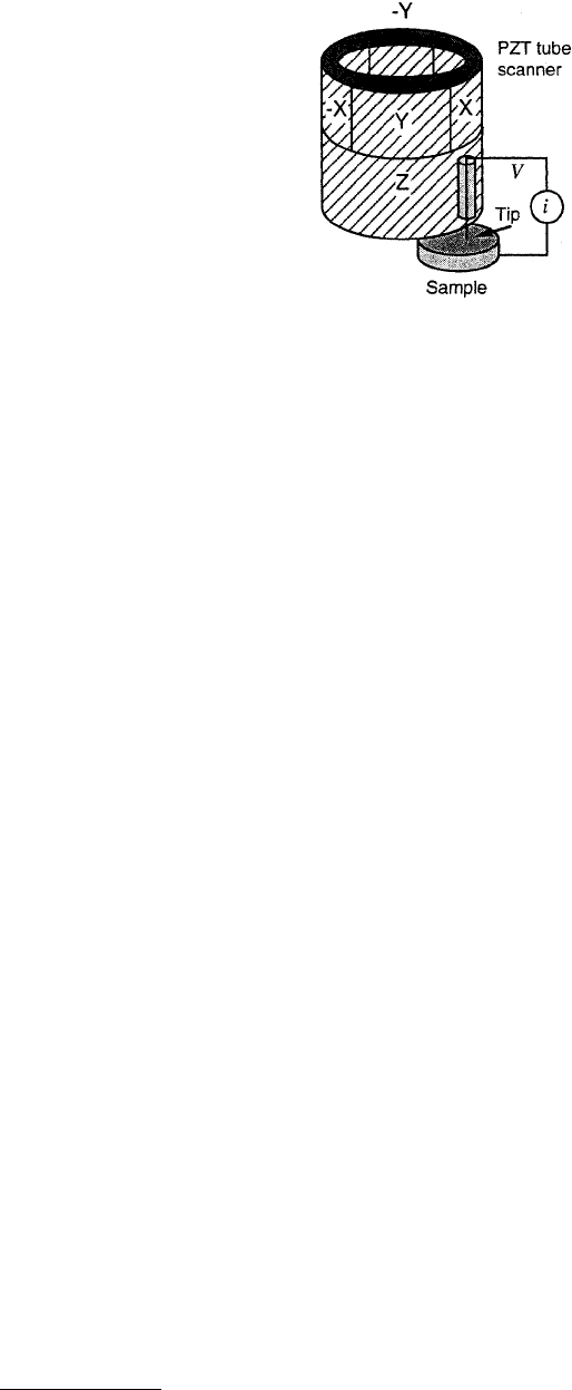
III STM for operation in ambient air, the sample is held in position while a piezoelectric crystal in the
form of a cylindrical tube scans the sharp metallic probe over the surface in a raster pattern while sensing
and outputting the tunneling current to the control station, Figure 1.5 (Anonymous, 1992b). The digital
signal processor (DSP) calculates the desired separation of the tip from the sample by sensing the
tunneling current flowing between the sample and the tip. The bias voltage applied between the sample
and the tip encourages the tunneling current to flow. The DSP completes the digital feedback loop by
outputting the desired voltage to the piezoelectric tube. The STM operates in both the constant-height
and constant-current modes depending on a parameter selection in the control panel. In the constant-
current mode, the feedback gains are set high, the tunneling tip closely tracks the sample surface, and
FIGURE 1.4 Principle of operation of the STM made by Binnig and Rohrer (1983).
© 1999 by CRC Press LLC
the variation in the tip height required to maintain constant tunneling current is measured by the change
in the voltage applied to the piezotube. In the constant-height mode, the feedback gains are set low, the
tip remains at a nearly constant height as it sweeps over the sample surface, and the tunneling current
is imaged. The following description of the instrument is almost exclusively based on Anonymous
(1992b).
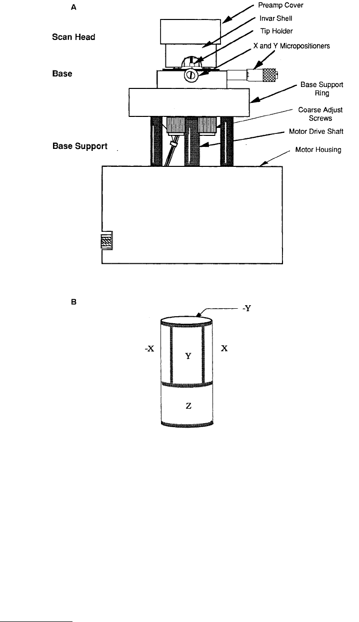
Physically, the Nanoscope STM consists of three main parts: the head which houses the piezoelectric
tube scanner for three-dimensional motion of the tip and the preamplifier circuit (FET input amplifier)
mounted on top of the head for the tunneling current, the base on which the sample is mounted, and
the base support, which supports the base and head, Figure 1.6A. The assembly is connected to a control
system that controls the operation of the microscope. The base accommodates samples up to 10 × 20 mm
and 10 mm in thickness. The different scanning heads mount magnetically on the tripod formed by the
front, coarse-adjust screws and the rear, find-adjust screws. Optional scan heads for the STM include 0.7
(for atomic resolution), 12, 75, and 125 µm square.
The scanning head controls the three-dimensional motion of tip. The removable head consists of a
piezotube scanner, about 12.7 mm in diameter, mounted into an Invar shell used to minimize vertical
thermal drifts because of good thermal match between the piezotube and the Invar. The piezotube has
separate electrodes for X, Y, and Z which are driven by separate drive circuits. The electrode configuration
(Figure 1.6B) provides X and Y motions, which are perpendicular to each other, minimizes horizontal
and vertical coupling, and provides good sensitivity. The vertical motion of the tube is controlled by the
Z-electrode which is driven by the feedback loop. The X and Y scanning motions are each controlled by
two electrodes which are driven by voltages of the same magnitudes, but opposite signs. These electrodes
are called –Y, –X, +Y, and +X. Applying complimentary voltages allows a short, stiff tube to provide a
good scan range without large voltages. The motion of the tip due to external vibrations is proportional
to the square of the ratio of vibrational frequency to the resonant frequency of the tube. Therefore, to
minimize the tip vibrations, the resonant frequencies of the tube are high, about 60 kHz in the vertical
direction and about 40 kHz in the horizontal direction. The tip holder is a stainless steel tube with a
300-µm inner diameter for 250-µm-diameter tips, mounted in ceramic in order to keep the mass on the
end of the tube low. The tip is mounted either on the front edge of the tube (to keep mounting mass
low and resonant frequency high) (Figure 1.5) or the center of the tube for large-range scanners, namely
75 and 125 µm (to preserve the symmetry of the scanning). This commercial STM will accept any tip
with a 250-µm-diameter shaft. The piezotube requires X–Y calibration which is carried out by imaging
an appropriate calibration standard. Cleaved graphite is used for the small-scan length head while two-
dimensional grids (a gold-plated ruling) can be used for longer-range heads.
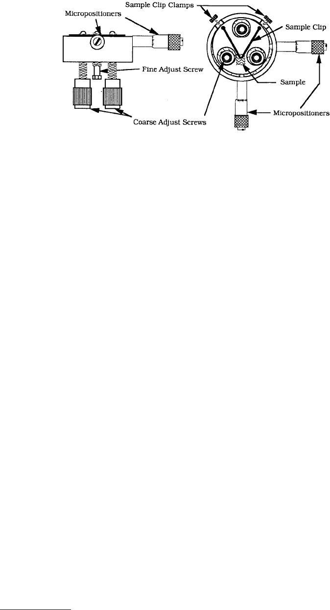
The Invar base holds the sample in position, supports the head, and provides X–Y motion for the
sample, Figure 1.6C. A spring-steel sample clip with two thumbscrews holds the sample in place. An X–Y
translation stage built into the base allows the sample to be repositioned under the tip. Three precision
FIGURE 1.5 Principle of operation of a commercial STM, a sharp tip attached to a piezoelectric tube scanner is
scanned on a sample.
© 1999 by CRC Press LLC
screws arranged in a triangular pattern support the head and provide coarse and fine adjustment of the
tip height. The base support consists of the base support ring and the motor housing. The base support
ring cradles the base allowing access to the adjustment screws. The stepper motor enclosed in the motor
housing allows the tip to be engaged and withdrawn from the surface automatically.
For measurements, the sample is placed under the sample-holding clip, with about half the sample
extending forward of the wire using an appropriate scanner, and a tip is inserted in the tip-holding tube
mounted on the piezotube. The tip is gripped with a tweezer near the sharp end and the blunt end of
the tip is inserted into the tip holder. For the tip to be held in the tube, it is necessary to put a small
bend in the tip before it is completely inserted. Next, the scanning head is placed on the three magnetic
FIGURE 1.6 Schematics of a commercial STM made by Digital Instruments, Inc.: (A) front view, (B) general
electrode configuration for piezoelectric tube scanner, and (C) front and top view of the STM base. (From Anonymous
(1992), “Nanoscope III Scanning Tunneling Microscope, Instruction Manual,” Courtesy of Digital Instruments, Inc.,
Santa Barbara, CA, 1992.)
© 1999 by CRC Press LLC
balls mounted on the threaded screws in the base. The tip is lowered with the coarse-adjustment screws
until there is only a slight gap, less than 0.25 mm (the tip will be damaged if brought into contact)
between the end of the tip and its reflected image visible on the sample. Next the scan parameters are
set, the motor is turned on, which engages the tip, and the scanning is initiated to form a desired image
of the sample surface.
Samples to be imaged with STM must be conductive enough to allow a few nanoamperes of current
to flow from the bias voltage source to the area to be scanned. In many cases, nonconductive samples
can be coated with a thin layer of a conductive material to facilitate imaging. The bias voltage and the
tunneling current depend on the sample. Usually they are set at a standard value for engagement and
fine-tuned to enhance the quality of the image. The scan size depends on the sample and the features of
interest. A maximum scan rate of 122 Hz can be used. The maximum scan rate is usually related to the
scan size. Scan rate above 10 Hz is used for small scans (typically 60 Hz for atomic-scale imaging with
a 0.7-µm scanner). The scan rate should be lowered for large scans, especially if the sample surfaces are
rough or contain large steps. Moving the tip quickly along the sample surface at high scan rates with
large scan sizes will usually lead to a tip crash. Essentially, the scan rate should be inversely proportional
to the scan size (typically 2 to 4 Hz for 1 µm, 0.5 to 1 Hz for 12 µm, and 0.2 Hz for 125 µm scan sizes).
Scan rate in length/time is equal to scan length divided by the scan rate in hertz. For example, for 10 ×
10 µm scan size scanned at 0.5 Hz, the scan rate is 20 µm/s. Typically, 256 × 256 data formats are most
commonly used. The lateral resolution at larger scans is approximately equal to scan length divided by 256.
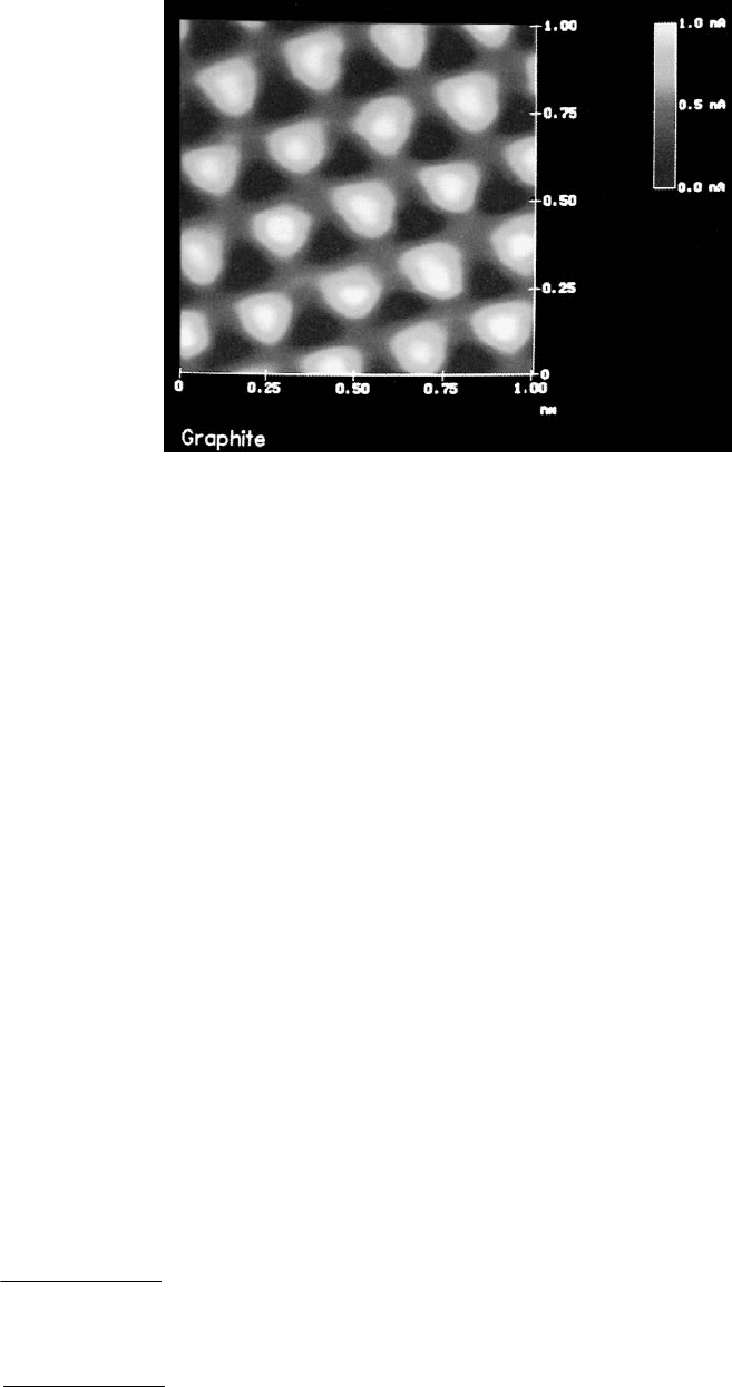
Figure 1.7 shows an example of an STM image of freshly cleaved, highly oriented pyrolytic graphite
(HOPG) surface taken at room temperature and ambient pressure (Binnig et al., 1986b; Park and Quate,
1986; Ruan and Bhushan, 1994b).
1.3.1.2.1 Electrochemical STM (ECSTM)
Electrochemical STM (ECSTM) allows the performance of the electrochemical reactions on the STM. It
includes a microscope base with an integral potentiostat, a short head with a 0.7-µm scan range, and a
differential preamp and the software required to operate the potentiostat and display the result of
electrochemical reaction.
1.3.1.2.2 Stand-Alone STM
The stand-alone STMs are available to scan large samples which rest directly on the sample. From Digital
Instruments, Inc., it is available in 12- and 75-µm scan ranges. It is similar to the standard STM except
the sample base has been eliminated. Two coarse- and one fine-adjustment screws used to position the
tip manually relative to the sample surface are mounted in the head shell.
1.3.1.3 Tip Construction
The STM cantilever should have a sharp metal tip with a low aspect ratio (tip length/tip shank) to
minimize flexural vibrations. Ideally, the tip should be atomically sharp, but, in practice, most tip
FIGURE 1.6(C)
© 1999 by CRC Press LLC
preparation methods produce a tip which is rather ragged and consists of several asperities with the one
closest to the surface responsible for tunneling. STM cantilevers with sharp tips are typically fabricated
from metal wires of tungsten (W), platinum–iridium (Pt–Ir), or gold (Au) and sharpened by grinding,
cutting with a wire cutter or razor blade, field emission/evaporator, ion milling, fracture, or electrochem-
ical polishing/etching (Ibe et al., 1990). The two most commonly used tips are made from either a Pt–Ir
(80/20) alloy or tungsten wire. Iridium is used to provide stiffness. The Pt–Ir tips are generally mechan-
ically formed and are readily available. The tungsten tips are etched from tungsten wire with an electro-
chemical process, for example, by using 1 mol KOH solution with a platinum electrode in an
electrochemical cell at about 30 V. In general, Pt–Ir tips provide better atomic resolution than tungsten
tips, probably due to the lower reactivity of Pt, but tungsten tips are more uniformly shaped and may
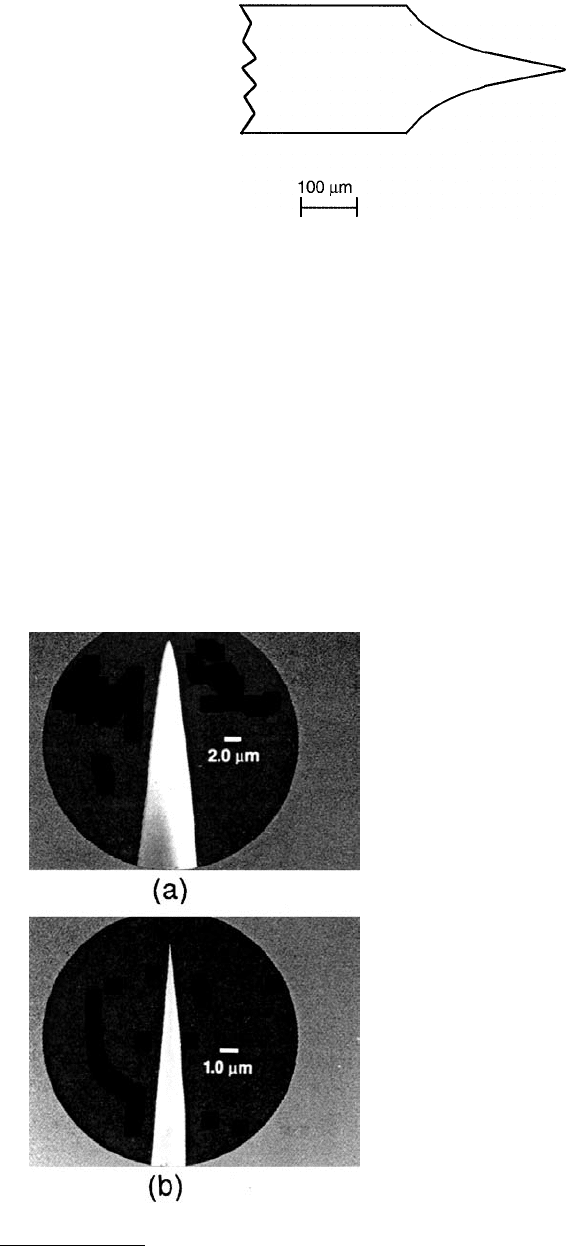
perform better on samples with steeply sloped features. The wire diameter used for the cantilever is
typically 250 µm with the radius of curvature ranging from 20 to 100 nm and a cone angle ranging from
10 to 60°, Figure 1.8. The wire can be bent in an L shape, if so required for use in the instrument. For
calculations of normal spring constant and natural frequency of round cantilevers, see Sarid and Elings
(1991).
Controlled geometry (CG) Pt-Ir probes are commercially available, Figure 1.9. These probes are
electrochemically etched from Pt-Ir (80/20) wire and polished to a specific shape which is consistent
from tip to tip. Probes have a full cone angle of approximately 15° and a tip radius of less than 50 nm.
For imaging of deep trenches (>0.25 µm) and nanofeatures, focused ion beam (FIB) milled CG probes
with an extremely sharp tip radius (<5 nm) are used. For electrochemistry, Pt-Ir probes are coated with
a nonconducting film (not shown in the figure). These probes are available from Materials Analytical
Services, Raleigh, NC.
Platinum alloy and tungsten tips are very sharp and have high resolution, but are fragile and sometimes
break when contacting a surface. Diamond tips were used by Kaneko and Oguchi (1990), Figure 1.10.
The diamond tip made conductive by boron ion implantation is found to be chip resistant. The diamond
FIGURE 1.7 Typical STM image of freshly cleaved, HOP graphite taken using a mechanically sheared Pt–Ir (80–20)
tip in constant-height mode (set point = 4 nA, bias = 16 mV, frequency = 20 Hz, 256 × 256 pixels, original scan size
3 × 3 nm). Bright spots correspond to the visible atoms.*
* Color reproduction follows page 16.
© 1999 by CRC Press LLC
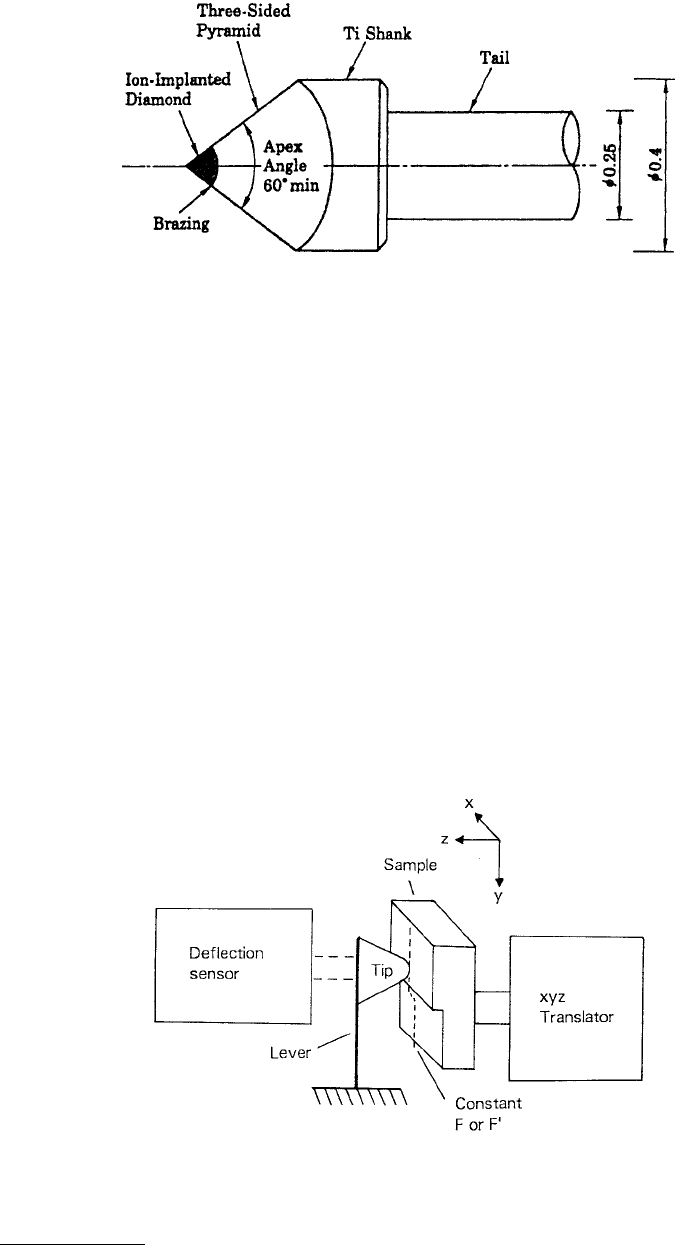
chip is brazed to a titanium shank having a tail diameter of 0.25 mm and total length of 10 mm. The
diamond is ground to the shape of a three-sided pyramid whose point is sharpened to a radius of about
100 nm. The smallest apex angle to achieve a sharp point without chipping is 60°. Finally, boron ions
are implanted in the diamond. Kaneko and Oguchi reported these tips as having a superior life.
1.3.2 Atomic Force Microscope
Like the STM, the AFM (a family of SFMs) relies on a scanning technique to produce very high resolution,
three-dimensional images of sample surfaces. AFM measures ultrasmall forces (less than 1 nN) present
between the AFM tip surface and a sample surface. These small forces are measured by measuring the
motion of a very flexible cantilever beam having an ultrasmall mass. While the STM requires that the
surface measured be electrically conductive, the AFM is capable of investigating surfaces of both con-
ductors and insulators on an atomic scale if suitable techniques for measurement of cantilever motion
are used. In the operation of high-resolution AFM, the sample is generally scanned instead of the tip as
FIGURE 1.8 Schematic of a typical tungsten cantilever with a sharp tip produced by electrochemical etching.
FIGURE 1.9 Schematics of (a) CG Pt–Ir probe and
(b) CG Pt–Ir FIB-milled probe.
© 1999 by CRC Press LLC
in STM, because AFM measures the relative displacement between the cantilever surface and the reference
surface, and any cantilever movement would add vibrations. However, AFMs are now available where
the tip is scanned and the sample is stationary. As long as the AFM is operated in the so-called contact
mode, little if any vibration is introduced.
The AFM combines the principles of the STM and the stylus profiler, Figure 1.11. In the AFM, the
force between the sample and tip is detected rather than the tunneling current to sense the proximity of
the tip to the sample. A sharp tip at the end of a cantilever is brought in contact with a sample surface
by moving the sample with piezoelectric scanners. During initial contact, the atoms at the end of the tip
experience a very weak repulsive force due to electronic orbital overlap with the atoms in the sample
surface. The force acting on the tip causes a lever deflection which is measured by tunneling, capacitive,
or optical detectors such as laser interferometry. The deflection can be measured to within ±0.02 nm, so
for a typical lever force constant at 10 N/m a force as low as 0.2 nN (corresponding normal pressure
~200 MPa for an Si
3
N
4
tip with a radius of about 50 nm against single-crystal silicon) could be detected.
This operational mode is referred to as the “repulsive mode” or “contact mode” (Binnig et al., 1986a).
An alternative is to use “attractive force imaging” or “noncontact imaging,” in which the tip is brought
in close proximity (within a few nanometers) to, and not in contact with, the sample (Martin et al.,
1987a). A very weak van der Waals attractive force is present at the tip-sample interface. Although in this
technique the normal pressure exerted at the interface is zero (desirable to avoid any surface deformation),
FIGURE 1.10 Schematic of a special diamond tip and shank with an overall length of 10 mm for use in STM.
(From Kaneko, R. and Oguchi, S. (1990), Jpn. J. Appl. Phys., 28, 1854–1855. With permission.)
FIGURE 1.11 Principle of operation of the AFM. (From McClelland, G. E. et al. (1987), Review of Progress in
Quantitative Nondestructive Evaluation, D. D. Thompson and D. E. Chimenti, eds., Vol. 6B, pp. 1307–1314, Plenum,
New York. With permission.

© 1999 by CRC Press LLC
it is slow and difficult to use and is rarely used outside research environments. In either mode, surface
topography is generated by laterally scanning the sample under the tip while simultaneously measuring
the separation-dependent force or force gradient (derivative) between the tip and the surface, Figure 1.11.
The force gradient is obtained by vibrating the cantilever (Martin et al., 1987a; McClelland et al., 1987;
Sarid and Elings, 1991) and measuring the shift of resonance frequency of the cantilever. To obtain
topographic information, the interaction force is either recorded directly or used as a control parameter
for a feedback circuit that maintains the force or force derivative at a constant value. The force derivative
is normally tracked in noncontact imaging. With an AFM operated in the contact mode, topographic
images with a vertical resolution of less than 0.1 nm (as low as 0.01 nm) and a lateral resolution of about
0.2 nm have been obtained (Albrecht and Quate, 1987; Binnig et al., 1987; Marti et al., 1987; Alexander
et al., 1989; Meyer and Amer, 1990a; Weisenhorn et al., 1991; Bhushan et al., 1993; Ruan and Bhushan,
1994b). With a 0.01-nm displacement sensitivity, 10 nN to 1 pN forces are measurable. These forces are
comparable to the forces associated with chemical bonding e.g., 0.1 µN for an ionic bond and 10 pN for
a hydrogen bond (Binnig et al., 1986a). For further reading, see Rugar and Hansma (1990), Sarid (1991),
Sarid and Elings (1991), Binnig (1992), Durig et al. (1992), Frommer (1992), Meyer (1992), Marti and
Amrein (1993), and Guntherodt et al. (1995) and dedicated issues of Journal of Vacuum Science Technology
(B9, 1991, pp. 401–1211) and Ultramicroscopy (Vols. 42–44, 1992).
Lateral forces being applied at the tip during scanning in the contact mode affect roughness measure-
ments (den Boef, 1991). To minimize effects of friction and other lateral forces in the topography
measurements in the contact-mode AFMs and to measure topography of soft surfaces, AFMs can be
operated in the so-called force modulation mode or tapping mode (Maivald et al., 1991; Radmacher
et al., 1992). In the force modulation mode, the tip is lifted and then lowered to contact the sample
(oscillated at a constant amplitude) during scanning over the surface with a feedback loop keeping the
average force constant. This technique eliminates frictional force entirely. The amplitude is kept large
enough so that the tip does not get stuck to the sample because of adhesive attractions. The modulation
mode can also be used to measure local variations in surface viscoelastic properties (Maivald et al., 1991;
Salmeron et al., 1993).
STM is ideal for atomic-scale imaging. To obtain atomic resolution with AFM, the spring constant of
the cantilever should be weaker than the equivalent spring between atoms. For example, the vibration
frequencies ω of atoms bound in a molecule or in a crystalline solid are typically 10
13
Hz or higher.
Combining this with the mass of the atoms m, on the order of 10
–25
kg, gives interatomic spring constants
k, given by ω
2
m, on the order of 10 N/m (Rugar and Hansma, 1990). (For comparison, the spring constant
of a piece of household aluminum foil that is 4 mm long and 1 mm wide is about 1 N/m.) Therefore, a
cantilever beam with a spring constant of about 1 N/m or lower is desirable. Tips have to be as sharp as
possible. Tips with a radius ranging from 20 to 50 nm are commonly available.
Atomic resolution cannot be achieved with these tips at the normal force in the nanonewton range.
Atomic structures obtained at these loads have been obtained from lattice imaging or by imaging of the
crystal periodicity. Reported data show either perfectly ordered periodic atomic structures or defects on
a larger lateral scale, but no well-defined, laterally resolved atomic-scale defects like those seen in images
routinely obtained with STM. Interatomic forces with one or several atoms in contact are 20 to 40 or
50 to 100 pN, respectively. Thus, atomic resolution with AFM is only possible with a sharp tip on a
flexible cantilever at a net repulsive force of 100 pN or lower (Ohnesorge and Binnig, 1993). Upon
increasing the force from 10 pN, Ohnesorge and Binnig (1993) observed that monatomic steplines were
slowly wiped away and a perfectly ordered structure was left. This observation explains why mostly defect-
free atomic resolution has been observed with AFM. We note that for atomic-resolution measurements
the cantilever should not be too soft to avoid jumps. We further note that measurements in the attractive-
force imaging mode may be desirable for imaging with atomic resolution.
The key component in AFM is the sensor for measuring the force on the tip due to its interaction
with the sample. A lever (with a sharp tip) with extremely low spring constants is required for high
vertical and lateral resolutions at small forces (0.1 nN or lower), but at the same time a high-resonant
frequency (about 10 to 100 kHz) in order to minimize the sensitivity to vibrational noise from the