Becker W. Advanced Time-Correlated Single Photon Counting Techniques
Подождите немного. Документ загружается.

94 5 Application of Modern TCSPC Techniques
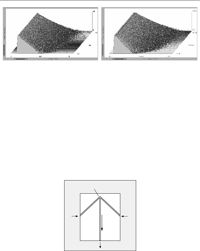
Fig. 5.34 Photochemical quenching transient of chlorophyll a. Sequence starts at the front.
Time per curve 100 us, 10,000 on/off cycles were accumulated. Left: Excitation at 650 nm.
Right: Excitation at 465 nm; the double peak is caused by an afterpulse of the laser
5.4.2 Continuous-Flow Mixing Techniques
Continuous-flow mixing is used to study the kinetics of chemical reactions, tran-
sient intermediate reaction products, protein folding, or conformational changes of
fluorophores. The principle is shown in Fig. 5.35. Two reagents are fed under high
pressure into separate inputs of a flow cell. They are combined in a mixing region
and then flow through the observation channel. The observation channel is illumi-
nated by a laser, and the absorption of the fluorescence along the channel is re-
corded. Because the velocity in the observation channel is known, the distance
along the channel represents the time scale of the reaction. With micromachined
channels, a resolution down to a few µs can be achieved [54].
IN 1
IN 2
OUT
Mixing Region
Flow
Fig. 5.35 Flow cell
By combining a continuous mixing experiment with fluorescence lifetime de-
tection changes in the concentration of a fluorophore can be separated from
changes in the quenching state. This is a clear benefit when observing protein
folding or other conformational changes of protein-dye complexes. The decay
functions along the channel can be obtained by TCSPC in combination with sin-
gle-point scanning or by simultaneous multipoint detection. The principles are
shown in Fig. 5.36.
5.4 Transient Fluorescence Lifetime Phenomena 95
Flow Cell
Laser
to
TCSPC
Scan
PMT
Photon
Pulses
Filter
Flow Cell
Laser
Cylinder Lens
Multi-Anode PMT Router
Channel
Photon
Pulses
to
TCSPC
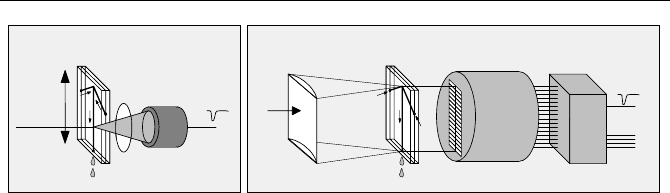
Fig. 5.36 Continuous-Flow mixer with TCSPC detection. Left: Single-point detection with
scanning. Right: Multipoint detection with a multianode PMT
Single-point detection is simple in terms of signal recording. A high-repetition-
rate laser is focused on the flow channel and the fluorescence is detected by a
PMT. The complete flow cell is mounted on a translation stage. A sequence of
fluorescence decay curves is recorded during a scan along the flow channel. Dif-
ferent optical systems can be used. The fluorescence can be detected from either
the back or the front of the flow cell. In either case, filters are required to block the
excitation light and to select the right detection wavelength interval. The benefit of
the single point design is that several detectors can be used to detect the fluores-
cence in different wavelength intervals or at 0° and 90° polarisation angle. The
drawback is that scanning takes time and consumes a large amount of the reagents.
Nevertheless, the setup is used to observe protein folding reactions on the micro-
second time scale [54]
Figure 5.36, right, shows a principle that uses the multidetector feature of
TCSPC. The laser beam is shaped into a line by a cylinder lens, and the whole
length of the flow channel is excited simultaneously. A fluorescence image of the
channel is transferred on the cathode of a multianode PMT. The photons are de-
tected simultaneously in all PMT channels (see Sect. 3.1, page 29). Multidetector
TCSPC is efficient in terms of reagent consumption. However, the system needs
careful calibration. The illumination along the channel can be nonuniform due to
speckle formation in the line-focused laser. Moreover, the efficiency of the indi-
vidual PMT channels is slightly different. Both effects are not necessarily stable
over a longer period of time, so that frequent recalibration is indicated.
5.4.3 Stopped-Flow Techniques
The stopped-flow technique uses the same mixing cell as described for the con-
tinuous flow (Fig. 5.35). However, the flow of the reagents is periodically
stopped, and the reaction is observed by recording the transient changes in absorp-
tion or fluorescence intensity. The principle of a TCSPC-based detection system
for stopped flow is shown in Fig. 5.37. An overview about the technique is given
in [440].
96 5 Application of Modern TCSPC Techniques
PMT
Photon
Pulses
Flow Cell
Laser
Valve
Valve
From
Pump
Filter
'Stop'
Experiment
Trigger
TCSPC Module
from Laser
start
stop
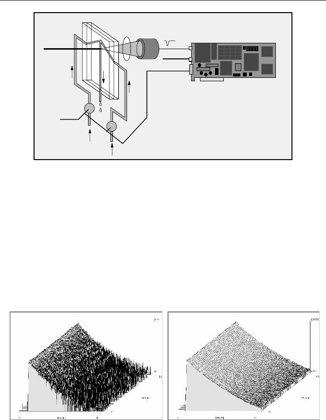
Fig. 5.37 Principle of a stopped-flow instrument with TCSPC
A laser is focused at the flow channel of the mixing cell shortly behind the mix-
ing region, and the fluorescence is detected by a PMT. The flow of the reagents
through the cell is periodically stopped by two valves. In commercial instruments
the flow is stopped within approximately one millisecond. Triggered by each stop
of the flow, the TCSPC device records a sequence of fluorescence decay curves.
To obtain a high signal-to-noise ratio, a large number of triggered sequences are
accumulated.
An example of a stopped-flow measurement with TCSPC is shown in Fig. 5.38.
A sequence recorded for a single flow-stop is shown left. The result accumulated
over 500 stops is shown right.
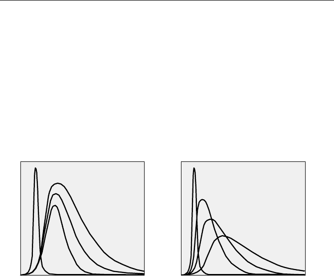
Fig. 5.38 Stopped-flow measurement by TCSPC, triggered sequential recording, 100 ms
per curve. Left: Recorded sequence after a single stop. Right: 500 stops accumulated. Data
courtesy of Osman Bilsel, University of Massachusetts Medical School, Worcester, MA
Although it is not directly visible in Fig. 5.38 the fluorescence lifetime shows
subtle changes over the time of the sequence. Figure 5.39, left is a zoom into
curves 10 (1 s after the stop) and 70 (7 s after the stop). Figure 5.39, right shows
the change of the mean lifetime over the time of the sequence.

5.5 Diffuse Optical Tomography (DOT) and Photon Migration 97
1234567 8
0
10
20
30
40
50
60
70
ps
s
Fig. 5.39 Left: curves 10 (1 s after the stop) and 70 (7 s after the stop). Right: change of the
mean lifetime over the time of the sequence
A stopped-flow setup can relatively easily be equipped with multispectral de-
tection. The fluorescence light is emitted from a small spot in the flow channel.
Therefore the flow cell can be placed directly in the input slit plane of a poly-
chromator. The spectrum is detected by a multianode PMT. The photons detected
in the spectral channels are recorded simultaneously by a TCSPC device and a
router. However, although the implementation is relatively simple, no spectrally
resolved TCSPC-based stopped-flow system has yet been described.
5.5 Diffuse Optical Tomography (DOT) and Photon
Migration
5.5.1 Principle of Diffuse Optical Tomography
Diffuse optical tomography (DOT) aims to resolve the spatial distribution of opti-
cal properties in highly scattering media. Biomedical applications of DOT are
based on illumination of thick tissue by NIR light, detection of diffusely transmit-
ted or reflected light, or the fluorescence of endogenous or exogenous fluoropho-
res [95, 251, 397, 542]. Typical applications of DOT techniques are optical mam-
mography, brain imaging, and noninvasive investigations of drug effects in small
animals.
The scattering in tissue is not isotropic. A considerably larger amount of light is
scattered forward rather than in reverse [126, 149, 367, 542]. For describing the
penetration of light into thick tissue it is, however, sufficient to assume isotropic
scattering with a reduced scattering coefficient. The reduced scattering coefficient,
µ´
s
, is
µ´
s
= µs (1 - g) (5.9)
with g being the average cosine of the scattering angle. For biological tissue g is
typically in the range from 0.7 to 0.9 [121, 367]. Reduced scattering coefficient of
breast tissue as a function of wavelength are shown in Fig. 5.40. Reduced scatter-
ing coefficients for various types of tissue are given in [367].
98 5 Application of Modern TCSPC Techniques
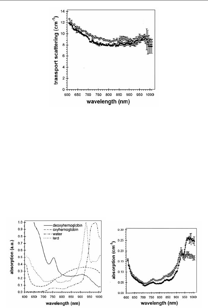
Fig. 5.40 Reduced scattering coefficients of breast tissue of two different patients measured
by diffuse reflection, from [121]
Typical values of µ´
s
are around 10 cm
-1
. Consequently, there are practically no
unscattered or „ballistic“ photons for tissue thicker than 1 cm [149]. Instead, the
photons must be considered to diffuse through the tissue. Consequently, the spatial
resolution of DOT images is extremely poor and cannot compete with positron
emission, X-ray and MRI techniques.
The absorption in tissue is dominated by oxy-haemoglobin, deoxy-haemo-
globin, lipids, and water [121]. The extinction coefficients of the tissue constitu-
ents are shown in Fig. 5.41, left. Absorption spectra of tissue measured in vivo are
shown right. There is an absorption window from approximately 650 to 900 nm.
Therefore, NIR light can be transmitted and detected through tissue layers as thick
as 10 cm. Absorption coefficients for various types of tissue are given in [367].
Fig. 5.41 Absorption coefficients in biological tissue [121]. Left: Absorption spectra of
tissue components in arbitrary units. Right: Absorption spectra of breast tissue of different
patients, measured by diffuse reflection
5.5 Diffuse Optical Tomography (DOT) and Photon Migration 99
In spite of the poor spatial resolution, DOT in the NIR has the benefit that the
measured absorption coefficients are related to the biochemical constitution of the
tissue, such as haemoglobin concentration and blood oxygenation [121, 346]. If
exogenous markers are used, the absorption or fluorescence delivers additional
information about blood flow, blood leakage, ion concentrations, or protein bind-
ing state [135, 369, 460].
Unfortunately it is hard to distinguish between the effects of scattering and ab-
sorption in simple steady state images. The situation is much better if pulsed or
modulated light is used to transilluminate the tissue and the pulse shape or the
amplitude and phase of the transmitted light are recorded. Figure 5.42 illustrates
the general effect of scattering and absorption on the shape of a pulse after migra-
tion through highly scattering tissue.
Input
Input
Absorption
1
2
3
Scattering1
2
3
Output for increasing
Output for increasing
time (ns)
time (ns)
Fig. 5.42 Effect of scattering and absorption on the shape of a pulse transmitted through
thick tissue
Increases in both scattering and absorption decrease the output intensity. How-
ever, increased scattering increases the pulse width while increased absorption
tends to decrease it [512]. Therefore, the shape of the „time-of-flight distribution“
of the photons can be used to distinguish between scattering and absorption.
Qualitatively, early photons are mainly influenced by scattering, whereas later
photons are increasingly influenced by absorption as well.
In diffuse reflection experiments, the depth of scattering and absorption
changes in the tissue can be derived from time-resolved data [481]. The first and
second moments of the time-of-flight distributions are especially sensitive to
changes in deep tissue layers [325, 328].
The advantage of time-resolved detection is obvious if fluorescence is to be de-
tected [90, 165, 174, 362, 388, 390]. The fluorescence lifetime of a fluorophore is
in first approximation independent of its concentration, but depends on the local
environment and the binding state. Unfortunately biological tissue does not show
any appreciable fluorescence of endogenous fluorophores for excitation in the
„spectroscopic window“ in the NIR. There are, however, a number of exogenous
fluorophores that are efficiently excited at NIR wavelengths [135, 369]. Fluores-
cence measurement in DOT aims at one of two things, either intensity and lifetime
changes induced by differences in the local environment parameters or the study
100 5 Application of Modern TCSPC Techniques
of blood content and blood-flow dynamics by fluorophores that stay exclusively in
the blood. With the progress in the development of new molecular probes [342],
fluorescence detection will become increasingly important.
It is extremely demanding to reconstruct tissue structures and optical properties
from time-resolved data. A number of different approaches are used to solve the
„inverse problem“ of DOT [12, 13, 97, 165, 176, 250, 387, 411, 506, 524]. All
approaches use a large number of time-resolved detection channels for different
wavelengths, varying source-detector distances, or varying transillumination an-
gles. The time-channel width required to quantify absorption and reduced scatter-
ing coefficient is of the order of 10 ps [387]. Low noise data are required to recon-
struct the tissue properties from the relatively small intensity and pulse shape
changes. A high signal-to-noise ratio can be obtained only by recording a large
number of photons.
The illumination intensity of DOT in human patients is limited by laser safety
regulations; on the other hand, the acquisition time for in vivo measurements must
be kept within reasonable limits and below the time scale of the physiological ef-
fects to be recorded. The only way to collect a large number of photons in a short
time is to increase the total detector area. However, using a large detector area with-
out sacrificing spatial resolution is made possible only by using a large number of
detector channels. Detection efficiency becomes even more important if fluores-
cence is to be detected in combination with normal DOT. Therefore, simultaneous
detection in a large number of time-resolved channels is an absolute requirement.
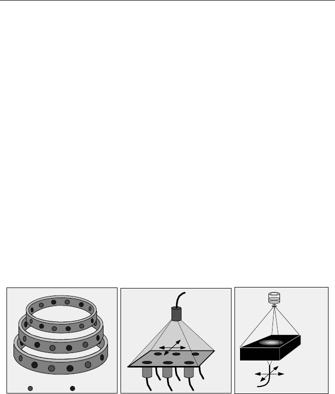
Three typical optical configurations of DOT systems are shown in Fig. 5.43.
Sources Detectors
Source
Detectors
scan
Y
X
scan
Y
X
Source Fibre
Camera System
Gated or
image intensifier
modulated
Fig. 5.43 Source-detector arrangements for optical tomography. Left: Classic tomography
setup. Circular arrangement of sources and detectors; sources are activated one after another;
light distribution is measured by all detectors. Middle: Scanning setup with one source and
several detectors. Source and detectors are scanned across the sample. Right: Camera setup;
source is scanned across the sample; light distribution is detected by a camera system
The traditional tomography setup is shown left. A large number of sources and
detectors are arranged around the sample. The light sources are switched on one
after another. For each source, the time-of-flight distributions of the photons (or,
in the frequency domain, the phase and amplitude of the signals) are recorded by
all detectors simultaneously [14, 222, 385, 443]. The setup is used for breast imag-
ing and infant brain imaging. Because the setup is compact and mechanically
simple, it is also used for optical tomography in conjunction with MRI imaging

5.5 Diffuse Optical Tomography (DOT) and Photon Migration 101
[384, 386]. When used for adult brain imaging, the detectors opposite to the
source do not detect reasonable signals. Therefore detectors and sources are ar-
ranged at only side of the head. The configuration can be considered a subset of
the arrangement shown in Fig. 5.43, left.
The setup shown in Fig. 5.43, middle, uses a scanning technique. Several lasers
of different wavelength are multiplexed into a single optical source. The light
source and the detector (or a number of detectors) scan simultaneously across the
sample. The scanning technique is successfully used for optical mammography
[124, 200, 201, 203, 412, 489, 490, 505, 506]. A scanning setup for small-animal
imaging is described in [174]. The benefit of scanning is that it obtains a high
spatial density of data points. Therefore the Nyquist condition can be fulfilled for
both spatial dimensions, and image artefacts are avoided. However, problems can
arise from edge effects. Not only can the detectors be damaged if the scan runs
over the edge of the sample, but also the reconstruction of the sample properties
has to cope with different photon migration near the edge [525].
The setup shown in Fig. 5.43, right, uses a time-resolved camera system for de-
tection. The camera uses a gated [125, 149] or modulated [460, 496] image inten-
sifier. The source is scanned across the sample, and for each source position a
sequence of images is taken at various gate delays or at several phase angles.
Optical tomography techniques for human medicine are currently at the stage of
clinical tests [204, 225, 489, 490]. Frequency domain instruments using modula-
tion techniques are competing with time-domain instruments using TCSPC.
A comprehensive overview of frequency-domain DOT techniques is given in
[88]. Particular instruments are described in [166, 347, 410]. It is commonly be-
lieved that modulation techniques are less expensive and achieve shorter acquisi-
tion times, whereas TCSPC delivers a better absolute accuracy of optical tissue
properties. It must be doubted that this general statement is correct for any particu-
lar instrument. Certainly, relatively inexpensive frequency-domain instruments
can be built by using sine-wave-modulated LEDs, standard avalanche photodi-
odes, and radio or cellphone receiver chips. Instruments of this type usually have a
considerable „amplitude-phase crosstalk“. Amplitude-phase crosstalk is a depend-
ence of the measured phase on the amplitude of the signal. It results from nonlin-
earity in the detectors, amplifiers, and mixers, and from synchronous signal pickup
[6]. This makes it difficult to obtain absolute optical tissue properties. A carefully
designed system [382] reached a systematic phase error of 0.5° at 100 MHz. A
system that compensates the amplitude-phase crosstalk via a reference channel
reached an RMS phase error of 0.2° at 100 MHz [370]. These phase errors corre-
spond to a time shift of 14 ps and 5.5 ps RMS, respectively.
Amplitude-phase crosstalk is intrinsically low in frequency-domain instruments
that use gain-modulated PMTs as detectors and mixers [166]. Results presented in
[98, 346] show that optical properties can be obtained with an accuracy compara-
ble to that of TCSPC-based instruments. The modulated-PMT technique is some-
what less efficient than TCSPC and does not work well at extremely low photon
rates. Nevertheless, the sensitivity is well within the range required for fluores-
cence detection in DOT.
TCSPC is superior in terms of efficiency and sensitivity. The effective detec-
tion bandwidth is much higher than for modulation systems. The IRF can be kept
102 5 Application of Modern TCSPC Techniques
stable within less than 2 ps peak-to-peak, or about 1 ps rms (see Fig. 7.34, page
296). The waveform is correctly sampled according to the Nyquist theorem. The
count rate of TCSPC is limited to a few MHz per TCSPC channel, which is often
considered a drawback. It should, however, be taken into account that TCSPC is
superior to any other technique in obtaining a superior signal-to-noise ratio (SNR)
from a given number of detected photons. Therefore, the limited count rate is less
important than commonly believed. The excellent results obtained with TCSPC-
based DOT instruments under clinical conditions demonstrate the applicability of
TCSPC [203, 204, 329, 489, 490].
Many DOT instruments are based on multiplexing several lasers and recording
in a single channel TCSPC channel of high count rate [119, 201, 202, 325, 414,
505]. Multidetector operation of up to eight detectors connected to a single
TCSPC channel was used in [120, 123, 124, 385, 386, 507]. A system with 32
fully parallel TCSPC channels based on NIM modules was described in [443] and
used for breast and brain imaging [222, 223, 225]. Recent TCSPC-based instru-
ments use packages of four parallel multidetector TCSPC devices operated in a
single PC [34, 328, 329, 412, 415, 489, 490, 506, 525, 526].
Optical mammographs and brain imagers are complex instruments with their
own control, data acquisition and data processing software. Nevertheless, TCSPC-
based instruments have a number of technical features in common. The general
technical approaches and some typical results are described below.
5.5.2 Scanning Mammography
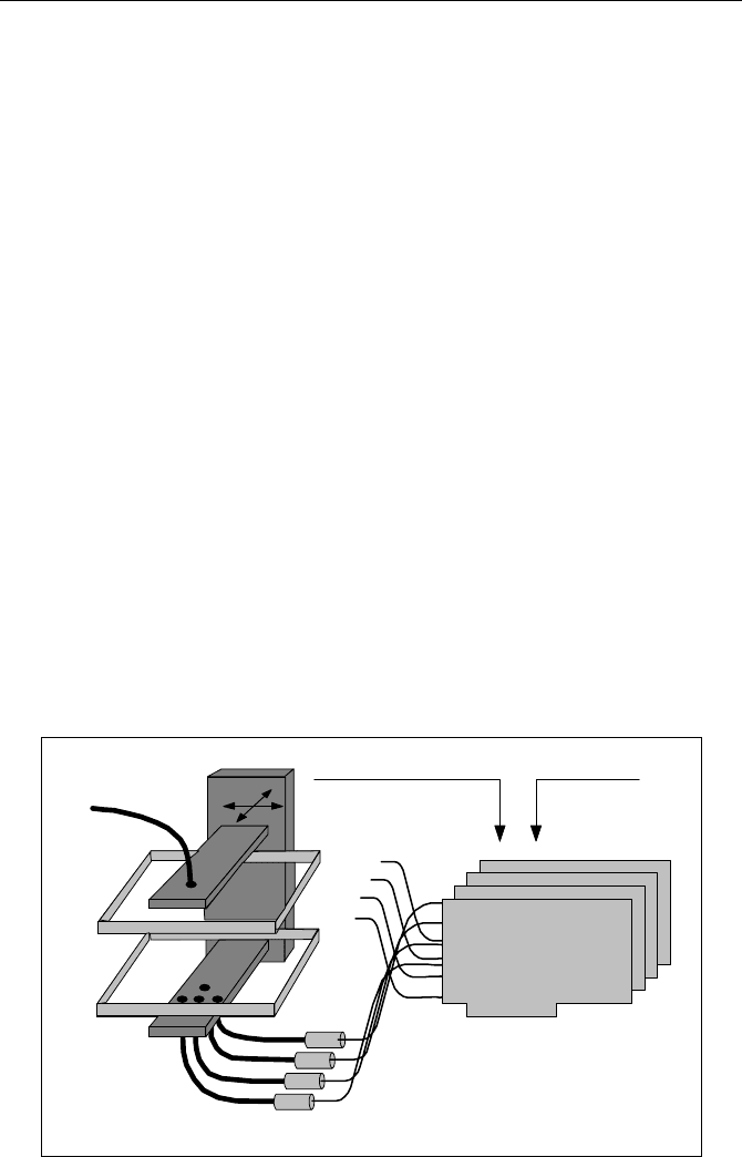
The principle of a typical TCSPC-based scanning mammograph is shown in
Fig. 5.44.
4 Module TCSPC Package
Y
scan
X
Detectors
Glass
Plate
Glass
Plate
Fibre
Bundles
Fibre
Laser
Scan Sync Pulses
from Scanner
Laser
Multiplexing
Signal
Reference
from
Laser
Fig. 5.44 Principle of a mammography scanning instrument

5.5 Diffuse Optical Tomography (DOT) and Photon Migration 103
The breast is slightly compressed between two glass plates. The laser light is de-
livered via an optical fibre. The transmitted light is collected by four fibre bundles
and fed to four detectors. Fibres and fibre bundles are certainly not a favourable
solution in terms of timing stability and detection efficiency. However, they de-
couple the electrical part of the system from the patient. The fibres are therefore an
important part of the system to satisfy safety regulations for medical instruments.
The complete fibre system is assembled on a common scanning stage and
scanned over the breast. One scan typically contains 1,000 to 5,000 pixels, which
is sufficient to avoid artefacts due to undersampling. The scan amplitudes and the
shape of the scanning area are different for different patients. To ensure that the
detectors are not damaged by overload, the scanner must be prevented from run-
ning beyond the edge of the breast. Therefore the scan is controlled by measuring
the detector output currents integrated over a time of a few milliseconds (see
Sect. 7.2.15, page 300). When the current exceeds a reasonable limit, the scan
direction is reversed.
Four PMTs detect the transmitted light under different projection angels. The
four detector signals are connected to the individual channels of a four-module
TCSPC system. In practice, variable neutral-density filters and long-pass filters
are placed in front of the detectors to compensate for different intensity at differ-
ent breast thickness and to reduce the daylight sensitivity.
Usually several laser wavelengths are multiplexed into a single source fibre.
The wavelengths can be multiplexed on a pulse-by pulse basis and recorded in the
same TAC interval [203, 412, 489, 490, 505, 506] or in intervals of 50 to 200 µs
(2,500 to 10,000 pulses) and recorded in different memory blocks by using the
routing capability of the SPC modules (please see Sect. 3.2, page 33). The benefit
of the second technique is that there is (almost) no crosstalk between the wave-
length channels, and signal distortion due to pile-up effects is reduced. A discus-
sion of laser multiplexing is given under Sect. 5.5.8, page 117.
For pulse-by-pulse multiplexing, the timing reference signal for the TCSPC
channels comes from one of the lasers. For pulse group multiplexing, the trigger
output signals of the lasers are combined in a reversed power splitter.
The recording in the TCSPC channels can be synchronised with the scanning
by software. In this case the time-of-flight distributions are read out from the
TCSPC modules for each individual pixel. The system can avoid devoting time to
readout during the scan by using sequential recording with memory swapping (see
Sect. 3.3, page 35); the data of each line are read during the scan of the next line.
Another convenient mode is hardware-controlled scanning (see Sect. 3.4, page
37). In this mode the TCSPC module holds the time-of-flight distributions of all
pixels of a scan in its memory. The data are read out when the scan is completed.
In both cases the data acquisition in the TCSPC channels is synchronised with
the scanning by clock pulses from the scan controller. It must, however, be taken
into account that the length of the lines of the scan varies since the return points of
the scan are controlled by the detector overload signals. Therefore, the scan soft-
ware must store the positions of the return points and the number of pixels be-
tween. These positions are used later to adjust the lines horizontally.
If fluorescence is to be detected in combination with DOT, detection efficiency
may become a crucial point. The detection efficiency increases with the total