Becker W. Advanced Time-Correlated Single Photon Counting Techniques
Подождите немного. Документ загружается.


124 5 Application of Modern TCSPC Techniques
Except for the different sizes, focal lengths and numerical apertures of the
lenses, the principle is the same as for a laser scanning microscope. The laser is
reflected into the system by a dichroic mirror, M1. The scan mirrors, M2 and M3,
deflect the laser beam. The scan lens system, L1 and L2, sends a parallel beam
through the imaging lens, L3. L3 focuses the laser on the sample surface. The
laser spot moves over the surface with the motion of the scan mirrors. The fluo-
rescence light is collected by L3 and goes back through L2 and L1. The scan mir-
rors descan the fluorescence into a stationary beam that is focused on the entrance
slit of a polychromator by L4. The spectrum of the fluorescence light is detected
by a multianode PMT, and photons of different wavelength are routed into differ-
ent memory segments. Details are described under Sect. 3.4, page 37, and
Sect. 3.1, page 29.
Many modifications of the optical system are possible. For example, the scan
mirrors can be placed between L1 and L2; L1 can be a negative lens, or L2 and L3
can be combined into one lens. L3 can be replaced with an endoscope. Moreover,
the focus of L3 can be scanned over the input plane of a gradient-index (GRIN)
lens (see Fig. 7.9, page 273). The GRIN lens can be used as a miniature endoscope
[320].
Biological tissue is highly scattering, a fact that has to be taken into account
when fluorescence decay functions are recorded. Scattering not only generates a
huge amount of backscattered excitation light but also results in photon migration
effects. Photon migration changes the shape of the fluorescence decay curves and
must be taken into account when tissue path lengths exceed a few millimeters.
Due to the high absorptivity of tissue in the ultraviolet and blue spectral range, the
effect on the decay curves is smaller than in the infrared. Nevertheless, optical
systems for time-resolved detection of tissue fluorescence should detect light only
from a small area around the excited spot. For fibre probes in direct touch with the
tissue this is automatically the case. In the system shown in Fig. 5.65 the detection
area is confined automatically by the entrance slit of the polychromator. It can be
further reduced by a field stop in the focus of L4.
Although macroscopic scanning systems for multispectral time-resolved fluo-
rescence imaging can be built with reasonable technical effort, no such system is
yet available. There are, however, microscopy systems for autofluorescence imag-
ing. These systems are described below.
5.6.2 Two-Photon Autofluorescence
Two-photon excitation by femtosecond NIR laser pulses can be used to obtain
clear images of tissue layers as deep as 1 mm [132, 278, 279, 344, 462, 495,
534]. The efficiency of two-photon excitation depends on the square of the
power density. It therefore works with noticeable efficiency only in the focus of
the laser beam. With a microscope lens of high numerical aperture a lateral reso-
lution around 300 nm and a longitudinal resolution of about 1 µm is obtained.
Two-photon laser scanning microscopy has therefore become a standard tech-
nique of tissue microscopy. Two-photon laser scanning can be combined with
5.6 Autofluorescence of Biological Tissue 125
multispectral detection [138, 473]. The technique is available in commercial in-
struments [48].
Dual-wavelength TCSPC detection in two-photon laser scanning microscopes
is relatively simple [37]. Multispectral TCSPC detection in a two-photon laser
scanning microscope requires a suitable relay optics between the objective lens
and the polychromator [35, 60]. Details are described under „TCSPC Laser Scan-
ning Microscopy“.
Applications of single-wavelength TCSPC imaging to autofluorescence of tis-
sue are described in [281, 282, 283, 428]. A commercial instrument for skin in-
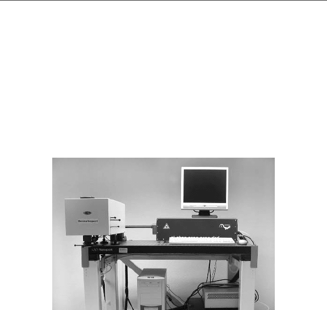
spection has been designed by Jenlab, Jena, Germany. The „Dermainspect“ is
based on a Ti:Sapphire laser, a fast optical scanner, and multidimensional TCSPC.
The instrument is shown in Fig. 5.66.
Fig. 5.66 „Dermainspect“ system of Jenlab, Jena, Germany
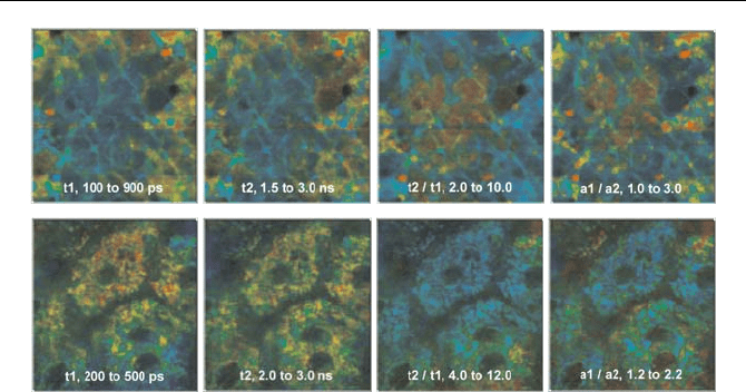
A typical result is shown in Fig. 5.67. It shows autofluorescence lifetime im-
ages of stratum corneum (upper row, 5 µm deep), and stratum spinosum (lower
row, 50 mm deep). The multiexponential decay was approximated by a double-
exponential model, and the decay parameters determined by a Levenberg-
Marquardt fit. The colour represents the fast lifetime component,
W
1
, the slow
lifetime component,
W
2
, the ratio of lifetime components,
W
1
/
W
2
, and the ratio of the
amplitudes of the components,
a
1
/ a
2
. The brightness of the pixels represents the
intensity.
The decay parameters show considerable variations throughout the image. Even
in a single-wavelength image, it can be expected that the decay parameters contain
a wealth of information. However, the biological meaning of the decay variations
still remains a subject of investigation.
126 5 Application of Modern TCSPC Techniques
Fig. 5.67 Time-resolved in-vivo autofluorescence images of human stratum corneum (up-
per row, 5 µm deep), and stratum spinosum (lower row, 50 mm deep). Left to right: fast
lifetime component,
W
1
, slow lifetime component,
W
2
, ratio of the lifetime components,
W
1
/
W
2
, and ratio of amplitudes, a
1
/ a
2
. The indicated parameter range corresponds to a
colour range from blue to red
5.6.3 Ophthalmic Imaging
Reflection and fluorescence imaging of the ocular fundus is an established tool of
diagnosing eye diseases [131, 474, 523, 434]. As usual, the fluorescence signals
contain components of several fluorophores which cannot be clearly separated in
intensity images. Moreover, intensity variations by oxygen quenching or different
binding states cannot be distinguished from concentration variations. Fluorescence
lifetime imaging is therefore considered a potential technique of diagnosing eye
diseases, especially age-related macular degeneration. A severe problem of meas-
urements in the eye is low photon flux due to the limitation of the excitation
power. Due to its near-ideal counting efficiency and its multiwavelength capabil-
ity, TCSPC imaging is clearly the best signal recording technique for detecting the
fluorescence decay functions [450, 452, 454].
Ophthalmic imagers often use a scanning technique with the pupil of the eye
placed in the exit pupil of the scanning system [461]. This reduces image distor-
tion and blurring due to the poor optical quality of the lens of the eye. Moreover,
confocal detection can be used to suppress reflection, scattering, and fluorescence
signals from the lens of the eye.
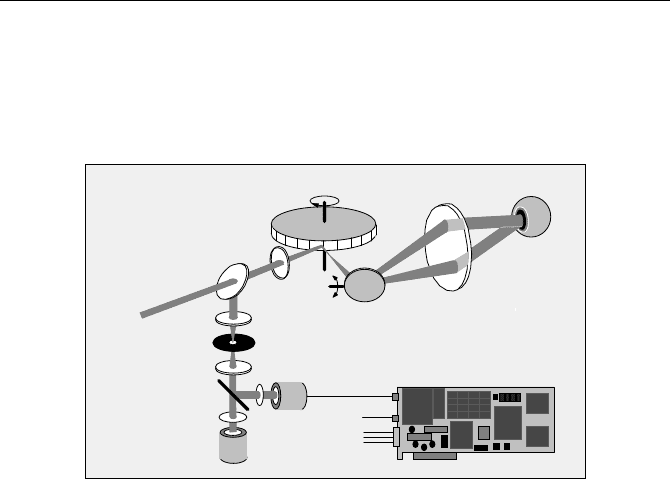
The general principle of an ophthalmic scanner is shown in Fig. 5.68. A 440 nm
picosecond diode laser is used for fluorescence excitation. The laser beam is de-
flected by an ultrafast scanner. The scanner consist of a fast-rotating polygon mirror
for x deflection and a galvanometer mirror for y deflection. A lens, L1, forms a
focus approximately in the middle of the polygon mirror and the galvanometer
mirror. A second lens, L2, sends a parallel beam of light into the eye. The beam
wobbles with the scanning, with a virtual pivot point in the pupil of the eye. The
5.6 Autofluorescence of Biological Tissue 127
lens of the eye focuses this beam on the retina. The light emitted by the retina leaves
the eye via the same beam path, travels back via the scan mirrors, and is separated
from the laser light by a beam splitter. It is focused into a pinhole by a third lens, L3,
and transferred to the detectors by another lens, L4. The optical setup may differ in
details for different scanners, but the general principle is the same.
Polygon mirror
Pinhole
L3
L4
Dichroic
Mirror
F1
F2
PMT
PMT
Galvanometer
Mirror
Beam
splitter
L1
L2
Eye
stop
from laser
start
scan clocks
from sanner
Laser
(fluorescence)
(reflection)
Fig. 5.68 Ophthalmic fluorescence lifetime imaging
Most ophthalmic scanners are designed for imaging the reflected light. They
contain a PMT that delivers an intensity-proportional signal of the light reflected
at the eye background. The signal is amplified to a video-compatible level, com-
bined with synchronisation pulses from the scanner, and displayed on a video
monitor.
A laser scanning ophthalmoscope can relatively easily be combined with the
TCSPC scanning technique (see Sect. 3.4, page 37). The fluorescence light from
the retina is split off by a dichroic mirror and detected by a second PMT. The
detection wavelength of the PMTs is selected by filters, F1 and F2. The photon
pulses from the fluorescence channel PMT are fed into the start input of the
TCSPC module. The stop pulses come from the diode laser.
To build up lifetime images, the TCSPC module needs scan synchronisation
pulses from the scanner. These can be either obtained directly from the scanner or
separated from the video signal of the reflection channel.
The count rates obtained from the autofluorescence of the eye background are
on the order of 500 to 10,000 s
-1
. Compared with autofluorescence of skin or tis-
sue this rate is very low. The laser power is limited to about 50 µW by eye safety
considerations. Moreover, a wavelength of 440 nm is far less efficient in exciting
autofluorescence than wavelengths around 400 nm or shorter. Although laser
diodes are available for 375 nm and 405 nm these wavelengths do not pass the
lens of the eye. Moreover, the optical quality of the lens of the eye is far from
being ideal. Therefore the fluorescence light returned into the scanner is not well
collimated. Because the effective aperture of the detection system is limited by the
size of the polygon facets a large fraction of the fluorescence signal is lost.
128 5 Application of Modern TCSPC Techniques
Because the excitation and the fluorescence light share the same optical path
good blocking of scattered excitation light is essential. Moreover, fluorescence of
materials in the excitation path must be carefully avoided. Especially fluorescence
from epoxy can be very similar to autofluorescence of tissue, both in the spectrum
and the decay profile. Problems can also arise from the emission of LEDs used to
control the pinhole wheel or the polygon mirror of the scanner. It may be neces-
sary to place an NIR blocking filter in front of the detector.
Due to the low count rate the acquisition times range from about 10 seconds for
single-exponential and 60 to 180 seconds for double-exponential decay analysis.
The long acquisition time causes problems with eye motion. It is impossible for a
patient to keep his eye fixed on a target point for longer than a few seconds. The
vision periodically wanders off from the fixed point and jumps back. The problem
is solved by acquiring a series of images with short acquisition times. The images
are inspected later; blurred images are discarded and the good images are centred
one over another and accumulated. The resulting data set contains enough photons
to run a fit procedure on the fluorescence decay data.
The application of an ophthalmic scanning TCSPC system to autofluorescence
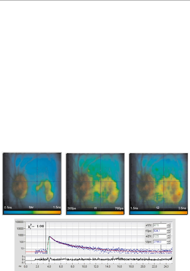
imaging at the fundus is described in [450, 451, 452, 453, 454]. A typical result is
shown in Fig. 5.69. A double-exponential deconvolution was run on the complete
pixel array. It delivered a fast lifetime component of the order of
W
1
= 400 ps and a
Fig. 5.69 TCSPC lifetime images of the human ocular fundus, obtained by a double-
exponential fit. The lifetime is colour-coded as indicated in the images. Upper left: Average
lifetime. Upper middle: Fast lifetime component. Upper right: Slow lifetime component.
Lower part: Decay curve in the selected spot and fit results. Courtesy of Dietrich
Schweitzer, Friedrich Schiller University Jena, Germany
5.7 TCSPC Laser Scanning Microscopy 129
slow one of about
W
2
= 3 ns, together with the amplitudes of the components, a
1
and
a
2
. A lifetime image of the average lifetime,
212211
/ aaaa
av
W
WW
(5.12)
is shown in the upper right part. The colour range from blue to red corresponds to
a lifetime range from 500 to 1,500 ps. Lifetime images of the fast lifetime compo-
nent,
W
1
, and of the slow lifetime component,
W
2
, are shown in the middle and right.
The fluorescence decay and the result of the fit for the pixel at the cursor position
are shown in the lower part of the figure.
The lifetimes are clearly dependent on the oxygen saturation, as has been
proved by oxygen breathing experiments. Moreover, significant differences in the
fast and slow lifetime components have been found for healthy volunteers and
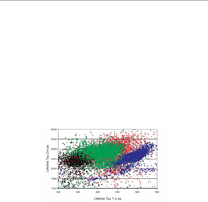
patients with age-related macular degeneration (AMD) [454]. Figure 5.70 shows a
scatter plot of the number of pixels versus the lifetime components,
W
1
and
W
2
, for
patients of different age, and a patient with dry AMD.
Fig. 5.70 Scatter plot of the pixels in a
W
1
versus
W
2
plane. Young healthy volunteer (black),
middle-aged healthy volunteer (green), patient with dry AMD (blue), isolated human lipo-
fuscin (red). From [454], courtesy of D. Schweitzer, Friedrich Schiller University Jena
5.7 TCSPC Laser Scanning Microscopy
Since their widespread introduction in the early 90 s, confocal [363], and two-
photon laser scanning microscopes [132, 343] have initiated a breakthrough in
biomedical imaging [316, 399, 543]. The high image quality obtained in these
instruments results mainly from the fact that out-of-focus light is strongly sup-
pressed or, in the case of two-photon excitation, not excited at all. As a result,
images of high contrast are obtained, and 3D imaging becomes feasible. More-
over, the scanning technique makes it relatively easy to perform detection in sev-
eral wavelength channels and multispectral detection [138]. In recent years more
features have been added, including excitation wavelength scanning, polarisation
imaging, and second-harmonic imaging. These multidimensional features make

130 5 Application of Modern TCSPC Techniques
laser scanning microscopes an almost ideal choice for steady-state fluorescence
imaging of biological samples [229, 278, 399, 401].
However, the fluorescence of organic molecules is not only characterised by the
intensity and the emission spectrum, it also has a characteristic lifetime. The life-
time can be used as an additional parameter to separate the emission of different
fluorophores, to probe ion concentrations and binding states in cells, and to investi-
gate interactions between proteins by fluorescence resonance energy transfer.
The application of the lifetime as a separation parameter is particularly useful
to distinguish the autofluorescence components in tissues. These components
often have poorly defined fluorescence spectra but can be distinguished by their
fluorescence lifetime [282, 339, 517]. FLIM has also been used to verify the laser-
based transfection of cells with GFP [501].
Furthermore, the fluorescence lifetime is a direct indicator of the quenching
rate due to interaction of the excited molecules with their local environment [308];
see Sect. 5.1.1, page 61. Unlike the fluorescence intensity, the fluorescence life-
time does not depend on the concentration of the fluorophore. It can therefore be
used to probe cell parameters such as ion concentrations or oxygen saturation [9,
17, 153, 185]. Fluorophores may also exist in a protonated and a deprotonated
form; the equilibrium between them is pH-dependent. If the protonated and the
deprotonated fluorophore have different lifetimes, the average lifetime is an indi-
cator of the local pH [9, 216, 307, 330, 439]. Moreover, the lifetime of many
fluorophores varies with whether they are bound to proteins, lipids, or DNA [271,
306, 398, 519]. There are a large number of other fluorophores and labelling pro-
cedures [217, 220], most of which have not yet been investigated for target-
induced lifetime changes. Lifetime variations have also been used as an indicator
of local refraction index changes [511].
The distance between two different fluorophore molecules can be probed by
fluorescence resonance energy transfer (FRET) [308]. The energy transfer rate
from the donor to the acceptor depends on the sixth power of the distance. FRET
becomes noticeable at distances on the order of a few nm and therefore occurs
only if the donor and acceptor are physically linked. With FLIM techniques,
FRET results are obtained from a single lifetime image of the donor [15, 32, 38,
61, 62, 63, 73, 80, 93, 147, 209, 405, 508].
The fluorescence lifetimes of typical fluorophores used in cell imaging are of
the order of a few ns. However, the lifetime of autofluorescence components and
of the quenched donor fraction in FRET experiments can be as short as 100 ps. In
cells, lifetimes of dye aggregates as short as 50 ps have been found [261]. The
lifetime of fluorophores connected to metallic nanoparticles [182, 183, 309, 337]
can be 100 ps and shorter.
The local environment, the binding or aggregation state, the quenching rate,
and the FRET efficiency of the fluorophore molecules in cells are normally inho-
mogeneous. Moreover, different fluorophores may overlap within the same pixel.
Therefore, the fluorescence decay functions found in cells are usually multiexpo-
nential. A FLIM technique should not only resolve lifetimes down to 50 ps, it
should also be able to resolve multiexponential decay functions.
Rough single-exponential lifetimes can be derived from data containing a few
hundred photons per pixel [187, 274]. This is not more than required for a medio-
5.7 TCSPC Laser Scanning Microscopy 131
cre steady state image. However, the resolution of several decay components re-
quires much more photons. Obtaining a large number of photons from the sample
means either long exposure or high excitation power. Therefore photobleaching
[140, 396] and photodamage [239, 277, 280] become a problem in precision FLIM
experiments. Consequently, recording efficiency is another key parameter of a
FLIM technique.
5.7.1 The Laser Scanning Microscope
The term „laser scanning microscope“ is used for a number of very different in-
struments. Scanning can be accomplished by galvano-driven mirrors in the beam
path, by piezo-driven mirrors, by a Nipkow disc, or by a piezo-driven sample
stage. This section refers to microscopes with fast beam scanning by galvano-
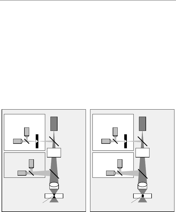
driven mirrors. Greatly simplified, the optical principle of these microscopes, is
shown in Fig. 5.71.
Laser
Dichroic
Detectors
Pinhole
Sample
Objective
Lens
Excited
Mirror
Scanner
Volume
Detectors
Non-Descanned
Detection
Descanned
Detection
Confocal
NUV / Visible
Dichroic
Mirror
Dichroic
Detectors
Pinhole
Sample
Objective
Lens
Mirror
Scanner
Detectors
Non-Descanned
Detection
Descanned
Detection
Confocal
Dichroic
Mirror
Excited
Laser
fs Pulses
Volume
NIR
Fig. 5.71 Optical principle of a laser scanning microscope. Left: One-photon excitation.
Right: Two-photon excitation
Laser-scanning microscopes can be classified by the way they excite and detect
fluorescence in the sample. One-photon microscopes use a NUV or visible CW
laser to excite the sample. Two-photon, or „Multiphoton“, microscopes use a fem-
tosecond laser of high repetition rate. The fluorescence light can be detected by
feeding it back through the scanner and through a confocal pinhole. The principle
is termed „confocal“ or „descanned“ detection. A second detection method is to
divert the fluorescence directly behind the microscope objective. The principle is
termed „direct“ or „nondescanned“ detection.

132 5 Application of Modern TCSPC Techniques
One-photon Excitation
Figure 5.71, left, shows the principle of a laser scanning microscope with one-
photon excitation. The laser is fed into the optical path via a dichroic mirror. It
passes the optical scanner, and is focused into the sample by the microscope ob-
jective. The focused laser excites fluorescence inside a double cone throughout the
complete depth of the sample. The fluorescence light is collected by the objective
lens. Detection of the fluorescence light can be accomplished by either descanned
or nondescanned detection.
Descanned Detection
The fluorescence light is fed back through the scanner, so that the motion of the
beam is cancelled. The fluorescence light is separated from the excitation light by
the dichroic mirror. The now stationary beam of fluorescence is passed through a
pinhole in the conjugate focus of the objective lens. In the plane of the pinhole,
light from outside the focal plane in the sample is substantially defocused. Out-of-
focus light is therefore suppressed by the pinhole. The principle is called „confo-
cal“ detection, and the microscope the „confocal microscope“.
The light passing through the pinhole is often split into several wavelength in-
tervals and detected by several PMTs. X-Y imaging is performed by scanning the
laser beam; optical sectioning or „Z stack“ recording is done by moving the sam-
ple up and down. The optical sectioning capability and the high contrast due to
out-of-focus suppression make confocal laser scanning microscopes superior to
conventional wide-field microscopes.
Nondescanned Detection
The fluorescence light can also be separated from the excitation light by a dichroic
mirror directly above the microscope objective lens, and fed into a detector. The
principle is termed „nondescanned“ or „direct“ detection, because the light does
not go back through the scanner. Nondescanned detection is normally used only in
conjunction with two-photon excitation (see paragraph below). For one-photon
excitation, nondescanned detection does not yield any depth resolution or out-of-
focus suppression. The image is therefore the same as in a wide-field microscope.
Nevertheless, nondescanned detection is sometimes used because of its simplicity,
high efficiency, and easy combination with TCSPC lifetime imaging.
Two-Photon Excitation
With a Ti:Sapphire laser or another high-repetition rate femtosecond laser, the
sample can be excited by simultaneous multiphoton absorption [132, 164, 278,
282, 343, 471, 472]. For biological specimens, three-photon or higher-order exci-
tation is rarely used. Nevertheless, such microscopes are normally called
„Multiphoton“ microscopes.
Two-photon excitation was predicted by Maria Göppert-Mayer in 1931 [189] and
introduced into laser microscopy by W. Denk, J.H. Strickler, and W.W.W. Webb
in 1990 [132]. The wavelength of two-photon excitation is twice the absorption
wavelength of the molecules to be excited. Because two photons of the excitation

5.7 TCSPC Laser Scanning Microscopy 133
light must be absorbed simultaneously, the excitation efficiency increases with the
square of the excitation power density. The high power density in the focus of a
microscope objective of high numerical aperture and the short pulse width of a
titanium-sapphire laser make two-photon excitation remarkably efficient. Excita-
tion occurs essentially in the focus of the objective lens. Consequently, depth
resolution is an inherent feature of two-photon excitation, even if no pinhole is
used. Moreover, since the scattering and the absorption at the wavelength of the
two-photon excitation are small, the laser beam penetrates through relatively thick
tissue. The loss on the way through the tissue can easily be compensated for by
increasing the laser power. The increased power does not cause much photodam-
age because the power density outside the focus is small. However, as long as
enough ballistic (nonscattered) excitation photons arrive in the focus, the fluores-
cence is excited.
Nondescanned Detection
Nondescanned (or „direct“) detection solves a problem endemic to fluorescence
scattering in deep sample layers. Fluorescence photons have a shorter wavelength
than the excitation photons and experience stronger scattering. Photons from deep
sample layers therefore emerge from a relatively large area of the sample surface.
To make matters worse, the surface is out of the focus of the objective lens. There-
fore the fluorescence cannot be focused into a pinhole.
Nondescanned detection splits off the fluorescence light directly behind the mi-
croscope lens and directs it to a large-area detector. Consequently, acceptable light
collection efficiency is obtained even for deep layers of highly scattering samples.
Two-photon imaging with nondescanned detection can be used to image tissue
layers several 100 µm (in extreme cases 1 mm) deep [85, 278, 344, 462, 534].
The absence of a pinhole in a two-photon microscope with nondescanned de-
tection makes the optical path relatively easy to align. Two-photon microscopes
can be built by upgrading a one-photon system with a Ti:Sapphire laser or by
attaching the laser and an optical scanner to a conventional microscope [136, 137].
Descanned Detection
For thin samples, such as single cells, scattering is negligible. In these cases two-
photon excitation is also used in conjunction with descanned detection. The pin-
hole is usually opened wide and used principally to suppress daylight leaking into
the objective lens.
Commercial Laser Scanning Microscopes
Commercial laser scanning microscopes use the same microscope body and the
same scan optics for one-photon and two-photon excitation. Most two-photon mi-
croscopes have lasers for one-photon excitation as well. They can switch between
both modes, and between descanned and nondescanned detection. Moreover, in
both the descanned and the nondescanned detection path, the light is split spectrally
by additional dichroic mirrors or dispersion prisms and several detectors are used to
record images in selectable wavelength ranges. The dichroic mirrors and filters are
assembled on motor-driven wheels and are changed on command. The laser power