Becker W. Advanced Time-Correlated Single Photon Counting Techniques
Подождите немного. Документ загружается.

144 5 Application of Modern TCSPC Techniques
Fig. 5.80 Mouse kidney sample stained with Alexa Fluor 488 WGA, Alexa Fluor 568
phalloidin, and DAPI. Recorded with two detectors connected to one TCSPC channel. Left:
488 nm channel, right: 535 nm channel. Colour represents lifetime, blue to red = 750 to
2,250 ps. From [37]
Detection can be achieved in 16, possibly even 32, wavelength channels by
splitting the light by a polychromator and detecting the spectrum by a multianode
PMT. The problem for multispectral FLIM is to transfer the fluorescence light
with high efficiency into the polychromator input slit. The most efficient method
of spectral detection would be descanned detection, i.e. to integrate the polychro-
mator into the scanning head. The pinhole would be placed directly in the input
image plane of the polychromator. Non-descanned spectral detection is more dif-
ficult because the detection light beam is not stationary. A practicable way to
transfer the light into the polychromator would be to project an image of the back
aperture of the microscope objective on the input slit of the polychromator.
In practice, the only feasible solution is often to transfer the light to the poly-
chromator slit plane by an optical fibre. The slit is removed, and the numerical
aperture at the input of the fibre is reduced to match the numerical aperture of the
polychromator. Because only moderate wavelength resolution is required, a rela-
tively thick fibre (up to 1 mm) can be used. Therefore a reasonably high coupling
efficiency with a single fibre can be obtained, even for nondescanned detection
systems. The fibre should be not longer than 50 cm to avoid broadening of the IRF
by pulse dispersion.
Another possibility is to use a fibre bundle. The input side of the bundle is
made circular, the out side is flattened to match the polychromator slit. The large
area of the fibre bundle makes it relatively simple to collect the light from the
nondescanned detection path of a scanning microscope. However, the aperture of
the microscope objective lens must be correctly imaged onto the input of the bun-
dle. Otherwise the illuminated spot scans over the bundle and causes the fibre
structure to appear in the image.
Multispectral TCSPC FLIM by using a polychromator at the fibre output of a
Zeiss LSM 510 NLO was demonstrated in [35]. One-photon excitation with a
frequency-doubled Ti:Sapphire laser was used. The spectrum was detected by a
5.7 TCSPC Laser Scanning Microscopy 145
16-channel detector head containing an R5900L16 PMT and the routing electron-
ics. An SPC730 TCSPC module recorded the photons into 16 wavelength and 64
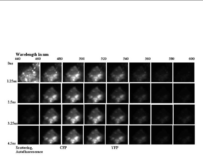
time channels. The image size was 64 u 64 pixels. Multispectral FLIM images of
an HEK (human embryonic kidney) cell transfected with CPF and YFP are shown
in Fig. 5.81.
Fig. 5.81 Multispectral FLIM images of an HEK cell transfected with CFP and YFP. Im-
ages in successive time and wavelength windows. Normalised for constant total intensity in
the time windows of the 500-to-520-nm channel
The 64 time channels were binned into four consecutive 1.25-ns intervals, the
16 wavelength channels into eight consecutive 20-nm intervals. The fluorescence
decay results in considerable intensity differences over the later time intervals. To
display the images in the time windows, the intensities were normalised. The same
normalisation factor was used for each row of images. The normalisation factor
was calculated so as to display equal total intensities of the images in the 500-to-
520-nm channel.
A similar system attached to a Zeiss LSM 410 microscope was used for track-
ing the metabolites of 5-ALA (5-aminolevulinic acid, an approved sensitiser for
photodynamic therapy) in living cells [438].
A two-photon microscope with multispectral FLIM and nondescanned detec-
tion is described in [60]. An image of the back aperture of the microscope lens is
projected into the input plane of a fibre. The fibre feeds the light into a polychro-
mator. The spectrum is detected by a PML16 multianode detector head, and the
time-resolved images of the 16 spectral channels are recorded in an SPC830
TCSPC module. Spectrally resolved lifetime images obtained by this instrument
are shown in Fig. 5.82.
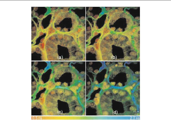
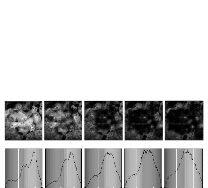
146 5 Application of Modern TCSPC Techniques
Fig. 5.82 Fluorescence lifetime images of a transverse section though the medulla of a
Cynomolgus monkey kidney acquired at the (a) 480-, (b) 510-, (c) 550-, and (d) 580-nm
wavelength components of the emission spectrum. The sample was stained with methyl
green and imaged by two-photon excitation at a wavelength of 920 nm. From [60], images
courtesy of Damian Bird, University of Wisconsin
5.7.5 High Count-Rate Systems
The count rates obtained for FRET and autofluorescence experiments in living
cells and tissue are rarely higher than a few 10
5
s
-1
(see Sect. Count Rate of FLIM
Experiments, page 159). These rates are well within the reach of a single TCSPC
module. Nevertheless, higher count rates may be obtained from fixed samples
stained with high concentrations of fluorophores of high quantum efficiency. To
exploit count rates in excess of 410
6
s
-1
, a multimodule TCSPC system can be
used. The light from the sample is split into several detection channels fed to sepa-
rate PMTs. Each PMT is connected to one channel of a multimodule TCSPC sys-
tem [39, 41]; see Fig. 5.83.
The TCSPC channels of this system have 100 ns dead time. The maximum use-
ful (recorded) count rate of each individual channel is 510
6
s
-1
. The system can be
used at a total recorded count rate up to 20
.
10
6
s
-1
, or a total detector count rate of
4010
6
s
-1
. A typical result is shown in Fig. 5.84.
5.7 TCSPC Laser Scanning Microscopy 147
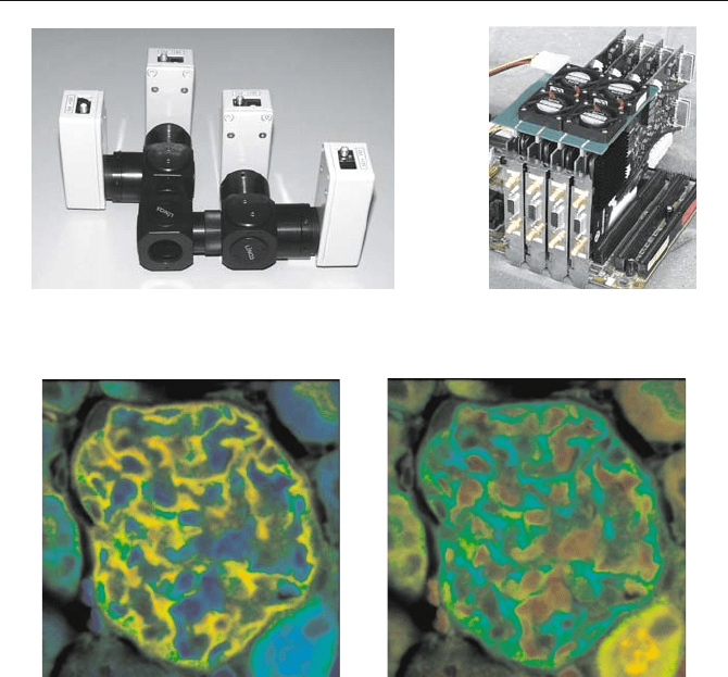
Fig. 5.83 Four-PMT detector assembly (left) and four-channel TCSPC imaging system
(right, on a Pentium motherboard)
Fig. 5.84 Mouse kidney sample stained with Alexa Fluor 488 wheat germ agglutinin, Alexa
Fluor 568 phalloidin, and DAPI, recorded by four detectors connected to separate TCSPC
channels. Left: Lifetime image; the colour represents the amplitude-weighted mean lifetime,
blue to red = 0.7 to 1.7 ns. Right: Colour represents the amplitude of the fast lifetime com-
ponent, blue to red = 0.1 to 0.9. From [39]
The sample was a mouse-kidney section (Molecular Probes, F24630) stained
with Alexa Fluor 488 WGA, Alexa Fluor 568 phalloidin, and DAPI. The excita-
tion wavelength was 860 nm. The laser power was about 400 mW at the input of
the microscope, and a 100 u NA=1.3 lens was used. Figure 5.84, left, shows a
lifetime image of the combined photon data of all four channels recorded within
ten seconds. A double-exponential Levenberg-Marquardt fit was applied to the
data. The colour of the image represents a single exponential approximation of the
lifetime obtained by weighting both lifetime components with their relative inten-
sities. Double-exponential lifetime analysis can by used to unmix fluorescence
components that appear in the same pixel. Figure 5.84 right, shows an image ob-
tained by using the amplitude of the fast lifetime component as colour. Images of
the amplitudes are related to the concentration ratio of the fluorophores emitting
the different lifetime components. They also separate the fractions of molecules of
148 5 Application of Modern TCSPC Techniques
a single fluorophore in different binding states, or the fractions of interacting and
noninteracting molecules in FRET experiments (see paragraph below).
Figure 5.84 shows that TCSPC can be used to obtain high-quality double expo-
nential lifetime images in 10 seconds or less. However, to obtain high count rates
a high excitation power has to be used, which can destroy the sample by photo-
damage or thermal effects. Figure 5.85 shows a sequence of recordings obtained
from the same specimen as shown in Fig. 5.84. The upper row shows a series of
intensity images, which were calculated from successive measurements by sum-
ming up fluorescence signals of the four detectors and of all time channels in each
pixel. The acquisition time of each image was 20 seconds. The initial total count
rate was 1410
6
s
-1
. The lower row shows the distribution of the mean lifetimes
over the image.
Fig. 5.85 Sequence of images recorded for 20 seconds each. The initial count rate was
1410
6
s
-1
. Upper row: Intensity images obtained from the recordings in all time channels.
Lower row: Distribution of the mean lifetime in the images (from 0.5 to 2.0 ns, normalised
on maximum). Photobleaching and thermal effects cause a progressive loss in intensity and
a variation in the lifetime distribution. From [39]
The sequence shows that the high excitation power causes photobleaching of
the sample. Since not all fluorophores are equally susceptible to photobleaching,
the distribution of the mean lifetime changes during the exposure. Usually the
longer lifetimes bleach more rapidly, so that the mean lifetime becomes shorter.
The results show that photobleaching at high excitation power can bias lifetime
measurements considerably.
A potential application of multimodule systems is high-speed two-photon mul-
tibeam scanning systems [53, 77]. FLIM systems with 4, 8 or even 16 beams and
the same number of parallel TCSPC channels appear feasible. The problem is to
direct the fluorescence signals from the individual beams to separate PMTs or
separate channels of a multianode PMT. If this problem is solved, two-photon
lifetime images can be recorded with unprecedented speed and resolution.

5.7 TCSPC Laser Scanning Microscopy 149
5.7.6 FRET Measurements by TCSPC FLIM
FRET measurements are an established technique used to determine distances in
cells on the 1-nm scale. The general principle of FRET [169, 230, 308] is shown
in Fig. 5.4, page 64. The fluorescence emission band of a donor molecule overlaps
the absorption band of an acceptor molecule. If both molecules are in close inter-
action, a radiationless energy transfer from the donor to the acceptor occurs. The
efficiency of the energy transfer increases with the 6th order of the reciprocal
distance.
The obvious difficulty of FRET measurements in cells is that the concentrations
of the donor and acceptor molecules are variable and unknown. Moreover, the
emission band of the donor extends into the emission band of the acceptor, and the
absorption band of the acceptor extends into the absorption band of the donor. A
number of different FRET techniques address these implications.
Steady-state FRET imaging uses the ratio of the donor and acceptor fluores-
cence intensities as an indicator of FRET [403]. The problem of the ratio tech-
nique is that the concentrations of the donor and acceptor may vary independently,
resulting in unpredictable errors.
The influence of the concentration can be largely avoided by calibrating the
crosstalk of the donor fluorescence in the acceptor detection channel and the
amount of directly excited acceptor fluorescence [159, 360, 402, 535]. The cali-
bration employs different cells, each containing only the acceptor and the donor,
and takes measurements at the donor and acceptor emission wavelength.
It is commonly accepted that the most reliable way to measure FRET in cells is
the acceptor-photobleaching technique. A donor image is recorded, then the ac-
ceptor is destroyed by photobleaching, and another donor image is recorded. The
FRET efficiency is obtained from the relative increase of the donor fluorescence
intensity [205]. The drawback of the technique is that it is destructive. It is there-
fore impossible to run successive FRET measurements in the same cell. It is also
difficult to use in living cells because the acceptor recovers after photobleaching
by diffusion effects.
FLIM-based FRET techniques avoid most of the problems of the steady-state
techniques. FLIM-FRET exploits the decrease in the donor lifetime with the effi-
ciency of the energy transfer. The lifetime does not depend on the concentration
and is therefore a direct indicator of FRET intensity. The FRET efficiency can, in
principle, be obtained from a single donor lifetime image. This is a considerable
advantage compared to steady-state techniques.
A general problem of FRET experiments in cells is that not all donor molecules
interact with an acceptor molecule. There are several reasons why a donor mole-
cule may not interact. The most obvious one is that the orientation of the dipoles
of the donor and acceptor molecules is random. The corresponding variation of the
interaction efficiency results an a distribution of the lifetimes. The effect on FRET
results is predictable and correctable [308].
A more severe problem is that an unknown fraction of donor molecules may
not be linked to an acceptor molecule. Some of the donor molecules may not be
linked to their targets, and not all of the targets may be labelled with an acceptor.
This can happen especially in specimens with conventional antibody labelling.
150 5 Application of Modern TCSPC Techniques
Surprisingly, the problem of incomplete labelling [305] is rarely mentioned in the
FRET literature and is normally not taken into account.
In cells expressing fusion proteins of the target proteins and variants of the
green fluorescent protein (GFP), however, the labelling can be expected to be
complete [513]. The components of the double exponential decay functions then
represent the fractions of interacting and noninteracting proteins. The resulting
donor decay functions can be approximated by a double exponential model, with a
slow lifetime component from noninteracting (unquenched) and a fast component
from the interacting (quenched) donor molecules. The effect on the donor decay
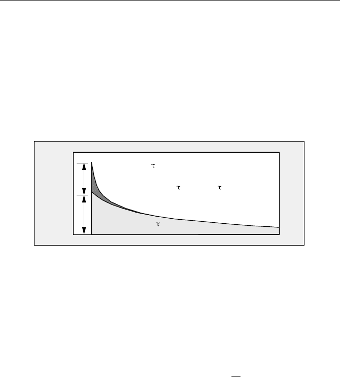
function is shown in Fig. 5.86.
Intensity
Time
unquenched
a
b
quenched
-t/
e
0
-t/
e
fret
f(t) = a
-t/
e
fret
-t/
e
0
+ b
Fig. 5.86 Composition of the donor-decay function
The decay analysis delivers the lifetimes of the interacting and noninteracting
donor molecules,
W
fret
and
W
0
, and the corresponding amplitudes, a and b. From
these parameters can be derived both the FRET efficiency,
E
fret
, the ratio of the
distance and the Förster radius,
r/r
0
, and the ratio of the number of interacting and
noninteracting donor molecules,
N
fret
/ N
0
:
0
/1
WW
fretfret
E
(5.13)
fretfret
rr
WWW
0
6
0
// or
1
1
/
6
0
fret
E
rr
(5.14)
baNN
fret
//
0
(5.15)
The predicted double exponential decay behaviour is indeed found in TCSPC
based [15, 32, 37, 38, 62, 80, 147, 405] and streak camera based FRET experi-
ments [61, 63]. The finding has implications for distance calculations based on
single-exponential FLIM-FRET [160, 230, 403, 520] and possibly even for
steady-state FRET techniques. Obviously, the distance between the donor and
acceptor molecules has to be calculated from
W
fret
, not from the average or „appar-
ent“ lifetime.
An application of TCSPC FLIM to CFP-YFP FRET is shown in Fig. 5.87 and
Fig. 5.88. The microscope was a Zeiss LSM 510 NLO two-photon laser scanning
microscope in the Axiovert 200 version. An excitation wavelength of 860 nm was
used. The nondescanned fluorescence signal from the sample was fed out of the
5.7 TCSPC Laser Scanning Microscopy 151
rear port of the Axiovert. A dual detector assembly with a dichroic beamsplitter
and two Hamamatsu R3809U MCPs was attached to this port. BG39 laser block-
ing filters and bandpass filters were inserted directly in front of the detectors. For
all measurements shown below, bandpass filters with 480 r 15 nm and
535 r 13 nm transmission wavelength were used. The filters were selected to
detect the fluorescence of the CFP and the YFP, respectively.
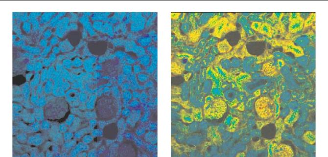
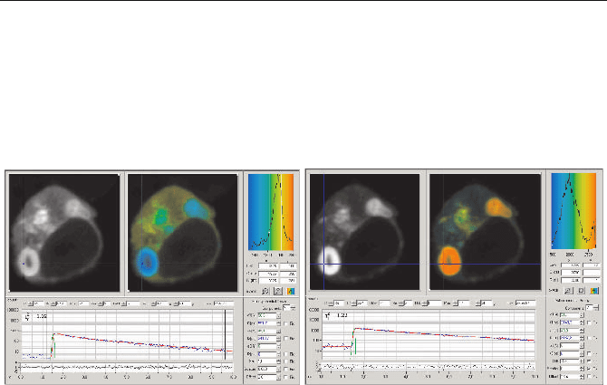
Fig. 5.87 HEK cell expressing two interacting proteins labelled with CFP and YFP. Single-
exponential lifetime images, lifetime distributions in a region of interest, and decay func-
tions in the selected spot. Left: CFP channel, blue to red corresponds to 1.5 to 2.2 ns. Right:
YFP channel, blue to red corresponds to 1.5 to 2.7 ns
Figure 5.87 shows FLIM results for a cultured HEK (human embryonic kidney)
cell expressing two interacting proteins labelled with CFP and YFP. FRET is to be
expected in the regions where the proteins are physically linked. The CFP (donor)
channel is shown in Fig. 5.87, left, the YFP (acceptor) channel in Fig. 5.87, right.
Both panels show an intensity image, a lifetime image with the average (ampli-
tude-weighted) lifetime,
W
m
, used as colour, the distribution of the lifetimes, and
the fluorescence decay functions in the selected spot.
The average donor lifetime varies from about 1.5 ns in the region of strong
FRET to about 1.9 ns in regions with weak FRET. The YFP intensity is highest in
the regions where the CFP lifetime is shortest. This is a strong indication that
FRET does indeed occur between CFP and YFP. The decay in the acceptor chan-
nel is a mixture of the FRET-excited acceptor fluorescence, a small amount of
directly excited acceptor fluorescence, and about 50% bleedthrough from the
donor fluorescence. Because YFP has a longer lifetime than CFP, regions of
strong FRET show an increased average lifetime.
The lower part of Fig. 5.87, left, shows the donor decay function in a selected
spot. The fluorescence decay is double-exponential, with a fast lifetime compo-
nent,
W
1
, of about 590 ps, and a slow component,
W
2
, of about 2.4 ns.
Figure 5.88 shows an image of the ratio of the amplitudes of the fast and slow
lifetime component,
a/b, and an image of the ratio of the lifetime components,
W
2
/
W
1
. As shown above in Fig. 5.86, a/b represents the ratio of the numbers of in-
teracting and noninteracting donor molecules,
N
fret
/ N
0
. The ratio of the lifetimes
is related to the FRET efficiency,
E
fret
.
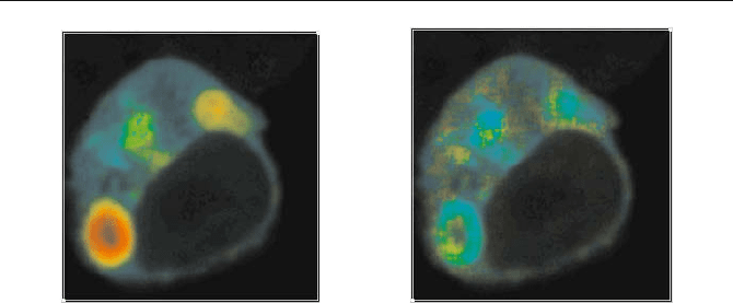
152 5 Application of Modern TCSPC Techniques
Fig. 5.88 Ratio of interacting and noninteracting donor molecules, N
fret
/ N
0
(left, blue to
red = 0.1 to 1.0), and ratio of the lifetime components,
W
2
/
W
1
(right, blue to red = 2.5 to 5).
The change in N
fret
/ N
0
is considerably larger than the change in E (please note the
different colour scales).
N
fret
/ N
0
varies from 0.38 to 1.0 in different regions of the
cell; the ratio of the lifetime components,
W
2
/
W
1
, varies only from 3.2 to 4. Conse-
quently, a large fraction of the variation of the average lifetime,
W
m
, (see Fig. 5.87)
results from changes in
N
fret
/ N
0
. The FRET efficiencies and distance ratios de-
rived from the double-exponential analysis and from the single-exponential (aver-
age) lifetimes in different regions of the cell are
E
fret
(r/r
0
)
6
Double-exponential,
W
fret
=
W
1
0.69 to 0.75 0.33 to 0.44
Single-exponential,
W
fret
=
W
m
0.28 to 0.43 1.3 to 2.53
The double-exponential analysis yields not only a substantially higher FRET effi-
ciency and a shorter distance, but also smaller variations in both values throughout
the cell. The change in
E
fret
corresponds to a distance variation of about 2%. A
variation this small may not be real but be introduced by crosstalk between the
lifetimes and the amplitudes of the two exponential components in the fitting rou-
tine. With CFP used as a donor, crosstalk may especially occur by the 1.3 ns-
decay component of CFP, see below. A double-exponential fit may partially
merge the 1.3-ns contribution into the fast decay component.
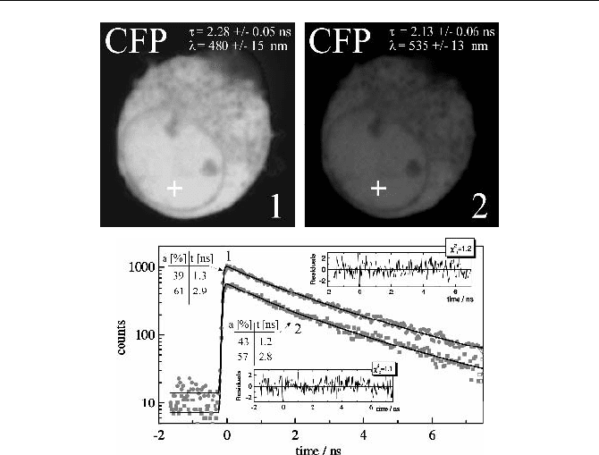
A remark appears indicated about the fluorescence of the CFP itself. Figu-
re 5.89 shows a cell transfected with CFP only. The two images were obtained in
the 480 nm channel and the 535 nm channel. Due to the long wavelength tail of
the CFP fluorescence spectrum [160] a considerable amount of CFP fluorescence
is detected in the 535 nm channel. A single-exponential fit over the whole images
delivers 2.28 ns and 2.13 ns, in close agreement with [400]. The decay functions
of a 3 u 3 pixel region around the indicated location are shown right. In agreement
with [508], the decay functions in both wavelength channels are double exponen-
tial. The lifetime components are 1.2 to 1.3 ns and 2.8 to 2.9 ns.
5.7 TCSPC Laser Scanning Microscopy 153
Fig. 5.89 HEK cell transfected with CFP only. Top left: 480 nm channel, top right: 535 nm
channel. The indicated lifetime forms a single-exponential fit over the whole cell. Bottom:
Decay curves in the selected spot of 3 u 3 pixels and double exponential lifetime compo-
nents
It might be expected that the 1.3 ns component has implications for FRET
measurements, and might even be confused with the lifetime of the quenched
donor fraction. The Levenberg-Marquardt fitting algorithm tends to merge closely
spaced lifetimes into a single one. If the FRET data are analysed with a double-
exponential model, the fit delivers a lifetime,
W
0
, of 2.3 to 2.6 ns for the un-
quenched donor fraction. This is close to the lifetime of the single exponential
approximation obtained for the cell containing only CFP (Fig. 5.89). It is likely
that
W
0
is actually a mixture of the 1.3-ns and 2.9-ns decay components found
there. Under these circumstances the double exponential model of FRET separates
the quenched and unquenched donor fractions correctly and the obtained
a/b and
r/r
0
can be considered to be correct.
Another solution is to fit the data by a triple-exponential model, with the life-
time components of the CFP used as a priori information. However, a triple-
exponential fit of the data recorded for the CFP-YFP cell with two slow decay
components fixed to 1.3 ns and 2.85 ns does not deliver significantly different
lifetimes and intensity coefficients for the short FRET lifetime component. The
F
2
of the fit is no better than for the double exponential fit. It is therefore not possible
to decide between the models within the photon statistics of the data.
It is an open question whether a lifetime image of the acceptor fluorescence can
be used to obtain additional information from a FRET experiment [242]. The