Becker W. Advanced Time-Correlated Single Photon Counting Techniques
Подождите немного. Документ загружается.


174 5 Application of Modern TCSPC Techniques
depends on the square of both the optical efficiency and the detector efficiency.
The design of the optical system and the selection of the detectors are therefore
crucial points of photon correlation experiments.
Detectors
Most photon correlation experiments use single photon avalanche photodiodes,
e.g. the SPCM-AQR detectors of Perkin Elmer [408]. These detectors have a
quantum efficiency that reaches 80% at 800 nm. However, single photon APDs
often experience timing shift and transit time jitter dependent on wavelength and
count rate. The changes can be of the order of 1 ns. Although the timing drift of
both detectors of a Hanbury-Brown-Twiss experiment may partially compensate,
it is difficult to obtain a time resolution below 0.5 ns or to investigate changes in
the correlation function versus intensity.
Timing problems can be avoided by using PMTs with high-efficiency GaAsP
photocathodes such as the Hamamatsu H742240 module [214]. The IRF of these
has a width of 200 to 350 ps. The IRF is almost independent of the wavelength
and remains stable up to count rates in the MHz range. The quantum efficiency
reaches 40% at 550 nm. Below 500 nm it is higher than for a single photon APD
(Fig. 6.17, page 231).
Recently new single-photon avalanche photodiodes have been introduced, see
Sect. 6.4.10 page 258. Compared with the SPCM-AQR the new devices have a
considerably improved timing behaviour but lower quantum efficiency in the NIR.
However, the efficiency below 600 nm is comparable or even better than for the
SPCM-AQR. It is likely though not proved that these detectors are superior to the
SPCM-AQR for correlation measurements in the visible spectral range.
Optical System
The collection efficiency of the optical system increases with the square of the
effective numerical aperture of the microscope objective lens. A good lens is
therefore essential in order to obtain a high coincidence rate. If the sample is
transparent the light can be collected from both sides, either by the condenser lens
of the microscope or by a second microscope lens. Theoretically, the collection
efficiency can be doubled and the coincidence rate increased by a factor of four.
Moreover, in a microscope with two aligned microscope lenses exciting and de-
tecting from both sides of the sample, the focal volume can be considerably de-
creased [64, 448]
Crosstalk
A troublesome effect in photon correlation experiments is light emission from
single photon APD detectors. When an avalanche is triggered in the APD, a small
amount of light is emitted. The effect and its implications for photon correlation
experiments and quantum key distribution are described in detail in [515] and
[299]. If the detectors are not carefully optically decoupled, false coincidence
peaks appear. An example is shown in Fig. 5.105.
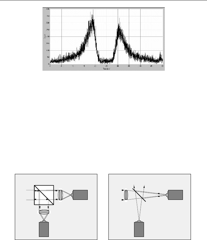
5.9 Picosecond Photon Correlation 175
Fig. 5.105 crosstalk between actively quenched APDs in a Hanbury-Brown-Twiss experi-
ment. Time scale 5 ns/div., optical configuration of Fig. 5.106, left
The probability of getting a crosstalk count in one detector is proportional to
the count rate in the other. Consequently, the total crosstalk count rate is propor-
tional to the sum of the count rates of both detectors, while the coincidence rate is
proportional to the product. Therefore, optical crosstalk becomes noticeable espe-
cially at low detector count rates.
The spectrum of the emission from the APDs extends from 600 to 1,000 nm
[299]. Therefore, decoupling often cannot be achieved by filters. The only way to
get reasonable results is with a carefully designed optical system. Features to be
strictly avoided are lenses focusing the emission of one diode into the other, and
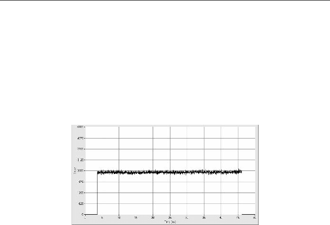
glass surfaces reflecting the emission into the other diode, as shown in Fig. 5.106.
Input
Detector 1
Detector 2
Beamsplitter Cube
Lens 2
Lens 1
Input
Detector 1
Detector 2
50% Mirror
Lens
SPAD SPAD
SPADSPAD
Fig. 5.106 Optical system with poor (left) and good (right) optical decoupling of the APDs.
The setup shown on the right does not focus light from one detector into the other
In Fig. 5.106, left, the light emitted by detector 1 is collimated by lens 1. 50%
of the light passes the beamsplitter cube, is partially reflected at the left plane of
the filter cube, reflected down to lens 2, and focused into detector 2. A better de-
coupling is achieved in Fig. 5.106, right. Light emitted by either detector is di-
rected out of the system or back to the light source. A small fraction of this light is
reflected at the surfaces of the lens, but the reflected light is strongly divergent and
not focused into the other detector. The decoupling can be further improved by
reducing the numerical aperture of the system, i.e. by reducing the diameter of the
lens or increasing its focal length. Unfortunately the lens in Fig. 5.106, right,
needs a relatively long focal length, which may result in a spot size larger than the
176 5 Application of Modern TCSPC Techniques
detector area. Therefore, the actual design may need to compromise between para-
sitic optical coupling of the detectors and counting efficiency.
A relevant question is whether PMTs show a similar emission effect as actively
quenched SPADs. In principle, a PMT may also emit light after detecting a photon,
e.g. by luminescence of the dynodes. However, a simple consideration shows that
light emission, if it exists, must be weak: A PMT works as a linear amplifier and does
not break down after detecting a photon. Therefore far less than one photon can be
emitted per detected photon. Indeed, no false correlation peak was found when two
H742240 PMTs were coupled directly cathode to cathode, see Fig. 5.107.
Fig. 5.107 Test of two H742240 PMT modules faced cathode to cathode. Time scale
5 ns/div. No light emission at a time scale of 50 ns is found
Consequently, problems with optical coupling are avoided if PMTs are used. The
optical system can be designed without compromising the efficiency, which may in
part compensate for the lower quantum efficiency of PMTs in the red and NIR range.
5.10 Fluorescence Correlation Spectroscopy
Fluorescence correlation spectroscopy (FCS) is based on exciting a small number
of molecules in a femtoliter volume and correlating the fluctuations of the fluores-
cence intensity. The fluctuations are caused by diffusion, rotation, intersystem
crossing, conformational changes, or other random effects. The technique dates
back to a work of Magde, Elson and Webb published in 1972 [335]. Theory and
applications of FCS are described in [51, 429, 430, 431, 456, 457, 497, 537, 556].
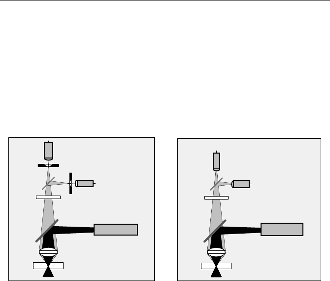
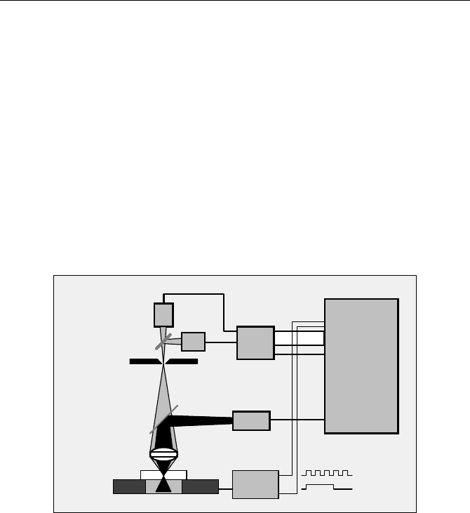
FCS measurements can be performed by one-photon excitation or by two-
photon excitation. An FCS system for one-photon excitation uses the confocal
detection principle, see Fig. 5.108, left. A continuous or high-repetition rate laser
beam is focused into the sample through a microscope objective lens. The fluores-
cence light from the sample is collected by the same lens, separated from the laser
by a dichroic mirror, and fed through a pinhole in the upper image plane of the
microscope lens. Fluorescence light from above or below the focal plane is not
focused into the pinhole and therefore is substantially suppressed. Only the fluo-
rescence light that passes the pinhole reaches the detectors. With a high-aperture
objective lens the effective sample volume is of the order of a femtoliter, with a
5.10 Fluorescence Correlation Spectroscopy 177
depth of about 1.5 µm and a width of about 400 nm. Typical FCS devices use one
detector or two detectors working in different wavelength intervals. More detec-
tors can be added to correlate photons in more than two different wavelength in-
tervals, or photons of different polarisation.
An FCS system with two-photon excitation is shown in Fig. 5.108, right [51,
457]. A femtosecond Ti:Sapphire laser of high repetition rate is used to excite the
sample. Because there is no appreciable excitation outside the focal plane of the
microscope lens a small sample volume is achieved without a confocal pinhole.
This makes the optical setup very simple. In terms of signal recording there is no
difference between one-photon and two-photon FCS.
Microscope Lens
NIR blocking filter
Dichroic
Laser
Sample
Visible / UV
Detector 1
Detector 2
Mirror
Dichroic
Mirror
Pinhole
Pinhole
CW or
high rep. rate pulsed
Microscope Lens
NIR blocking filter
Dichroic
Laser
Sample
Femtosecond
NIR
Detector 1
Detector 2
Mirror
Dichroic
Mirror
Fig. 5.108 One-photon FCS (left) and two-photon FCS (right)
Classic FCS correlators detect the individual photons of the fluorescence signal
and correlate the detection times [544]. If several detectors are used in different
wavelength intervals, it is possible to obtain the autocorrelation of the signals of
the individual detectors or the cross-correlation between the signals of different
detectors [455, 456]. The correlation function is often calculated directly in the
correlator hardware. However, storing the photon detection times and calculating
the correlation off-line offers more flexibility in the data processing [156]. Auto-
and cross-correlation functions can be calculated without the limitations [273] of
the typical correlator hardware, and within any time window of the total recorded
time interval. It is thus possible to exclude from the calculation artefacts caused by
fluorescence of impurities or glitches of the laser power. Moreover, photon-
counting histograms, BIFL results, and higher-order correlation can be calculated
from the same data (see Sect. 5.12, page 191).
5.10.1 Combined FCS/Lifetime Experiments by TCSPC
FCS and fluorescence lifetime experiments are often used in combination to ex-
plore the fluorescence dynamics of dye-protein complexes. The traditional ap-
proach is to acquire FSC and lifetime data in separate experiments [226, 458].
178 5 Application of Modern TCSPC Techniques
However, almost all advanced TCPCS devices are able to record lifetime data
and FCS data simultaneously [25, 65]. The advantage compared to the traditional
approach is that FCS and lifetime data originate from the same sample, from the
same spot of a sample, or even from the same molecules. TCSPC data can there-
fore be used to distinguish between different types of molecules, different quench-
ing states, or different binding or conformation states of dye-protein complexes; it
is also possible to include lifetime variations in the correlation [498, 548]. The
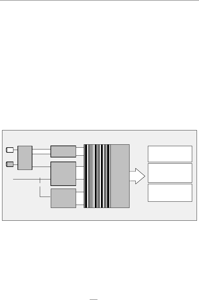
principle of TCSPC-based FCS is shown in Fig. 5.109.
The single-photon pulses of the detectors are fed into a router (see Sect. 3.1,
page 29). For each photon detected in any of the detectors, the router delivers a
single-photon pulse and the number of the detector that detected the photon. The
TCSPC module determines the time of the photon in the laser pulse sequence
(„micro time“) and the time from the start of the experiment („macro time“). The
detector number, the micro time, and the macro time are written into a first-in-
first-out (FIFO) buffer (see Sect. 3.6, page 43). The output of the FIFO is continu-
ously read by the computer, and the photon data are written in the main memory
of the computer or on the hard disc.
Start
Stop
from Laser
Micro Time
Channel
Detectors
Detector
Timing
Router
TCSPC Module
T
readout
to hard
disc
Histogram of t:
Fluorescence decay
Autocorrelation of T:
Fluorescence
Cross-Correlation of T:
Fluorescence
Cross Correlation
Correltation
t
Channel / Wavelength
Macro Time
n
FIFO
number
Buffer
Reference
Fig. 5.109 Combined FCS/Lifetime recording by TCSPC
By analysing the photon data fluorescence decay curves and FCS curves are
obtained. The decay curves of the individual detectors are obtained by building up
the histograms of the micro times. Fluorescence correlation curves of the individ-
ual detectors are obtained by correlating the macro times of the photons of these
detectors. Cross-correlation curves are obtained by correlating the macro time of
the photons of different detectors. The (unnormalised) autocorrelation function
G(
W
) of an analog signal I(t) is
dttItI
T
G
T
T
T
³
fo
)()(
2
1
lim)(
WW
(5.23)
5.10 Fluorescence Correlation Spectroscopy 179
For photon counts N in successive, discrete time channels
G(
W
) becomes
¦
)()()(
W
tNtNtG (5.24)
In practice, the count rate of FCS measurements is 10
3
to 10
5
photons per sec-
ond, and the photon times are measured with a resolution of the order of 10 to
100 ns. Interpreting such data as a continuous waveform and applying one of the
formulas above would result in exceedingly long calculation times. Surprisingly,
this obvious fact is only rarely mentioned in the FCS literature [156, 532].
In typical TCSPC time-tag data, the clock period of the macro time, T, is
shorter than the dead time of the TCSPC device. Therefore only one photon can be
recorded at a particular macro time. Consequently,
N(t) and N(t +
W
) can only be 0
or 1. The multiplication in the autocorrelation function becomes a simple compare
(or an exclusive-or) operation, and the integral of the autocorrelation becomes a
shift, compare, and histogramming procedure. The calculation of FCS from
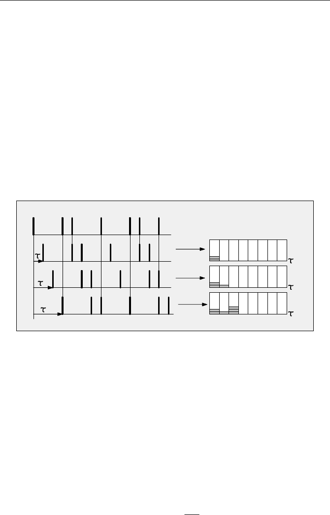
TCSPC data is illustrated in Fig. 5.110.
t
t
t
t
Number of coincidences
Photon times
2T
3T
T
G
Fig. 5.110 Calculation of the autocorrelation function from TCSPC time-tag data
The times of the individual photons are subsequently shifted by one macro time
clock period,
T, and compared with the original detection times. The coincidences
found between the shifted and the unshifted data are transferred into a histogram
of the number of coincidences,
G, versus the shift time,
W
. The obtained G(
W
) is the
autocorrelation function.
The cross-correlation function between two signals is obtained by a similar
procedure. However, the photon times of different signals are compared.
The result obtained by the shift-and-compare procedure is not normalised.
Normalisation can be interpreted as the ratio of the number of coincidences found
in the recorded signal to the number of coincidences expected for an uncorrelated
signal of the same count rate. The normalised autocorrelation and cross-
correlation functions are
2
)()(
P
T
n
N
n
GG
WW
(5.25)
180 5 Application of Modern TCSPC Techniques
with
n
T
= total number of macro time intervals, Np = total number of photons, and
21
)()(
PP
t
crossncross
NN
n
GG
WW
(5.26)
with
N
p1
= total number of photons in signal 1, N
p2
= total number of photons in
signal 2.
The described procedure yields
G(
W
) in equidistant
W
channels equal to the
macrotime clock period,
T. There is no distortion of the correlation function or of
the photon statistics by binning. This is an advantage if a model has to be fit to the
obtained function. The downside is that the number of
W
channels is extremely
high, especially if
G(
W
) is calculated up to large values of
W
, and that the noise in
the individual
W
channels does not decrease with increasing
W
. The curves therefore
have a different appearance than the results of the multi-W algorithm commonly
used in hardware correlators. One way to obtain a similar result as the multi-W
algorithm is to apply progressive binning to the calculated
G(
W
) data. Another way
is to periodically apply binning steps to the photon data during the
G(
W
) calcula-
tion [532].
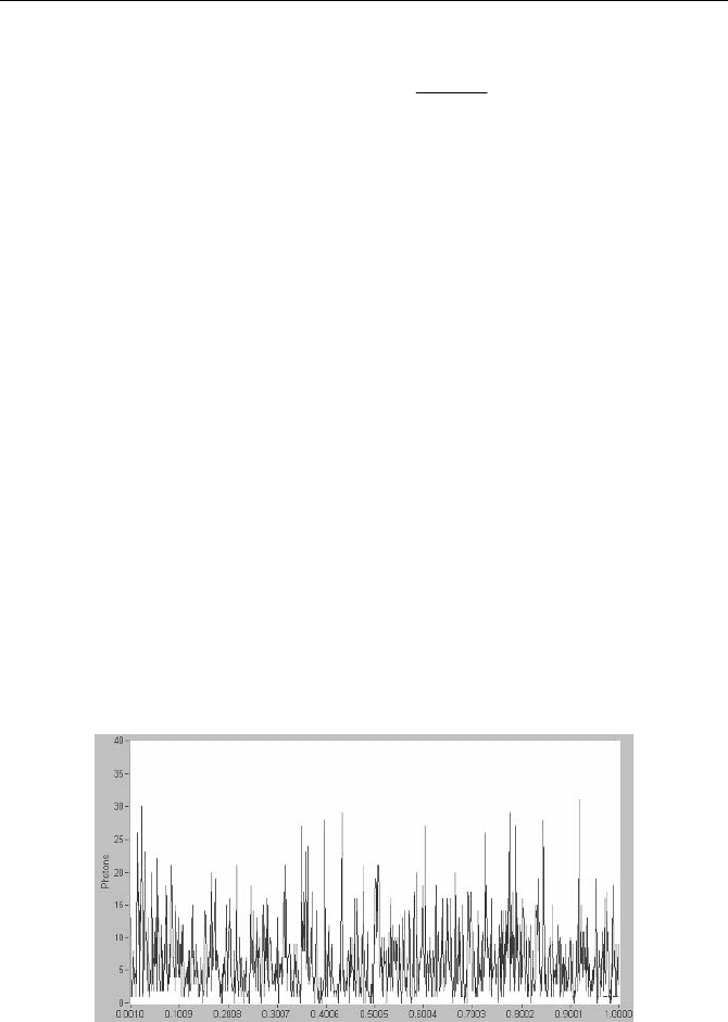
A typical result of TCSPC FCS is shown in Fig. 5.111 and Fig. 5.112. A GFP
solution was excited by a Coherent MIRA femtosecond Ti:Sapphire laser. The
detector was an SPCM-AQR module from Perkin Elmer, the TCSPC module an
SPC830 from Becker & Hickl. The count rate integrated in 1 s intervals fluctu-
ated between 5 and 7 kHz. The acquisition time was 980 seconds.
Figure 5.111 shows the fluctuations of the photon flux integrated in 1 ms inter-
vals over an interval of 1 second. Due to the diffusion of the GFP molecules, the
intensity fluctuations are clearly larger than the Poisson statistics of the average
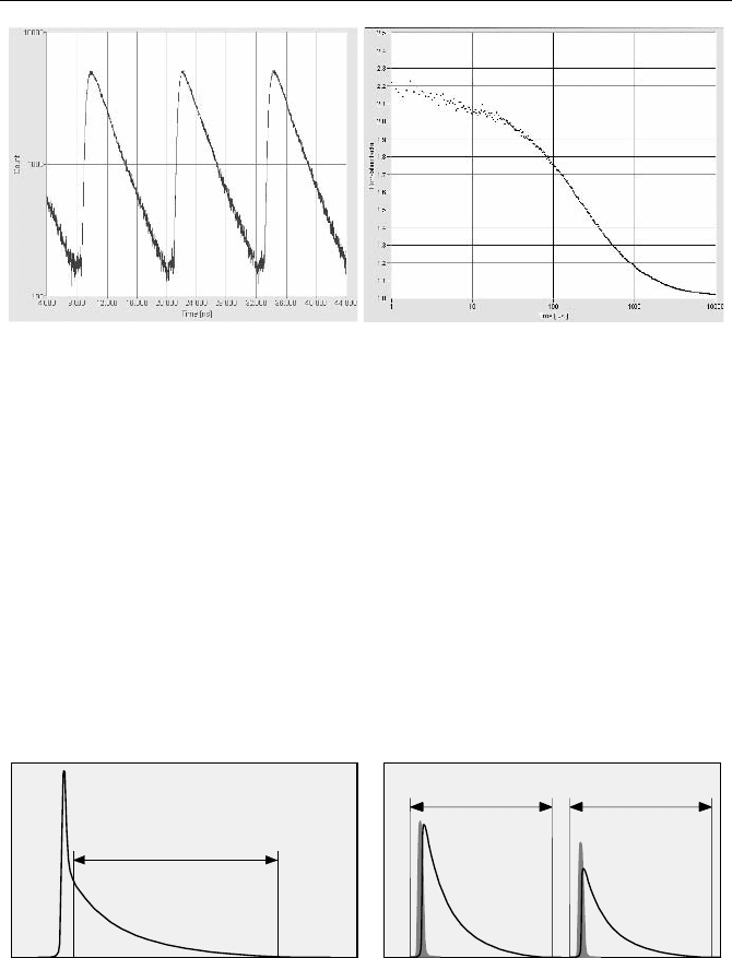
photon number. Figure 5.112 shows the fluorescence decay function over several
laser periods and the FCS function.
Fig. 5.111 GFP solution excited by Ti:Sapphire laser. Fluctuation of the photon counts in
1 ms intervals over 1 s of the data stream
5.10 Fluorescence Correlation Spectroscopy 181
Fig. 5.112 Fluorescence decay curve (left) and FCS curve (right) of a GFP solution. Count
rate 5 to 7 kHz , acquisition time 980 s. From [42], courtesy of Zdenek Petrasek and Petra
Schwille, Biotec TU Dresden
For fast TCSPC modules with large FIFO buffers, FCS and cross-FCS func-
tions can be calculated and displayed on-line during the measurement. Simultane-
ously, the complete FIFO data stream is written to the hard disc. This file can be
used for later off-line calculation. Off-line calculation has the benefit that correla-
tion functions in any time intervals within the total experiment time can be calcu-
lated and the binning of the data points can be selected. Time intervals with tem-
porarily increased photon flux, e.g. with fluorescence of impurities, can be
excluded from the calculation [156].
Gated FCS detection has been described in [310]. In a more general way, the
micro times of the photons can be used to suppress Raman light in one-photon
FCS, or to distinguish the fluorescence excited by two lasers of different wave-
length and interlaced pulses. The principle is shown in Fig. 5.113.
Laser 1
Laser 2
Time Window 1 Time Window 2
Fluorescence
Fluoresccene
Fluorescence
Raman Peak
Time Window
Fig. 5.113 Correlating TCSPC data within selectable time windows. Left: Suppression of
Raman light. Right: Separation of the fluorescence excited by two lasers of different wave-
length and interlaced pulse trains
Another, more challenging, application of micro times would be to include
them into the correlation to obtain information about the homogeneity of the life-
time. A simple, yet not very efficient way to use the lifetime information is to
correlate photons in different micro time windows. [44] uses a filter algorithm
which separates the FCS curves of molecules of different lifetime [65]. An ex-
182 5 Application of Modern TCSPC Techniques
periment that uses micro times to obtain correlation down to the picosecond scale
is described under Sect. 5.11.3, page 189.
5.10.2 FCS in Laser Scanning Microscopes
The optical configuration of an FCS instrument (Fig. 5.108, page 177) is almost
identical to the configuration of a laser scanning microscope (Fig. 5.71, page 131).
A laser scanning microscope with TCSPC-FLIM by multidimensional TCSPC
can, in principle, be used for FCS as well. The general requirements are a TCSPC
module that can be operated in the „Scan Sync In“ and in the FIFO mode, and a
microscope with a beam parking function. In practice the stability of the beam
position can be a problem. The mirror drivers and the driving electronics are de-
signed for maximum scanning speed, not necessarily for minimal beam jitter.
However, the slightest amount of beam jitter, even if not noticeable in normal
imaging operation, makes the recorded data useless for correlation. Therefore not
all laser scanning microscopes are suitable for FCS experiments [537]. Moreover,
the detectors in TCSPC FLIM microscopes are usually selected for high time
resolution, not for highest efficiency and low afterpulsing. FCS recording can
therefore require a relatively long acquisition time. A good compromise for com-
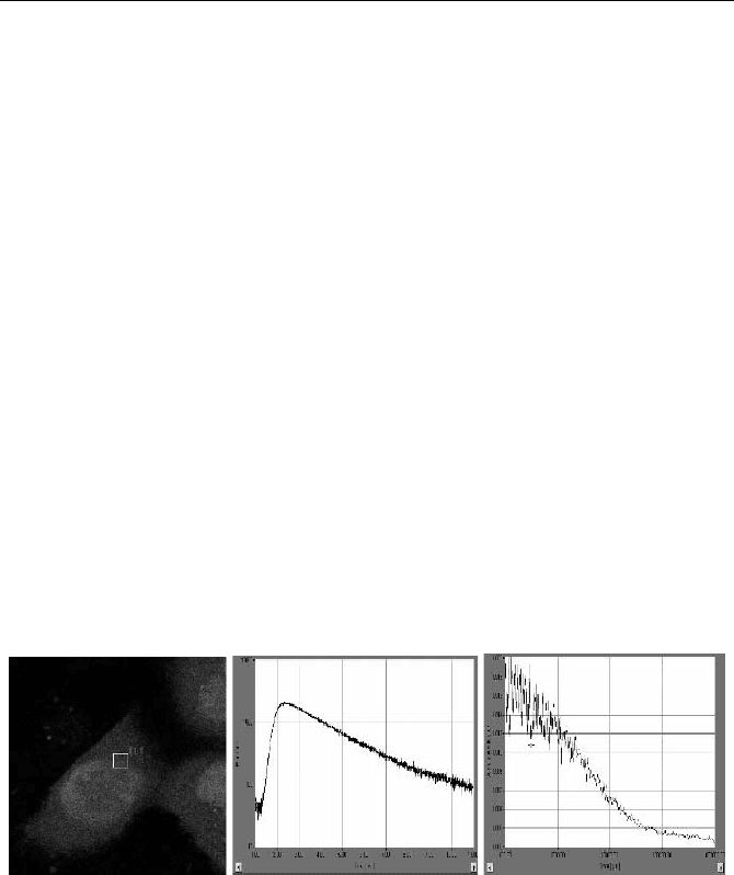
bined FLIM and FCS operation is the H742240 detector. Figure 5.114 shows an
FSC recording obtained in a laser scanning microscope [42]. The FLIM image is
shown at left. A fluorescence decay curve and an FCS curve in a selected spot are
shown in the middle and at right.
Fig. 5.114 FLIM image (left), decay curve (middle) and FCS curve (right) obtained in a
TCSPC laser scanning microscope. [42] and courtesy of Zdenek Petrasek and Petra
Schwille, Biotec TU Dresden
It should be noted that FCS measurements in cells are more difficult than in so-
lution. Especially in transfected cells the fluorophore concentration cannot be
accurately controlled. It is usually much higher than required for FCS. The num-
ber of molecules in the focus can easily be of the order of 100, resulting in an
extremely small amplitude of the correlation function. Moreover, there is usually
motion in living cells that shows up in the FCS curves at a time scale above
100 ms.
5.10 Fluorescence Correlation Spectroscopy 183
The potential for using different fluorescence techniques in a single instrument
[435] has not yet fully been explored. The TCSPC technique has the benefit that
high-quality lifetime images, FCS and FCCS data, lifetime variations, and photon
counting histograms (see page 191) can be recorded in a single instrument and
from the same sample [42].
A frequently asked question is whether or not FCS data recording and scanning
a sample can be combined. It is generally impossible to scan a sample at a scan
rate of the order of the photon times to be correlated. The frame rate must be faster
than the shortest correlation time, which is practically impossible. FCS is therefore
incompatible with the pixel dwell times and frame rates used in laser scanning
microscopes.
It has been shown that FCS with millisecond resolution can be obtained from a
circular line scan. FCS images of moderate pixel numbers can be obtained by
scanning a sample at a pixel dwell time much longer than the longest time to be
correlated. A possible solution is shown in Fig. 5.115.
Laser
Detector 1
Microscope Lens
Dichroic
Pinhole
Dichroic
Detector 2
HRT-41
HRT-81
HRT-82
SPC-630
SPC-830
SPC-134
Routing
CFD
Sync
FIFO Mode
Sample
Scan Stage
Scan
Controller
Pixel
Line
Routing
Fig. 5.115 Combination of FCS with scanning
A scan stage is used to scan the sample in one or two directions. The scan con-
troller delivers two control signals – a „pixel“ signal that changes its state at the
transition to the next pixel, and a „line“ signal that changes its state at the transi-
tion to the next line. These signals are fed into two of the unused routing input bits
of the TCSPC module. The scan rate is made slow enough to get a full FCS re-
cording in each pixel. This requires a time of several seconds. In this time a large
number of photons is recorded so that all transitions of the „pixel“ and „line“ sig-
nals appear in the data stream. Therefore, the scanning action can be tracked by
analysing the state of the pixel and line signals in the data file, and separate FCS
functions for the individual pixels can be calculated.
Theoretically, the same procedure can also be used in a commercial confocal or
two-photon laser scanning microscope if a sufficiently slow scan speed can be
selected. To distinguish the individual pixels of the scan, the short scan control