Becker W. Advanced Time-Correlated Single Photon Counting Techniques
Подождите немного. Документ загружается.

104 5 Application of Modern TCSPC Techniques
detection area and, therefore, with the number of detectors. Furthermore, addi-
tional detectors yield data under additional projection angles. Therefore it can be
advantageous to use more than the four detectors shown in Fig. 5.44. In that case,
several detectors are connected to one TCSPC channels via a router.
Due to the different signal recording techniques, instruments, and data analysis
techniques used by different workgroups, the results obtained for equivalent in
vivo measurements can differ considerably. The instruments are therefore tested
by comparing results obtained for „phantoms“, i.e. artificial samples with inclu-
sions of known scattering and absorption properties [130, 188, 200, 222, 413, 444,
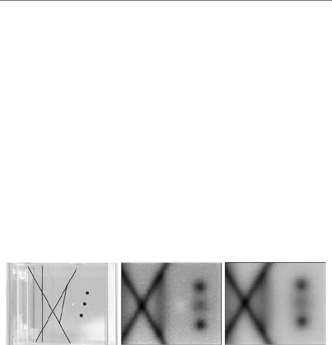
527]. A phantom consisting of a rectangular cuvette with several black wires of
1.7 mm diameter and one transparent and three black spheres of 8 mm diameter is
shown in Fig. 5.45, left [34]. The inner thickness of the cuvette is 6.8 cm. A mix-
ture of whole milk and water with the addition of a small amount of black ink is
used as the scattering liquid. At 670 nm the reduced scattering and absorption
coefficients are about 10 cm
-1
and 0.04 cm
-1
, respectively, and thus typical of the
optical properties of breast tissue. Figure 5.45, centre and right, shows images
obtained from a 65 by 53 pixel scan with a step size of 2.5 mm. Fig. 5.45, centre,
was calculated from an early time window that was adjusted to contain 10% of the
photons in an arbitrarily selected reference pixel. Figure 5.45, right, was calcu-
lated from all photons in the time-of-flight distributions. The acquisition time was
100 ms per pixel.
Fig. 5.45 Phantom (left) and images obtained from earliest 10% of the photons (centre) and
all photons (right) of a TCSPC scan. The glass sphere shows up dimly in the image of the
early photons. From [34]
Images in early time-windows are particularly sensitive to changes in the scat-
tering coefficient, whereas images in late time windows show mainly changes in
the absorption. Therefore the glass sphere shows up in the early time-window
(centre image), although this has a lower signal-to-noise ratio.
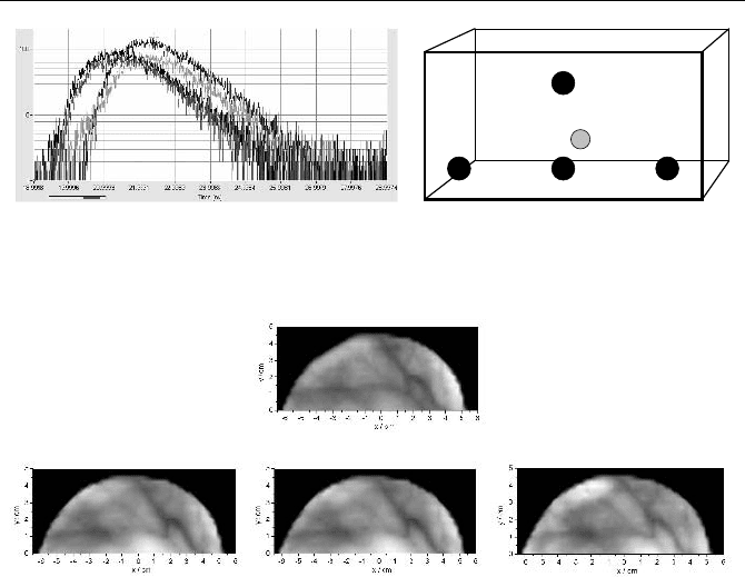
Figure 5.46, left, shows the time-of-flight distributions for four detectors detect-
ing at different projection angles at a single point of a breast scan. The acquisition
time was 100 ms. The source-detector geometry is shown at right. The time offsets
between the curves are due to different delays in the fibre bundles, detectors, and
TCSPC channels. Mammograms were calculated from the photons in the 8th of 10
equidistant time windows spread over the time-of-flight distribution. A result [34]
is shown in Fig. 5.47.
5.5 Diffuse Optical Tomography (DOT) and Photon Migration 105
D1D2 D3
D4
Source
Fig. 5.46 Left: Time-of-flight distributions in one pixel of a breast scan. Different projec-
tion angels, acquisition time 100 ms per pixel. Right: Detector and source configuration. D1
is the direct detector, D2, D3 and D4 are offset by 2 cm
Fig. 5.47 Mammograms of a healthy volunteer recorded simultaneously at four projection
angles. The images were generated from photon counts in a late time window. The ar-
rangement of the mammograms corresponds to that of the detectors D1-D4 (s. Figure 5.46).
From [34]
Images in early and late time windows show qualitatively the scattering and the
absorption in the tissue [118]. They can therefore be used to distinguish tumours
in breast tissue. Most (though not all) tumours have increased absorption due to
increased haemoglobin content and blood leakage. Most tumours are therefore
prominent in the late time window. An additional benefit of the late time window
is that the images are almost free of edge effects [201]. Cysts have usually de-
creased scattering and are visible in early time windows.
Quantitative data of the absorption and reduced scattering coefficients require
the application of an appropriate analytical model [96, 97, 524]. The modelled
distribution is convoluted with the IRF and fitted to the measured time-of-flight
distributions in the individual pixels at several wavelengths [201, 203, 477, 506].
A comparison of the accuracy of scattering and absorption coefficients obtained
by the time-window technique and by fitting a homogeneous diffusion model to
the data is given in [118]. The authors find that the diffusion model yields higher
accuracy for the scattering, whereas the late time window yields higher accuracy
for the absorption.
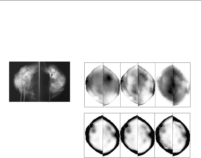
A typical mammography result is shown in Fig. 5.48. The figure shows X-ray
mammograms (left) and optical mammograms recorded at 685 nm, 785 nm, and
915 nm (right). The upper row of mammograms are absorption images, the lower
row scattering images. The absorption, µ
a
, is derived from the intensity in a late
D4
D1D2
D3
106 5 Application of Modern TCSPC Techniques
time window, the scattering, µ´
s
, from a diffusion model. Increased absorption is
shown dark, increased scattering bright. The tumor shows up by its increased
haemoglobin concentration, which causes increased absorption at 685 m and
785 nm. At 915 nm the image is dominated by the absorption of water, which is
slightly reduced at the tumor position. The cysts have decreased scattering at all
wavelengths and are therefore visible in the µ´
s
images.
Late
gate
P’
s
685 785 915
cysts
tumour
R L R L R L
R LR LR L
Fig. 5.48 Left: X-ray mammograms showing benign lesions (cysts) on the right breast and a
tumour on the left breast. Top right: Optical mammography images in a late time window
showing the absorption at 685 nm, 785 nm, and 915 nm. Bottom right: Optical mammogra-
phy images showing the scattering coefficient at 685 nm, 785 nm, and 915 nm. The tumor
is detected in the absorption images, the cysts in the scattering images. Images courtesy of
Alessandro Torricelli, Politecnico di Milano
5.5.3 Brain Imaging
The typical principle of a brain imager for newborn infants is shown in Fig. 5.49.
Brain imagers for adults can be considered a subset of the setup shown.
The instrument outlined in Fig. 5.49 uses four diode lasers of different wave-
lengths, which are electronically multiplexed. Typical wavelengths are 685 nm,
785 nm, 830 nm, and sometimes 760 nm. As in scanning mammography, the la-
sers can be multiplexed pulse by pulse, or in groups of 2,500 to 10,000 pulses, i.e.
in intervals of 50 to 200 µs per wavelength. Pulse group multiplexing gives less
pile-up errors and is generally to be preferred (see Sect. 5.5.8, page 117).
5.5 Diffuse Optical Tomography (DOT) and Photon Migration 107
32 Detector Positions
32 Detectors
32 Fibre Bundles
4 Eight-Channel Routers
Four TCSPC Channels
4 Lasers
1:32 Fibre Switch
32 Fibres
to
Source
Positions
Fig. 5.49 TCSPC tomography setup for brain imaging
Picosecond diode lasers are by far the most economic light sources for DOT.
Fibre lasers [223] and Ti:Sapphire lasers [222, 443] have been used as well. These
lasers have high power and short pulse width, but a high price. Wavelength multi-
plexing can be achieved only by synchronising several lasers and at a pulse-by-
pulse basis.
Multiplexed laser light is distributed to 32 source positions by a fibre switch.
The light from 32 detection positions is collected by 32 fibre bundles. A system
described in [443] uses four eight-channel MCP-PMTs and 32 individual NIM-
based TCSPC systems for detection. Due to its fully parallel architecture, the sys-
tem achieves exceptionally high data throughput, and low pile-up error. Of course,
an instrument like this is very large and transportation is a problem due to its sheer
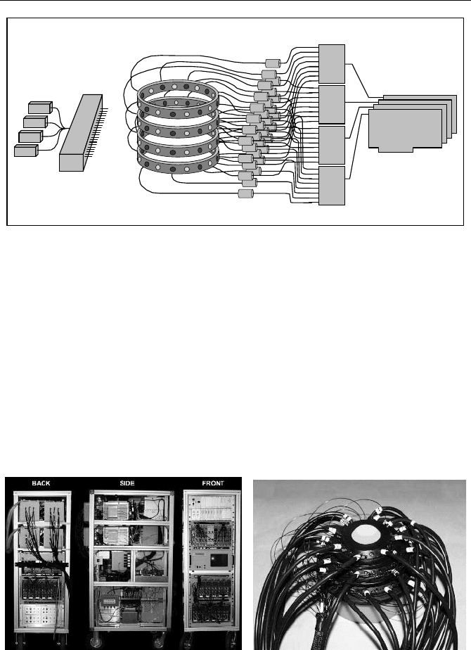
weight. Figure 5.50 gives an impression of the complexity of the system.
Fig. 5.50 Left: Photograph of the main rack of the parallel 32 channel instrument described
in [443]. Right: Optical interface to the patient, with the source and detection fibres, from
[222]
The main rack contains all components apart from the laser source and MCP-
PMT cooler unit. Most of the electronics and the control PC are housed on the
front of the 19-inch rack. Four variable optical attenuator boxes containing eight

108 5 Application of Modern TCSPC Techniques
units each and the fibre switch are located at the back, while the MCP-PMT cool-
ers can be seen in the top part of the side view.
An optical patient interface is shown right. The system can be used for brain
and breast imaging. Applications are described in [222, 223, 224, 225, 234, 444].
Advanced TCSPC techniques can reduce the size and the weight of the instru-
ment considerably. As shown in Fig. 5.49, the detector signals are divided into
four groups of eight signals and connected to four routers. The routers are con-
nected to individual channels of a four-channel TCSPC system. The TCSPC sys-
tem is then reduced to the size of an industrial or even a standard PC.
Static Brain Imaging
Structural tomography data are acquired by switching the multiplexed lasers con-
secutively through all source positions. For each source position time-of-flight
distributions are recorded for all laser wavelengths in all detector channels. Con-
sequently, there is enough time to read out the time-of-flight distributions for each
source position. The number of waveform memory blocks in the TCSPC modules
is the number of detector positions multiplied by the number of laser wavelengths.
Consequently, the TCSPC system in Fig. 5.49 needs a total of 128 waveform
memory blocks, or 32 blocks per TCSPC channel. This is no problem for any
modern TCSPC module.
The count rates in the individual detector channels may differ over a wide range
depending on the distance from the source. For channels directly adjacent to the
current source position, the rate can be as high as several MHz. The PMTs can
even be driven into overload. On the other hand, channels opposite to the source
position may not collect any reasonable number of photons at all. Except for PMT
overload and possible PMT damage, the huge dynamic range poses no major
problem in a fully parallel TCSPC system. An overloaded channel simply does not
record reasonable data. However, in a system with routers, the overloaded detector
may block the router to which it is connected and thus prevent other signals from
being recorded. Therefore, if the count rate of a detector becomes too high, either
this detector must be switched off or an automatic intensity regulator of some kind
must be placed in front of the detectors. As with scanning mammography,
information about overloads can be obtained by monitoring the detector output
current in the preamplifiers (see Sect. 7.2.15, page 300).
The wide dynamic range of the input signals should be taken into account for
the connecting scheme of the detectors to the routers. The total count rate proc-
essed per router and per TCSPC module should be distributed as uniformly as
possible.
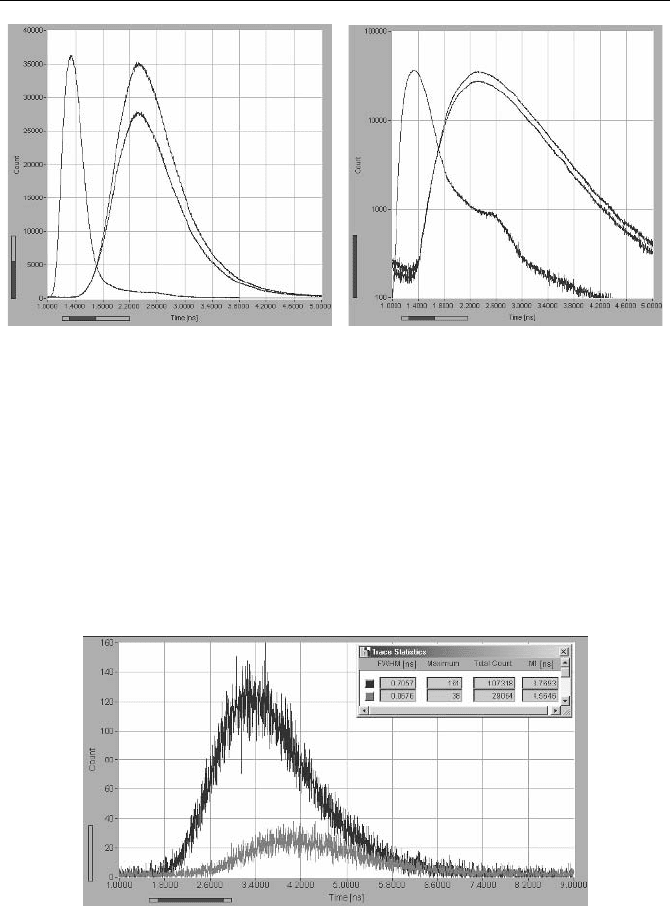
Typical time-of-flight distributions are shown in Fig. 5.51. The curves were re-
corded with a diode laser of 2.5 mW power, 785 nm wavelength, and 50 MHz
repetition rate. The detector was an H577320. The curves show the time-of-flight
distribution for two different locations at the forehead, with the instrument re-
sponse function. The source-detector distance was 6 cm. The acquisition time was
20 s, the count rates were between 800 kHz and 1 MHz.
5.5 Diffuse Optical Tomography (DOT) and Photon Migration 109
Fig. 5.51 Time-of-flight curves for two different source and detector positions at the fore-
head, and instrument response functions. Left linear scale, right logarithmic scale. Source-
detector distance 6 cm, Laser 2.5 mW, 785 nm, 50 MHz. Acquisition time 20 s, ADC reso-
lution 4,096 channels, time scale 400 ps/division
The obtained count rates drop dramatically with the source-detector distance.
With a laser power of a few mW, meaningful signals can be recorded diametri-
cally through an infant head. For an adult head this is impossible. However, weak
signals can be detected from temple to temple, as shown in Fig. 5.52. With a
2.5 mW, 785 nm laser and a H577320 detector a total number of photons of
29,000 and 107,000 was acquired within an acquisition time of 60 s. The count
rates were 896 s
-1
and 2,688 s
-1
, respectively.
Fig. 5.52 Time-of flight curves detected form temple to temple through an adult head.
Slightly different source and detector positions. Laser power 2.5 mW, wavelength 785 nm,
H577320 detector, acquisition time 60 s ADC resolution 4,096 channels, time scale
800 ps/division

110 5 Application of Modern TCSPC Techniques
Dynamic Brain Imaging
Dynamic changes in the time-of-flight distributions are caused by the heart beat,
variable oxy- and deoxyhemoglobin concentration induced by brain activity, and
effects of associated physiological regulation. The haemodynamic response to
brain stimulation is on the time scale of a few seconds [150, 171, 172, 502, 503].
Another, much faster signal has a typical rise time of 100 ms and is termed „event
related optical signal“, or EROS. The fast signal was found by [173, 194, 197,
198, 199, 480, 545]. All these experiments have been performed with CW or fre-
quency-domain instruments.
Recently Liebert et al. have demonstrated that advanced TCSPC is able to re-
cord effects of brain activity with 50 ms time resolution, clear separation of scat-
tering and absorption, and probably better depth resolution than CW or frequency-
domain techniques [324, 327, 328]. A system of four parallel TCSPC modules
with four individual detectors and several multiplexed laser diode lasers is used. A
fast sequence of time-of-flight distributions is recorded in consecutive time inter-
vals of 50 to 100 ms. Variations of the optical properties in the brain are derived
from the intensity and the first and second moments of the time-of-flight distribu-
tions [325].
Quaresima et al. used a single TCSPC channel and a multianode PMT to record
sequences of time-of-flight curves in eight parallel channels [421]. The acquisition
time per step of the sequence was 166 ms. The data of five steps were averaged.
Values of µ’
s
and µ
a
were calculated from the averaged data by using a standard
model of diffusion theory.
In principle, the recorded sequence could be triggered with the stimulation
event and accumulated. However, in practice there are is a strong variation in the
data due to heart beat [197, 502] and respiration. The response of the brain to the
stimulation can more reliably be separated from other effects by recording the full
sequence over a large number of stimulation events. To record a virtually unlim-
ited sequence the TCSPC channels are operated in the „continuous flow“ mode
(see Fig. 3.9, page 36).
The setup shown in Fig. 5.49 can, in principle, be used to record fast changes in
the brain at 4 laser wavelengths and 32 detector positions. However, the limited
speed of the fibre switch normally allows one to record sequences only for one or
two source positions at a time. The result is a total number of 128 to 256 wave-
forms each 50 to 100 ms or 32 to 64 per TCSPC module. The corresponding read-
out rate in the memory swapping mode is well within the range of currently used
TCSPC modules. However, improved fibre switches may allow one to multiplex a
larger number of source positions at a rate of 100 s
-1
or faster. The data transfer
rate then exceeds 10 Mbyte/s, and precautions have to be taken to sustain this rate
over a longer time.
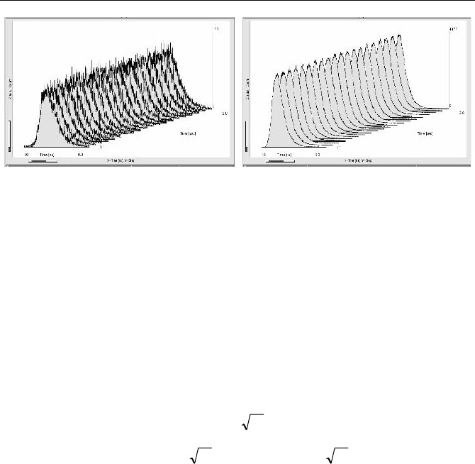
Figure 5.53 shows 20 time-of-flight curves selected from a continuous-flow se-
quence recorded with a single H577320 detector. The acquisition time was 100 ms
per curve, the ADC resolution 1,024 channels. The light source was a diode laser of
2.5 mW average power, 785 nm wavelength, and 50 MHz repetition rate. The left
sequence was detected with a source-detector distance of 8 cm, the right one with
a distance of 5 cm. The count rates where 1.810
5
s
-1
and 4.510
6
s
-1
, respectively.
5.5 Diffuse Optical Tomography (DOT) and Photon Migration 111
Fig. 5.53 20 steps of a TOF sequence recorded at an adult human head by TCSPC memory
swapping. Acquisition time 100 ms per curve, ADC resolution 1,024 channels. Diode laser
785 nm, 2.5 mW, detector H577320. Left: Source-detector distance 8 cm, count rate
1.810
5
s
-1
. Right: Source-detector distance 5 cm, count rate 4.510
6
s
-1
It should be noted that the a count rate of 4.510
6
s
-1
is close to the limit of a
single channel in currently available TCSPC devices. Intensity measurements at
rates this high require a correction for counting loss (see Sect. 7.9.2, page 338).
The moments of the time-of-flight distributions are not influenced by counting
loss. Certainly, there is a small pulse-shape error due to classic pile-up. However,
because only small changes in the moments are of interest, the pile-up-error is not
substantial.
The standard deviation of the total photon number and of the first moment can
be estimated as follows:
N
N
V
(5.10)
N
tofM
/
1
VV
or NT
fwhmM
/5.0
1
#
V
(5.11)
V
N
= standard deviation of the number of photons
V
M1
= standard deviation of the first moment
N = number of photons in a single TOF distribution
V
tof
= standard deviation of the times of flight of the photons
T
fwhm
= Full width at half maximum of the time-of-flight distributions
With
N = 10
5
photons per time-of-flight distribution, and a width of T
fwhm
= 2 ns the
standard deviation of the photon number is 316, or 0.316%. The standard devia-
tion of the first moment is 3.16 ps. Changes induced by variable oxy- and deoxy-
hemoglobin concentration are of about the same size. Recording these changes
requires accumulation of only some 10 stimulation periods. The accuracy required
to record the fast (EROS) signal is estimated to be 0.1% in the intensity and 220 fs
in the phase [173]. With TCSPC at a count rate of 2 MHz and 50 ms acquisition
time, standard deviations on these levels can be obtained by accumulating about
10 and 1,000 stimulation periods, respectively. The fast signal is therefore well
within the reach of TCSPC DOT.
112 5 Application of Modern TCSPC Techniques
5.5.4 Muscle and Bone Studies
Time-resolved DOT is sometimes used for optical biopsy of bone tissue, and to
track heamodynamics and oxygen kinetics in muscle tissue. The instruments typi-
cally use the same setup as brain imaging [443], see Fig. 5.49, page 107. An appli-
cation to DOT at the human forearm is described in [234]. The number of source
and detection channels can often be reduced from the number in the setup shown
in Fig. 5.49.
Tracking of the heamodynamics and oxygen kinetics in muscles requires sev-
eral multiplexed laser wavelengths and sequential recording in several detector
channels. Instruments described in [120, 123, 507] use two lasers multiplexed
pulse-by-pulse and eight detection channels routed into a single TCSPC channel.
An instrument described in [23] uses supercontinuum generation in a photonic
crystal fibre and multiwavelength TCSPC to obtain absorption and scattering
coefficients simultaneously at 16 wavelengths.
An instrument for optical biopsy of bones based on a diode laser and a single
TCSPC channel is described in [151, 152]. Other instruments use a tuneable syn-
chronously pumped dye laser and a Ti:Sapphire laser [414]. The lasers are
switched into a single source fibre by a fibre switch. A single TCSPC channel
records the diffusely reflected light and a reference signal split off from the source
fibre.
5.5.5 Exogenous Absorbers
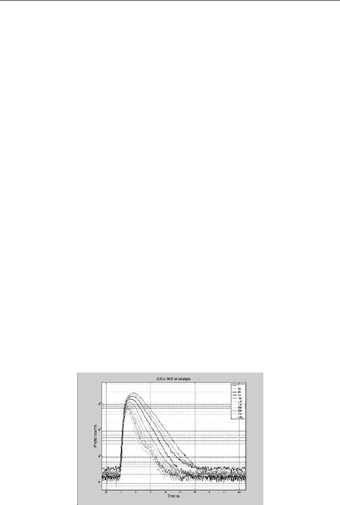
Exogenous chromphores can be used by detecting either their absorption or their
fluorescence. The only endogenous chromophore currently approved for use at
human patients is indocyanine green, ICG, a blood-pool agent [135, 369]. It
absorbs strongly between 650 and 850 nm and therefore clearly shows up in the
time-of-flight distributions of DOT. An example is given in Fig. 5.54.
Fig. 5.54 Time-of-flight distributions of a scattering solution for different ICG concentra-
tion

5.5 Diffuse Optical Tomography (DOT) and Photon Migration 113
ICG can be used to detect blood-flow dynamics. In the brain the absorption
change after an ICG bolus shows differences in superficial and deeper blood ves-
sels [327, 328, 329], and may be useful to indicate occlusion of vessels and areas
of increased stroke risk. ICG dynamics have also been used for breast tumor iden-
tification. Tumors usually have increased blood content, an increased number of
blood vessels, and increased leakage of dye from the vessels into the tissue. Even
with ICG injection, however, the contrast between tumour tissue and healthy tis-
sue remains low. Moreover, the dwell time of ICG in the tissue is only about 10
minutes so that short acquisition times are required.
5.5.6 Fluorescence
For a given number of recorded photons, fluorescence detection in general yields a
better intrinsic SNR than an absorption measurement. However, compared to the
diffusely transmitted or reflected intensity the fluorescence intensity is much
lower. The SNR actually obtained depends on the efficiency of the optics and the
detection system, the tissue thickness, the fluorophore concentration and quantum
yield, and the acceptable acquisition time.
Fluorescence applications in DOT are based either on the accumulation of a
fluorophore in the blood or on intensity and lifetime changes induced by the local
environment parameters or the binding state to proteins or lipids [342, 460]. Such
changes can be particularly strong if two dye molecules with different, but over-
lapping spectra are attached to the ends of a protein or lipid chain. Depending on
the folding state of the link between the fluorophores, the fluorescence is then
either unquenched or quenched by fluorescence resonance energy transfer
(FRET). Time-resolved detection has clear benefits in these applications.
Currently DOT fluorescence techniques are mainly used for small-animal imag-
ing. Fluorescence DOT as a diagnostic tool of human medicine is in an early
stage, mainly because only indocyanine green (ICG) is approved for application in
human patients [135, 369]. ICG in water has a fluorescence quantum yield of
about 4% [135]. The fluorescence lifetime of ICG bound to human serum albumin
(HSA) in water was determined to be double exponential, with contributions of
84% of 615 ps and 16% of 190 ps [182, 337]. The lifetime is clearly dependent on
the solvent. The results shown in Fig. 5.12, page 74, yield lifetimes of about
550 ps and 190 ps for ethanol and water, respectively. Lifetimes this short are
difficult to separate from the time-of-flight distribution in thick, inhomogeneous
tissue. It is therefore difficult to exploit possible lifetime changes of ICG for tissue
characterisation.
The typical shape of the fluorescence signals in scattering media is shown in
Fig. 5.55 and Fig. 5.56. The fluorescence of beads stained with IRD38 (Li-Cor,
Inc.) embedded in agarose phantoms was recorded by TCSPC. A femtosecond
titanium-sapphire laser was used for excitation, and a H742250 PMT module for
detection. The IRF width of the system is about 300 ps. Figure 5.55 shows the
variation in the recorded fluorescence decay data with the pH for beads at the
surface of the phantom.