Becker W. Advanced Time-Correlated Single Photon Counting Techniques
Подождите немного. Документ загружается.

84 5 Application of Modern TCSPC Techniques
A far better way to obtain time- and wavelength-resolved fluorescence data is
to record a sequence of fluorescence decay curves during the wavelength scan [28,
389]. Currently all advanced TCSPC devices have sequential recording modes
implemented so that there is actually no argument for using gated spectral re-
cording. The recording sequence in the TCSPC device can be controlled the same
way as for gated detection, i.e. either by software or by a hardware sequencer and
a trigger pulse from the monochromator drive. An example of a wavelength-
resolved sequence is shown in Fig. 5.21.
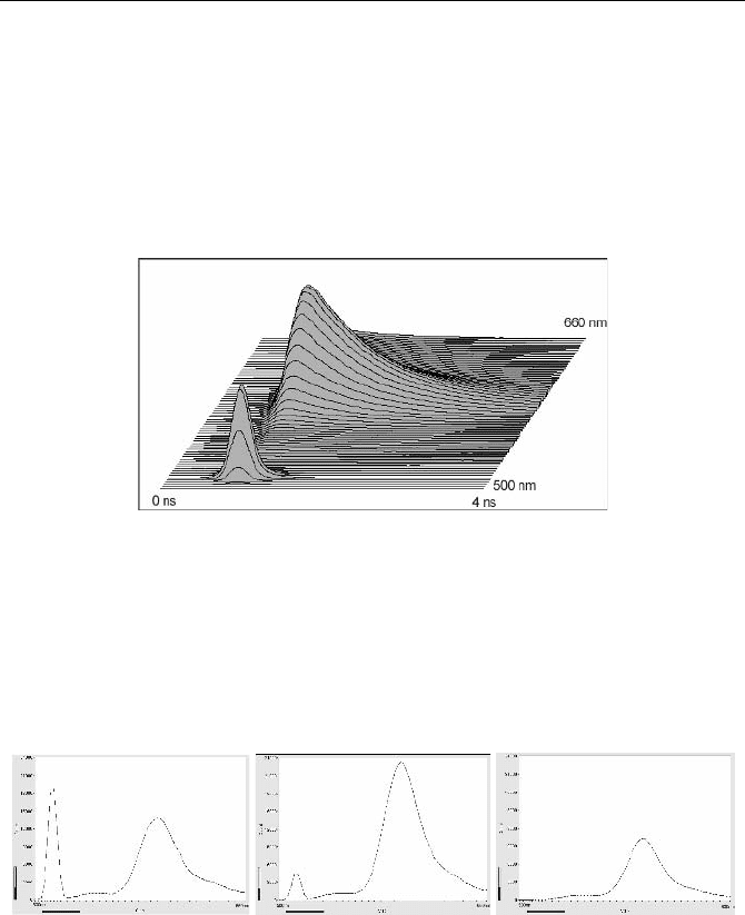
Fig. 5.21 Wavelength-resolved decay profile of DODCI (3,3´-diethyloxacarbocyanin-iodid)
in ethanol. Excitation by mode-locked argon laser at 514 nm. Scattered excitation light
forms a peak at the excitation wavelength
If time-resolved spectra are required, they can easily be calculated from the re-
corded sequence of decay curves. Three spectra obtained from the data of
Fig. 5.21 are shown in Fig. 5.22. Time-resolved spectra obtained from deconvo-
luted sequential decay measurements are presented in [285].
Fig. 5.22 Time resolved spectra calculated from the data of Fig. 5.21. Left to right: Maxi-
mum of excitation pulse, maximum of fluorescence, 1 ns after maximum of excitation pulse
5.2 Multispectral Fluorescence Lifetime Experiments
Biomedical applications of time-resolved fluorescence often preclude the scanning
of a spectrum by a monochromator. The total excitation dose may be limited by
photobleaching, or the investigated systems may show dynamic changes in their
fluorescence behaviour. For applications in human patients, the laser power is
5.2 Multispectral Fluorescence Lifetime Experiments 85
limited by laser safety regulations. To record a reasonable number of photons
within a reasonable acquisition time, it is essential to have high recording effi-
ciency. A spectrally resolved fluorescence-lifetime technique for biomedical ap-
plications should therefore record the decay curves in a large number of wave-
length intervals simultaneously.
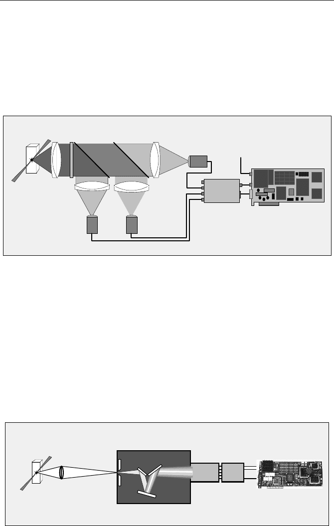
Multiwavelength operation of a TCSPC device can be achieved by using a sys-
tem of dichroic mirrors and a corresponding number of individual detectors (see
Sect. 3.1, page 29). The general optical setup is shown in Fig. 5.23.
Detector 3
Dichroic
Beam Splitters
Detector 1 Detector 2
Filter
Lens 1
Lens 4
Lens 2
Lens 3
Laser
Sample
TCSPC module
Router
from Laser
Fig. 5.23 Multiwavelength detection system with dichroic beamsplitters
The fluorescence light is collected and collimated by lens 1. A filter blocks the
scattered excitation light. The fluorescence light is split spectrally by several di-
chroic mirrors and focused onto several detectors via lens 2 through lens 4. The
numerical aperture of lens 1 can be made relatively high so that a high collection
efficiency is achieved. The setup is often used for time-resolved laser scanning
microscopy [37, 38] and single-molecule spectroscopy [419]. The drawback is that
the number of wavelength channels is very limited and the wavelength intervals
are fixed.
A more detailed fluorescence spectrum is obtained by using a polychromator
(or spectrograph) and recording the spectrum with a multianode PMT. The princi-
ple of a multiwavelength fluorescence experiment is shown in Fig. 5.24.
Polychromator
16 Ch. PMT
Routing
electronics
TCSPC Module
Transfer
Lens
Laser
Sample
Fig. 5.24 Multiwavelength fluorescence experiment
86 5 Application of Modern TCSPC Techniques
The sample is excited in the usual way by a high-repetition rate pulsed laser.
The fluorescence light from the sample is transferred by a lens to the input slit of
the polychromator. The polychromator splits the light spectrally and projects a
fluorescence spectrum on the cathode of a 16-channel or 32-channel multianode
PMT with routing electronics. For each photon, the routing electronics generate a
timing pulse and a digital data word that indicates in which channel the photon
was detected [35, 40]. These signals are used in the TCSPC module to build up the
photon distribution over time in the fluorescence decay and the wavelength (see
Sect. 3.1, page 29). Consequently, all photons detected by the PMT are used to
build up the result, and the maximum possible signal-to-noise ratio is obtained for
a given number of photons emitted by the sample.
The crucial parts of the system are the polychromator and the transfer optics.
Polychromators and monochromators are usually optimised for high spectral reso-
lution. This requires keeping the optical aberrations on the path through the poly-
chromator smaller than the slit width. The result is a relatively low f-number,
typically 1:3.5 to 1:8. The f-number limits the fraction of the fluorescence light
that can be transferred into the entrance slit (see Sect. 7.2.4, page 279). Moreover,
the efficiency of any grating is far less than 100%. Therefore some loss of photons
on the way from the sample to the detector in unavoidable. A multiwavelength
system based on a polychromator is less efficient than a system based on dichroic
beamsplitters, but by far more efficient than a system that scans the spectrum by a
monochromator.
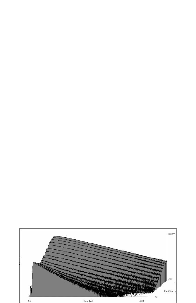
Figure 5.25 shows decay curves of a mixture of rhodamine 6G and fluorescein,
both at a concentration of 5
.
10
4
mol/l. The fluorescence was excited by a 405 nm
picosecond diode laser at a repetition rate of 20 MHz. The detector was a
R5900-L16 (Hamamatsu) in a PML-16 (Becker & Hickl) detector head. The de-
tector head contains the routing electronics, i.e. delivers the detector channel num-
ber and the timing pulse to the TCSPC module. The fluorescence signal was
spread spectrally by a polychromator (MS 1258M, Polytec) over the cathode area
of the R5900-L16. High concentration caused some reabsorption of the Rhodamin
6G fluorescence, resulting in a clearly visible change in the shape of the decay
curves and an increased lifetime.
Fig. 5.25 Fluorescence of a mixture of Rhodamin 6G and fluorescein, simultaneously re-
corded over time and wavelength

5.3 Excitation-Wavelength Multiplexing 87
Applications of multiwavelength TCSPC to laser scanning microscopy have
been demonstrated in [35, 60]. Spectrally resolved detection in diffuse optical
tomography is described in [23]. A multianode MCP PMT and an SPC330
TCSPC module were used to resolve the luminescence of alkali halides under N,
Ar, Kr, and Xe ion irradiation [266].
Another, yet more complicated way to record the spectrally split signal is posi-
tion-sensitive detection by a delay-line-anode PMT [510], or a resistive-anode
PMT [262].
It should be mentioned that multispectral detection can also be achieved by
placing a linear variable interference filter in front of the multianode PMT. How-
ever, the filter does not split the spectrum as a polychromator does. It rather blocks
the majority of the photons, transmitting only a small interval around a centre
wavelength that varies over its length. The efficiency is therefore low. Neverthe-
less, multiwavelength detection through a linear variable filter can be a solution to
TCSPC detection at low pulse repetition rates. Low-repetition-rate experiments
suffer from pile-up problems rather than from low efficiency. Because multidetec-
tor operation reduces pile-up distortions, count rates can be used that are higher
than those for a single detector.
5.3 Excitation-Wavelength Multiplexing
Biological samples contain a wide variety of endogenous fluorophores [282, 339,
432, 434, 452, 517, 555]. Moreover, a wide variety of exogenous [220] fluoropho-
res are available. The different fluorophores cannot always be excited at only one
wavelength [184, 517, 555]. In this case dual- or better multiwavelength excitation
yields additional information. In fact, the wonderful fluorescence images pre-
sented by the microscope companies are in a large part obtained by multiwave-
length excitation. Several wavelengths are also used to distinguish absorbers in
diffuse optical tomography (see Sect. 5.5, page 97).
Of course, measurements at different excitation wavelengths can by performed
consecutively, i.e. by recording fluorescence data for one wavelength, and then for
another. Excitation wavelength scanning is possible by using computer-controlled
tuneable Ti:Sapphire lasers, such as the „Mai Tai“ of Spectra Physics or the
„Chameleon“ of Coherent. However, for biological samples consecutive meas-
urements at different excitation wavelengths are not always feasible. Exposure to
the first wavelength may induce changes in the sample so that consecutive meas-
urements are not directly comparable, or transient effects may preclude consecu-
tive measurements altogether.
The lasers must then be multiplexed at a rate faster than the changes expected
in the sample. One way to multiplex lasers is to synchronise their pulse periods
and delay the pulses of different lasers by different fractions of the pulse period.
The fluorescence signals are recorded simultaneously in the same TAC range of a
TCSPC device. The principle is shown in Fig. 5.26.
88 5 Application of Modern TCSPC Techniques
Laser 1 Laser 2 Laser 1 Laser 2
Fluores-
Stop pulses to TCSPC
Fluorescence 1
Fluorescence 2
Recorded Signal:
cence 2
Fluores- Fluores- Fluores-
cence 1cence 1 cence 2
Fig. 5.26 Pulse-by-pulse multiplexing
Pulse-by-pulse multiplexing was used very early for dual wavelength TCSPC
measurements [540], and is still used in diffuse optical tomography [120, 124,
203, 412, 443] and single-molecule spectroscopy. The technique is useful for
combined fluorescence correlation and lifetime experiments because correlation
functions are obtained for times down to a few laser pulse periods [66]. It has,
however, the drawback that the effective stop rate of the TCSPC module is re-
duced in proportion to the number of laser wavelengths. The reduction of the stop
rate increases the pile-up errors, so that the maximum count rate is correspond-
ingly low. In particular, pile-up in one fluorescence signal reduces the amplitudes
of all successive ones. It is therefore often better to multiplex the lasers at a longer
time scale, and to use the multiplexing capability of TCSPC to get the correspond-
ing fluorescence signals separated. The principle is shown in Fig. 5.27.
Fluorescence 1 Fluorescence 2
Stop pulses
Laser 1
Laser 2
to TCSPC
Routing Signal = 1 Routing Signal = 2
Result
Fluorescence 2
Fluorescence 1
Memory 1
Memory 2
Fig. 5.27 Pulse group multiplexing. Please note the higher stop pulse rate compared to
Fig. 5.26
For a long time the most difficult problem of pulse-group multiplexing has
been the lasers. Conventional high-repetition rate lasers cannot be turned off and
on at sub-µs speed, and simple optical switches of the required speed and on-off
ratio do not exist. Picosecond diode lasers, however, can be switched on within
less than a microsecond and switched off within one or two pulse periods. Diode
lasers with multiplexing capability are currently available for 375 nm, 405 nm,
5.3 Excitation-Wavelength Multiplexing 89
440 nm, 473 nm, and for a large number of wavelengths from 635 nm to above
1,000 nm.
Due to the different excitation wavelengths blocking of the excitation light can
cause problems in multiwavelength excitation fluorescence experiments. The
usual long-pass filters cannot be used because the filter for the laser of long wave-
length would block most of the fluorescence excited by the laser of shorter wave-
length. Usually the problem can be solved by using bandpass filters in front of the
detectors. If the fluorescence excited by both lasers is to be detected in a single
detector, notch filters can be used to block the laser wavelengths.
Excitation-wavelength multiplexing in combination with dual-wavelength de-
tection is particularly useful to record the fluorescence of plants. Green leaves
show the typical chlorophyll fluorescence around 700 nm, and a blue-green fluo-
rescence which originates from flavinoids [232]. The problem is that the excitation
light induces changes in the fluorescence lifetime of chlorophyll. The effect is
termed „Kautski effect“ or „Chlorophyll Transients“ [192, 193, 345]. It is caused
by changes in the rate of photochemical and nonphotochemical quenching. The
typical time constants of the effects are about 1 ms and several seconds, respec-
tively (see Sect. 5.4.1, page 90). Multiplexing on the microsecond scale is required
to obtain constant nonphotochemical quenching for both excitation wavelengths.
Keeping the photochemical quenching constant requires multiplexing rates faster
than 100 µs.
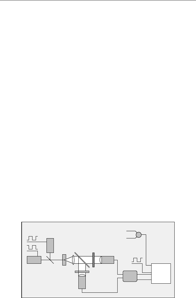
A suitable experiment setup is shown in Fig. 5.28. The sample is excited by two
lasers at 405 nm and at 650 nm. The lasers are multiplexed by a TTL signal from a
pulse generator. The light from the sample is split by a dichroic mirror into a
short-wavelength and a long-wavelength component. These components are re-
corded simultaneously by two PMTs detecting through different bandpass filters.
The PMTs are connected to the TCSPC module via a router. One routing bit is
required to separate the photons of both detectors. A second routing bit is used to
separate the photons excited by the two lasers. The stop signal for the TCSPC
module comes from the synchronisation outputs of the lasers. Because only one
laser is active at a time, the pulses can be combined by a simple power combiner.
Laser 1
Laser 2
Sample
Dichroic
mirror
Bandpass
Bandpass
filter 695 nm
filter 515 nm
405 nm
650 nm
Detector 1
Detector 2
Multiplexing
signal
TCSPC
Router
Multiplexing
signal
Power combiner
Synchronisation
from lasers
routing
start
stop
Fig. 5.28 Dual-wavelength excitation and detection experiment
90 5 Application of Modern TCSPC Techniques
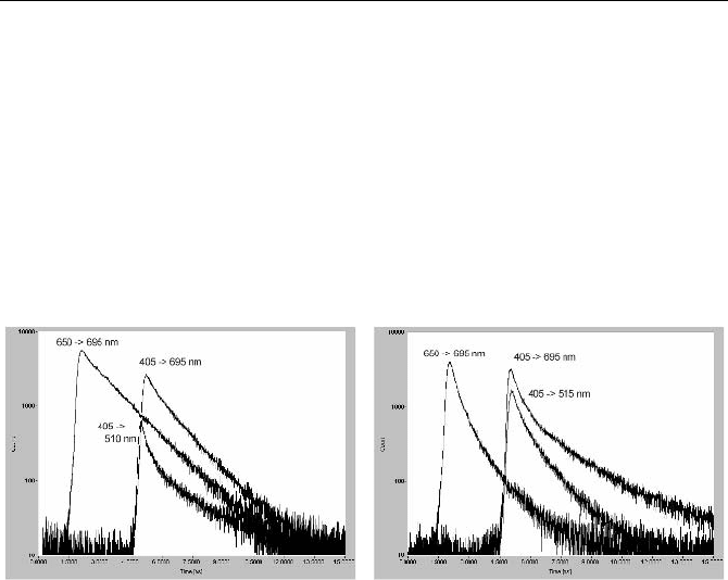
A result for a leaf from a rubber plant is shown in Fig. 5.29. Results for a fresh
leaf are shown left, results for a dry leaf right. The synchronisation signal of the
650 nm laser was delayed by 3 ns to make the curves better distinguishable. The
multiplexing period was 50 ms. At this rate the lifetime is modulated by photo-
chemical quenching, but not by nonphotochemical quenching. Therefore, different
lifetimes of the 695 nm emission are obtained for both wavelengths. No such ef-
fect is seen in the 695 nm emission of the dry leaf. The green emission at 515 nm
from the fresh leaf has a considerably lower intensity, a shorter lifetime, and a
multiexponential decay profile. This indicates that a strong, nonuniform quench-
ing process is at work. Both the intensity and the lifetime of the green emission
increase in the dry leaf.
Fig. 5.29 Dual-wavelength excitation and dual-wavelength detection of the fluorescence of
a fresh leaf (left) and a dry leaf (right). Multiplexed excitation at 405 nm and 650 nm, dual-
detector recording at 515 nm and 695 nm
5.4 Transient Fluorescence Lifetime Phenomena
Transient changes in the fluorescence lifetime can by driven by the excitation light
itself, by stimulating a system by an intense laser flash, by adding a chemical re-
agent, or by changes in the conformation of dye-protein or dye-DNA complexes
[353]. The time scale of the lifetime changes can extend from nanoseconds to hours.
Typical examples are excitation-driven fluorescence transients of chlorophyll in
living plants, photobleaching experiments, flash photolysis, continuous flow mixing
techniques, stopped flow experiments, and experiments for photodynamic therapy.
5.4.1 Chlorophyll Transients
When a dark-adapted leaf or a living plant cell is exposed to light, the intensity of
the chlorophyll fluorescence shows characteristic changes. These changes were
found by Kautsky and Hirsch in 1931 [259] and have been termed fluorescence
induction, fluorescence transients, or Kautsky effect [192, 193, 345]. The general
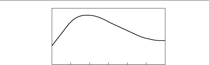
behaviour of the fluorescence intensity is shown in Fig. 5.30.
5.4 Transient Fluorescence Lifetime Phenomena 91
time (s)
-1-3 -2 0 1 2
10 10 10 1010 10
Intensity
a
b
Fig. 5.30 General behaviour of the chlorophyll a fluorescence after exposing a dark-adapted
leaf to light. The fluorescence intensity first increases due to a decrease of photochemical
quenching (a) and then decreases due to an increase of nonphotochemical quenching (b)
When the light is switched on, the fluorescence intensity starts to increase. Af-
ter a steep rise, the intensity falls again and finally reaches a steady-state level.
The rise time is of the order of a few milliseconds to one second; the fall time can
be from a few seconds to minutes. The initial rise of the fluorescence intensity is
attributed to the progressive closing of reaction centres in the photosynthesis
pathway. Therefore the quenching of the fluorescence by the photosynthesis de-
creases with the duration of illumination, and the fluorescence intensity increases
correspondingly. The quenching by the photosynthesis pathway is called „photo-
chemical quenching“.
The slow decrease of the fluorescence intensity at later times is termed „non-
photochemical quenching“. Nonphotochemical quenching seems to be essential in
protecting the plant from photodamage, or may even be a result of moderate
photodamage. The processes that lead to nonphotochemical quenching are often
referred to as „photoinhibition“.
A large number of experimental setups are used to measure the chlorophyll
transients [191, 275, 276]. Often a continuous light of variable intensity is applied
simultaneously with a weak modulated light of constant intensity. By detecting
only the fluorescence signal at the modulation frequency, the fluorescence effi-
ciency is recorded as a function of intensity and time. A second technique uses an
intense flash of light to close the reaction centres and records the fluorescence
intensity before and after the flash. In all these experiments, changes in the quan-
tum efficiency are hard to distinguish from changes in the number of fluorescing
molecules.
This difficulty is easily avoided by fluorescence lifetime detection. By using
the sequential recording capability of multidimensional TCSPC, the fluorescence
transients can be directly observed. A simple setup for recording the nonphoto-
chemical quenching is shown in Fig. 5.31.
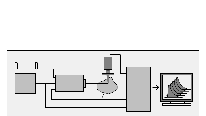
92 5 Application of Modern TCSPC Techniques
Start
f(t,T) mode
start
TCSPC
Module
stop
Experiment
Diode Laser Module
Sync Out
Power
Filter
Picosecond
635 or 650 nm
Detector
700nm
Pulse Period 20 to 50 ns
+12V
Trigger
Fig. 5.31 Recording the nonphotochemical quenching transient of chlorophyll a. The
TCSPC module records a single sequence of fluorescence decay curves starting with the
switch-on of the laser
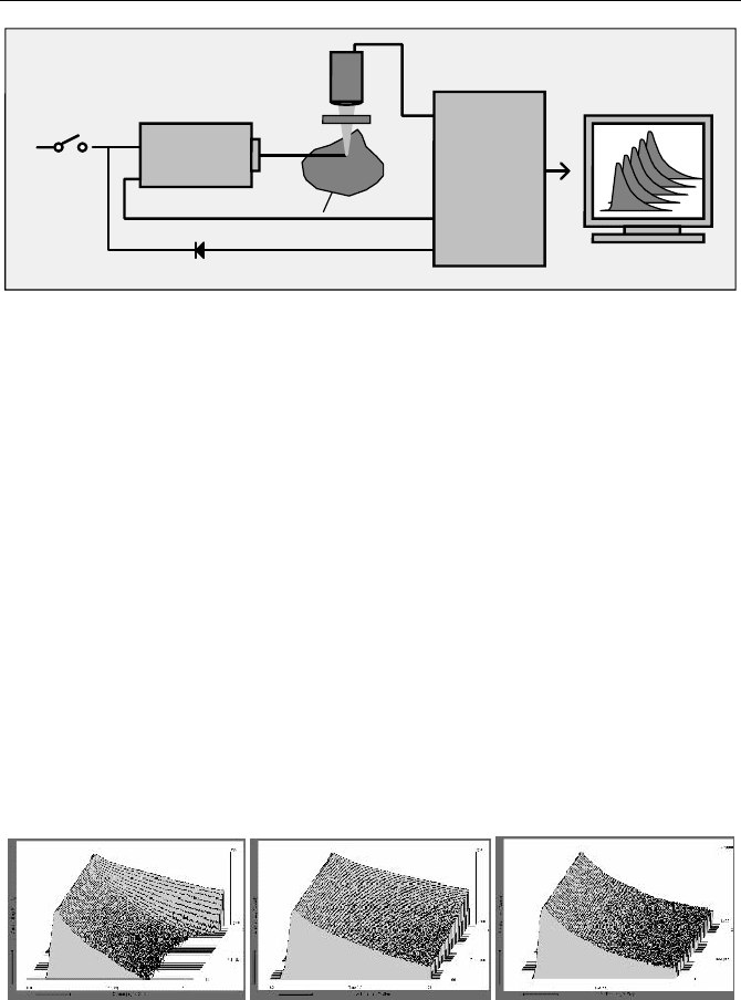
The fluorescence of the chlorophyll in a leaf is excited by a picosecond diode
laser. The fluorescence is separated from the scattered excitation light by a band-
pass filter and detected by a PMT. The photon pulses from the PMT are used as
start pulses of the TCSPC module, the reference pulses from the laser as stop
pulses. When the laser is switched on, a recording sequence in the TCSPC module
is triggered. This is done by connecting a diode from the operating voltage input
of the laser to the TTL-compatible experiment trigger input of the TCSPC module.
For the results shown below, a Becker & Hickl BHL600 laser module was
used, with a wavelength of 650 nm, 80 ps pulse duration, and 50 MHz repetition
rate. The incident power density at the surface of the leaf was approximately
1 mW/mm
2
. The measurement wavelength was selected by a 700 r 15 nm band-
pass filter. The fluorescence decay curves were recorded in one TCSPC channel of
a Becker & Hickl SPC134 system. One fluorescence decay curve was recorded
each 2 seconds, at a count rate of about 210
6
s
-1
. Dead time compensation was
used to avoid the influence of counting loss on the recorded intensity. Typical
results are shown in Fig. 5.32.
Fig. 5.32 Sequences of fluorescence decay curves of leaves after start of illumination. Left
to right: Fresh leaf, faded leaf, dried leaf. Time per curve 2 seconds, logarithmic intensity
scale. Sequence starts from the back
To make the lifetime changes more visible, the sequence starts at the back. In a
fresh leaf the fluorescence lifetime decreases considerably in the first few seconds
of illumination. In a faded leaf the effect is slower and less pronounced. A dry leaf
does not show any noticeable lifetime changes.
5.4 Transient Fluorescence Lifetime Phenomena 93
The recording of the rising part of the fluorescence transient (the decrease of
photochemical quenching) requires a time resolution of the order of 100 µs per
curve. A sequence this fast cannot be recorded in a single-shot experiment. There-
fore, the recording of the sequence must be repeated and the data accumulated
until enough photons have been collected. A suitable setup is shown in Fig. 5.33.
start
TCSPC
Module
stop
Trigger
Diode Laser
Sync Out
Power
Filter
Picosecond
Detector
700nm
Laser On
500ms
15ms
on
off
Pulse
Generator
+12V
Experiment
Fig. 5.33 Recording the photochemical quenching transient of the chlorophyll a fluores-
cence. The laser is periodically switched on for 10 ms. A fast recording sequence is started
at the beginning of each „laser on“ interval, and a large number of recording cycles is ac-
cumulated
The setup uses a picosecond diode laser with fast on/off capability. For the re-
sults shown below a BDL405 and a BHLP700 laser (both Becker & Hickl)
were used for excitation at 405 nm and 650 nm, respectively. The laser is con-
trolled by a pulse generator that turns the light periodically on and off. The „on“
duration is 10 ms, the period 500 ms. Within the „on“ phases, the laser delivers
picosecond pulses at the normal pulse period of 20 ns. The leaf is excited by this
pulse sequence. The fluorescence photons are detected by a PMT and recorded in
the TCSPC module. The TCSPC module runs a hardware-controlled sequence of
recordings, with a time per curve of 100 us and an overall number of 100 curves.
The start of the sequence is triggered with the rising edge of the „laser on“ signal,
and a large number of such cycles is accumulated.
Each „laser on“ period initiates a normal transient of the chlorophyll fluores-
cence. Photochemical quenching decreases with its typical time constant within
the 10 ms „on“ period. In the subsequent „off“ period, photochemical quenching
recovers to its initial state. Due to the low duty cycle of the „laser on“ signal, the
average excitation intensity is low and does not cause much nonphotochemical
quenching. Therefore, the change of photochemical quenching can be recorded
independently, if only the duty cycle of the laser on/off control signal is kept low
enough. Typical results are shown in Fig. 5.34. 10,000 on/off cycles were accumu-
lated. The sequence starts at the front.