Voet D., Voet Ju.G. Biochemistry
Подождите немного. Документ загружается.

site and binds as a single strand to the exonuclease site.
Thus, the formation of a Watson–Crick base pair facilitates
the binding of the primer strand to the polymerase site
preparatory for the next round of chain extension, whereas
a mismatched base pair greatly slows the polymerase reac-
tion while promoting the binding of the primer strand to
the exonuclease site. Comparison of the editing complex
with those of the Klentaq1 ⴢ DNA complexes suggests that
the transfer of the primer strand from the polymerase to
the editing sites of Klenow fragment requires that the
dsDNA translocate backward (toward the 3¿ end of the
template strand) by several angstroms along the helix axis.
h. Pol I Functions Physiologically to Repair DNA
For some 13 years after Pol I’s discovery, it was generally
assumed that this enzyme was E. coli’s DNA replicase
because no other DNA polymerase activity had been de-
tected in E. coli. This assumption was made untenable by
Cairns and Paula De Lucia’s isolation, in 1969, of a mutant
E. coli whose extracts exhibit ⬍1% of the normal Pol I ac-
tivity (although it has nearly normal levels of the 5¿S3¿
exonuclease activity) but which nevertheless reproduce at
the normal rate. This mutant strain, however, is highly sus-
ceptible to the damaging effects of UV radiation and
chemical mutagens (substances that chemically induce
mutations; Section 32-1A). Pol I evidently plays a central
role in the repair of damaged (chemically altered) DNA.
Damaged DNA, as we discuss in Section 30-5, is de-
tected by a variety of DNA repair systems. Many of them
endonucleolytically cleave the damaged DNA on the 5¿
side of the lesion, thereby activating Pol I’s 5¿S3¿ exonu-
clease.While excising this damaged DNA, Pol I simultane-
ously fills in the resulting single-strand gap through its
polymerase activity. In fact, its 5¿S3¿ exonuclease activity
increases 10-fold when the polymerase function is active.
Perhaps the simultaneous excision and polymerization ac-
tivities of Pol I protect DNA from the action of cellular
nucleases that would further damage the otherwise
gapped DNA.
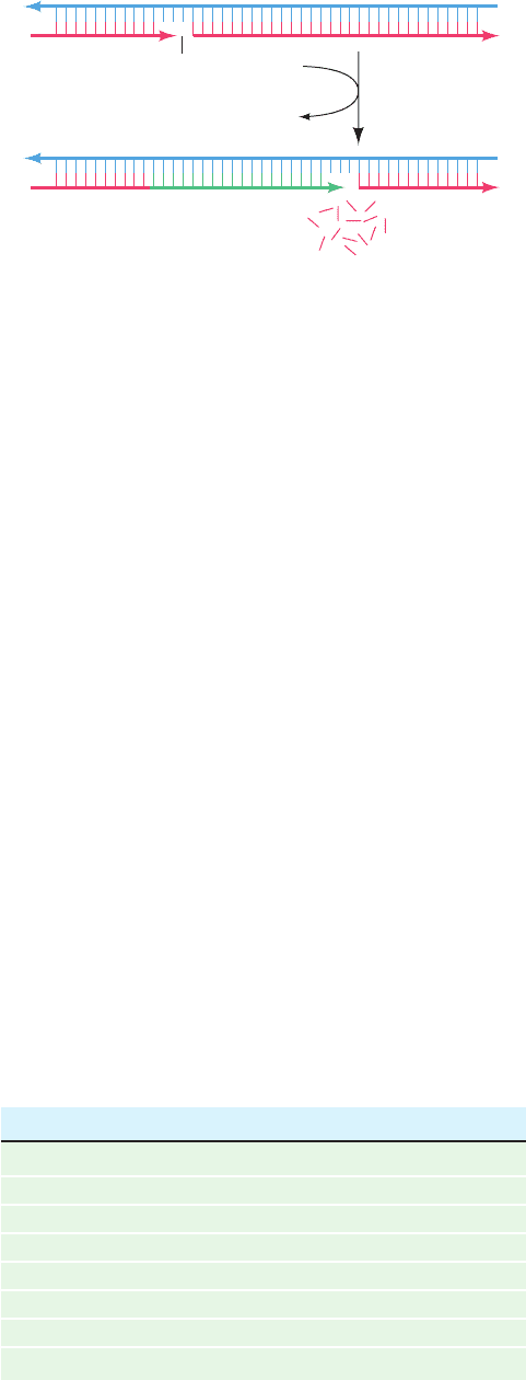
i. Pol I Catalyzes Nick Translation
Pol I’s combined 5¿S3¿ exonuclease and polymerase
activities can replace the nucleotides on the 5¿ side of a sin-
gle-strand nick on otherwise undamaged DNA.These reac-
tions, in effect, translate (move) the nick toward the DNA
strand’s 3¿ end without otherwise changing the molecule
(Fig.30-11).This nick translation process, in the presence of
labeled deoxynucleoside triphosphates, is synthetically em-
ployed to prepare highly radioactive DNA (the required
nicks may be generated by treating the DNA with a small
amount of pancreatic Dnase I).
j. Pol I’s 5ⴕ S 3ⴕ Exonuclease Functions
Physiologically to Excise RNA Primers
Pol I’s 5¿S3¿ exonuclease also removes the RNA primers
at the 5¿ ends of newly synthesized DNA while its DNA poly-
merase activity fills in the resulting gaps (Fig. 5-34). The im-
portance of this function was demonstrated by the isolation
of temperature-sensitive E. coli mutants that are neither
viable nor exhibit any 5¿S3¿ exonuclease activity at the re-
strictive temperature of ⬃43°C (the low level of polymerase
activity in the Pol I mutant isolated by Cairns and De Lucia
is apparently sufficient to carry out this essential gap-filling
process during chromosome replication). Thus Pol I has an
indispensable role in E. coli DNA replication although a
different one than was first supposed.
B. DNA Polymerase III
The discovery of normally growing E. coli mutants that have
very little Pol I activity stimulated the search for an addi-
tional DNA polymerizing activity. This effort was rewarded
by the discovery of two more enzymes, designated, in the or-
der they were discovered, DNA polymerase II (Pol II) and
DNA polymerase III (Pol III). The properties of these en-
zymes are compared with that of Pol I in Table 30-1. Pol II
and Pol III had not previously been detected because their
combined activities in the assays used are normally ⬍5%
that of Pol I.
A mutant E. coli lacking measurable Pol II activity grows
normally. However, Pol II has been implicated as a partici-
pant in repairing DNA damage via the SOS response
(Section 30-5D),as have two additional E. coli enzymes that
Section 30-2. Enzymes of Replication 1181
Figure 30-11 Nick translation as catalyzed by Pol I.
Nick
Mononucleotides
DNA polymerase I
dNTPs
PP
i
5'
3'5'
3'
5'
3'5'
3'
Table 30-1 Properties of E. coli DNA Polymerases
Pol I Pol II Pol III
Mass (kD) 103 90 130
Molecules/cell 400 ? 10–20
Turnover number
a
600 30 9000
Structural gene polA polB polC
Conditionally lethal mutant ⫹⫺ ⫹
Polymerization: 5¿S3¿ ⫹⫹ ⫹
Exonuclease: 3¿S5¿ ⫹⫹ ⫹
Exonuclease: 5¿S3¿ ⫹⫺ ⫺
a
Nucleotides polymerized min
⫺1
ⴢ molecule
⫺1
at 37°C.
Source: Kornberg,A. and Baker, T.A., DNA Replication (2nd ed.), p. 167,
Freeman (1992).
JWCL281_c30_1173-1259.qxd 8/10/10 9:10 PM Page 1181
were more recently discovered: DNA polymerase IV
(Pol IV) and DNA polymerase V (Pol V) (Section 30-5Db).
a. Pol III Is E. coli’s DNA Replicase
The cessation of DNA replication in temperature-
sensitive polC mutants above the restrictive (high) temper-
ature demonstrates that Pol III is E. coli’s DNA replicase.
Its Pol III core has the subunit composition ␣ε where ␣,
the polC gene product (Table 30-2), contains the poly-
merase function. The catalytic properties of Pol III core re-
semble those of Pol I (Table 30-1) except for Pol III core’s
inability to replicate primed ssDNA or nicked dsDNA.
Rather, Pol III core acts in vitro at single-strand gaps of
⬍100 nucleotides, a situation that probably resembles the
state of DNA at the replication fork. The Pol III 3¿S5¿ ex-
onuclease function, which resides on the enzyme’s ε sub-
unit, is DNA’s primary editor during replication; it enhances
the enzyme’s replication fidelity by up to 200-fold. How-
ever, the Pol III 5¿S3¿ exonuclease acts only on single-
stranded DNA, so it cannot catalyze nick translation. is an
accessory protein that stimulates the editing function of ε.
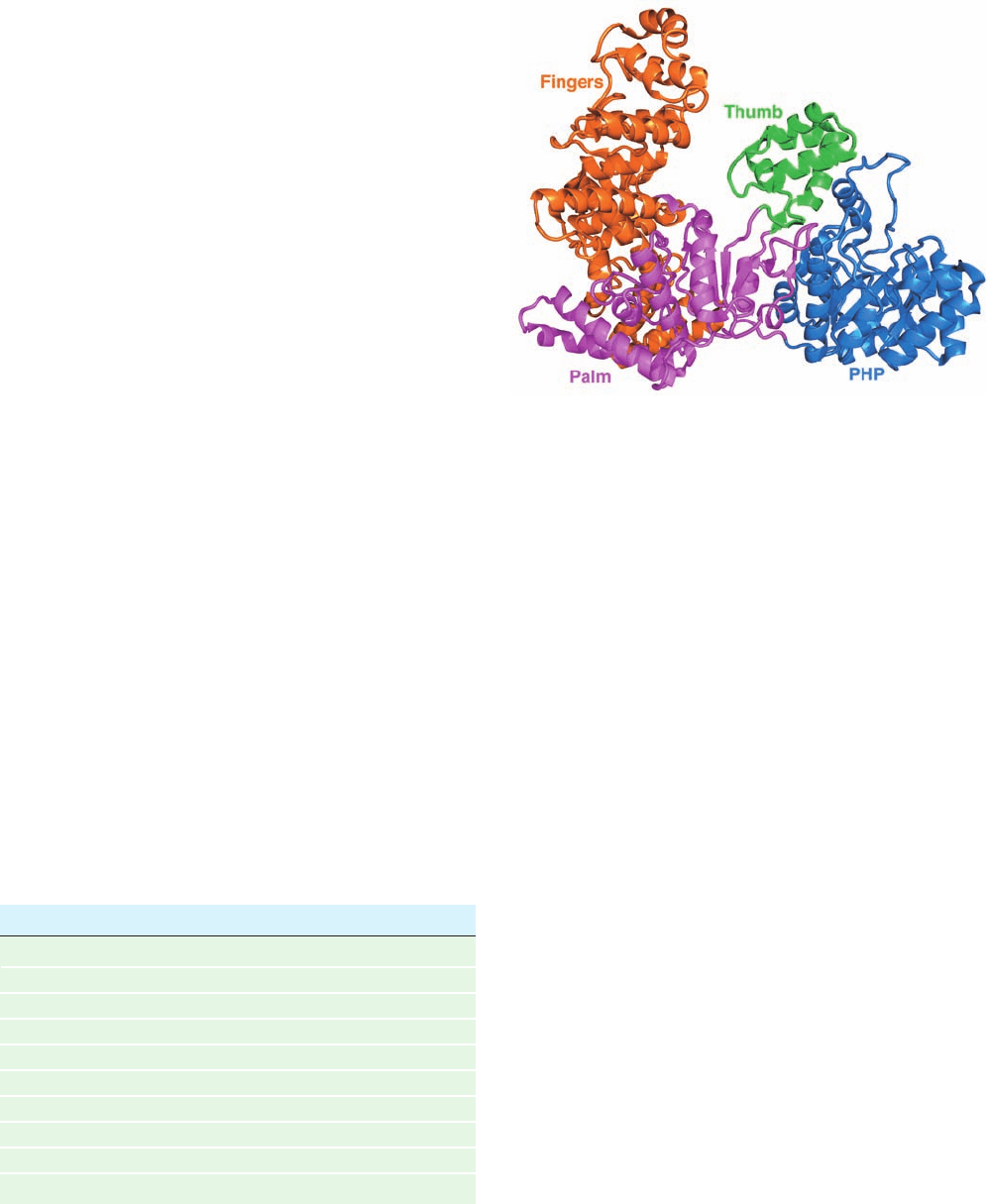
The X-ray structure of residues 1 to 917 of the 1160-
residue E. coli Pol III ␣ subunit, determined Mike O’Donnell
and John Kuriyan, reveals that this protein has the
expected thumb, fingers, and palm domains (Fig. 30-12).
Nevertheless, it exhibits no significant sequence similarity
and a different fold from all but two other DNA poly-
merases of known structure (both from gram-positive bac-
teria). In addition, Pol III ␣ has an N-terminal PHP (for
polymerases and histidinol phosphatase) domain that but-
tresses both the palm and thumb domains.
Pol III core (␣
ε
)functions in vivo as part of a compli-
cated and labile multisubunit enzyme, the Pol III holoen-
zyme, which consists of at least 10 types of subunits (Table
30-2). The latter 7 subunits in Table 30-2 act to modulate
Pol III core’s activity. For example, Pol III core has a
processivity of 10 to 15 residues; it can only fill in short
single-stranded regions of DNA. However, Pol III core is
rendered processive by association with the  subunit in
the presence of the 7-subunit ␥ complex (␥
2
␦␦¿⌿). As-
sembly of the processive enzyme is a two-stage process in
which the ␥ complex transfers the  subunit to the primed
template in an ATP-dependent reaction followed by the
assembly of Pol III core with the  subunit on the DNA
(Section 30-3Cc). The  subunit confers essentially unlim-
ited processivity (⬎5000 residues) on the core enzyme
even if the ␥ complex is subsequently removed. In fact, the
 subunit is very strongly bound to the DNA, although it
can freely slide along it.
b. The  Subunit Forms a Ringlike Sliding Clamp
The observation that a  subunit clamped to a cut circu-
lar DNA slides to the break and falls off suggests that the 
subunit forms a closed ring around the DNA, thereby pre-
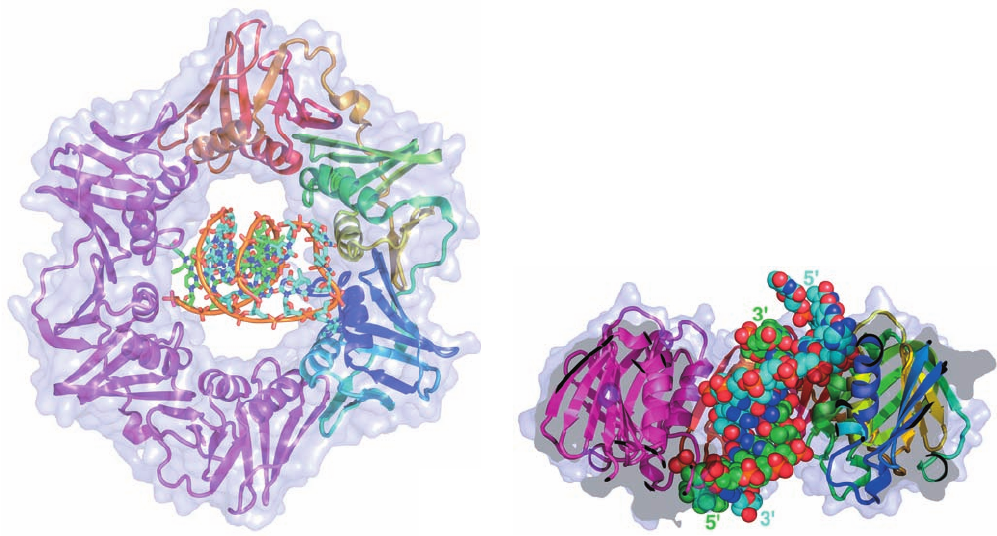
venting its escape. Kuriyan and O’Donnell determined the
X-ray structure of the  subunit in complex with a
primer–template DNA (dsDNA with a single-stranded ex-
tension on the 5¿ end of one of its strands, the template
strand). The protein forms a homodimer of C-shaped, 366-
residue monomer units that associate to form an ⬃80-Å-
diameter doughnut-shaped structure (Fig. 30-13a) that is
equivalently known as the sliding clamp and the  clamp.
The sliding clamp’s central hole is ⬃35 Å in diameter,
which is larger than the 20- and 26-Å diameters of B- and
A-DNAs (recall that the hybrid helices which RNA
1182 Chapter 30. DNA Replication, Repair, and Recombination
Table 30-2 Components of E. coli DNA Polymerase III
Holoenzyme
Subunit Mass (kD) Structural Gene
␣
a
130 polC (dnaE)
ε
a
27.5 dnaQ
a
10 holE
b
71 dnaX
c
␥
b
45.5 dnaX
c
␦
b
35 holA
␦¿
b
33 holB
b
15 holC
b
12 holD
 40.6 dnaN
a
Components of the Pol III core.
b
Components of the ␥ complex.
c
The ␥ and subunits are encoded by the same gene sequence; the ␥
subunit comprises the N-terminal end of the subunit.
Sources: Kornberg,A. and Baker, T.A., DNA Replication (2nd ed.),
p. 169, Freeman (1992); and Baker, T.A. and Wickner, S.H., Annu. Rev.
Genet. 26, 450 (1992).
Figure 30-12 X-ray structure of the E. coli Pol III ␣ subunit.
The protein is drawn in ribbon form with its thumb, PHP, palm,
and finger domains green, blue, magenta, and orange,
respectively. Note the handlike shape of the protein but its
entirely different fold from that of Klentaq1 (Fig. 30-9).
[Based on an X-ray structure by Mike O’Donnell,The
Rockefeller University, and John Kuriyan, University of
California at Berkeley. PDBid 2HQA.]
JWCL281_c30_1173-1259.qxd 8/10/10 9:10 PM Page 1182
primers make with DNA have A-DNA-like conforma-
tions; Section 29-1Bc). Each  subunit consists of six tan-
dem ␣ motifs of identical topology, which associate in
pairs to form three pseudo-2-fold symmetric domains of
very similar structures (although with ⬍20% sequence
identity). The dimeric ring therefore has the shape of a 6-
pointed star in which the 12 helices line the central hole
and the  strands associate in six  sheets that form the
protein’s outer surface. Electrostatic calculations indicate
that the interior surface of the ring is positively charged,
whereas its outer surface is negatively charged.
The ␣ helices lining the protein’s central hole are all ap-
proximately perpendicular to their radially adjacent seg-
ments of the sugar–phosphate backbone. These helices
therefore span the major and minor grooves of the DNA
rather than entering into them as do many helices that
make sequence-specific interactions with dsDNA (e.g.,
Section 31-3Da). Since A- and B-DNAs have 11 and 10.5 bp
per turn, whereas the sliding clamp has a pseudo-12-fold
symmetry, it appears that the sliding clamp largely mini-
mizes its associations with its threaded DNA, which facili-
tates the unencumbered passage of the DNA through the
sliding clamp. Nevertheless, the primer-template DNA’s
helix axis is inclined to the homodimeric protein’s 2-fold
axis by ⬃22° such that its ssDNA segment is in van der
Waals contact with a specific portion of the  clamp’s inner
wall (Fig. 30-13b; in previous model building studies based
on the structure of the sliding clamp alone, these axes were
assumed to be coincident). In fact, primer–template DNA
binds to the sliding clamp ⬃4-fold more tightly than does
dsDNA and the mutation of the residues that interact with
the ssDNA segment significantly reduces the efficiency of
DNA replication. Indeed, as is explained in Section 30-3Cc,
the interaction between the ssDNA and the sliding clamp
is physiologically significant.
C. Unwinding DNA: Helicases and Single-Strand
Binding Protein
Pol III holoenzyme, unlike Pol I, cannot unwind dsDNA.
Rather, three proteins, DnaB protein (the product of the
dnaB gene; proteins may be assigned the name of the gene
specifying them but in roman letters with the first letter cap-
italized), Rep helicase, and single-strand binding protein
Section 30-2. Enzymes of Replication 1183
Figure 30-13 X-ray structure of the  subunit of E. coli Pol III
holoenzyme in complex with DNA. (a) The homodimeric sliding
clamp is drawn in ribbon form embedded in its semitransparent
surface diagram and viewed along its 2-fold axis with one
subunit magenta and the other colored in rainbow order from
its N-terminus (blue) to its C-terminus (red).The DNA, which
consists of a 10-bp double-stranded segment with a 4-nt single-
stranded extension at the 5¿ end of one of the strands, is drawn in
stick form with the C atoms of the template strand cyan, those of
the primer strand green, N blue, O red, and P orange, and with an
orange rod connecting successive P atoms in each strand.
(b) Cutaway diagram of the structure in Part a rotated 90° about
the horizontal axis.The DNA, which is shown in space-filling
form, is inclined by ⬃22° to the protein’s 2-fold axis, which is
vertical in this diagram. Note that in a replicating system, the
primer strand would be extended toward the top of the diagram.
[Based on an X-ray structure by John Kuriyan, University of
California at Berkeley, and Mike O’Donnell,The Rockefeller
University. PDBid 3BEP.]
(a)
(b)
JWCL281_c30_1173-1259.qxd 9/2/10 9:01 AM Page 1183
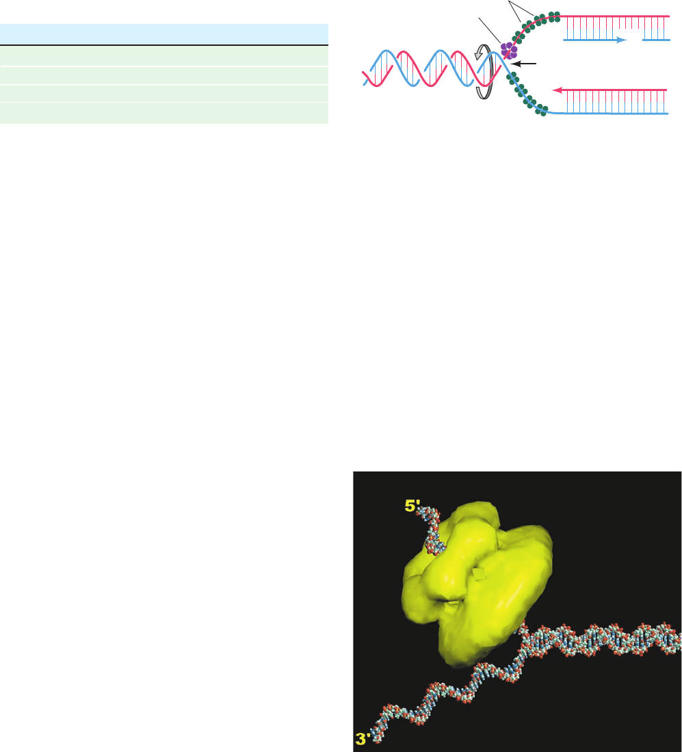
(SSB) (Table 30-3), work in concert to unwind the DNA be-
fore an advancing replication fork (Fig. 30-14) in a process
that is driven by ATP hydrolysis.
a. Hexameric Helicases Mechanically Separate the
Strands of dsDNA by Climbing Up One Strand
Access to the genetic information encoded in a double
helical nucleic acid requires that its base-paired strands be
separated. The proteins that do so, which are known as
helicases, form a diverse group of enzymes that facilitate a
variety of functions including DNA replication, recombi-
nation, and repair, as well as transcription termination
(Section 31-2Da), RNA splicing, and RNA editing (Section
31-4A). Indeed, all forms of life contain helicases, 12 vari-
eties of which occur in E. coli. A helicase functions by
translocating along one strand of a double helical nucleic
acid so as to separate the strands in its path.This, of course,
requires free energy, and hence helicases are driven by
NTP hydrolysis. Helicases have been classified into six su-
perfamilies that vary in their characteristics, including their
direction of translocation along their bound single strand
(5¿S3¿ or 3¿S5¿) and whether they function as hexameric
rings or dimers.
E. coli DnaB protein, a hexameric helicase of identical
471-residue subunits, separates the strands of dsDNA by
translocating along the lagging strand template in the 5¿S
3¿ direction while hydrolyzing ATP (it can also use GTP
and CTP but not UTP). Electron microscopy and X-ray
studies reveal that DnaB forms a hexameric ring that, de-
pending on conditions, exhibits C
3
or C
6
symmetry and
which encloses an ⬃30-Å-diameter central channel. Hexa-
meric DnaB binds three primase molecules via the latter’s
helicase binding domain (Section 30-2E). Similarly, the
bacteriophage T7 gene 4 helicase/primase (bacterio-
phage T7 infects E. coli) forms a two-tiered hexagonal
ring (Fig. 30-15) whose N-terminal domains (residues
1–271) contain its primase activity and whose C-terminal
domains (residues 272–566) carry out its helicase function.
T7 gene 4 helicase/primase (also called T7 gp4; gp for gene
product) translocates along ssDNA in the 5¿S3¿ direction
while preferentially hydrolyzing dTTP (but also hydrolyzes
dATP and ATP).
Leemor Joshua-Tor determined the only known X-ray
structure of a hexameric helicase in complex with DNA,
that of the E1 protein of bovine papillomavirus, which
translocates along ssDNA in the 3¿S5¿ direction (the
opposite direction of DnaB and T7 gp4).The protein in the
structure, which contains the C-terminal 274 residues of
the 605-residue E1 protein, consists of two domains: a 74-
residue N-terminal oligomerization domain and a 200-
residue C-terminal AAAⴙ domain (AAA⫹ for ATPases
associated with cellular activities; a functionally diverse
protein family). There are two families of hexameric heli-
cases, the RecA family and the AAA⫹ family. RecA-family
hexameric helicases (RecA catalyzes homologous recom-
bination; Section 30-6Ab), such as DnaB and T7 gp4,
1184 Chapter 30. DNA Replication, Repair, and Recombination
Table 30-3 Unwinding and Binding Proteins of E. coli
DNA Replication
Protein Subunit Structure Subunit Mass (kD)
DnaB protein Hexamer 50
SSB Tetramer 19
Rep protein Monomer 68
PriA protein Monomer 76
Source: Kornberg,A. and Baker, T.A., DNA Replication (2nd ed.), p. 366,
Freeman (1992).
Leading strand
Lagging strand
DnaB protein
SSB
Fork movement
5'
3'
5'
3'
Figure 30-14 Unwinding of DNA by the combined action of
DnaB and SSB proteins. The hexameric DnaB protein moves
along the lagging strand template in the 5¿S3¿ direction.The
resulting separated DNA strands are prevented from
reannealing by SSB binding.
Figure 30-15 Electron microscopy–based image
reconstruction of T7 gene 4 helicase/primase. In this two-tiered
hexameric ring (yellow), the smaller lobe of each subunit forms
the N-terminal primase domain and the larger lobe forms the
C-terminal helicase domain.The protein is postulated to interact
with DNA as is depicted by this model of a DNA fork consisting
of a 30-bp duplex segment and two 25-nt single-stranded
segments with the 5¿ tail threaded through the hexameric ring.
The way in which the 3¿ tail interacts with the protein, if at all, is
unknown. [Courtesy of S.S. Patel and K.M. Picha, University of
Medicine and Dentistry of New Jersey.]
JWCL281_c30_1173-1259.qxd 8/10/10 9:11 PM Page 1184
translocate in the 5¿S3¿ direction and occur mainly in
eubacteria and their phages, whereas AAA⫹ hexameric
helicases, such as the E1 protein, translocate in the 3¿S5¿
direction and occur mainly in archaea, eukaryotes, and
their viruses.
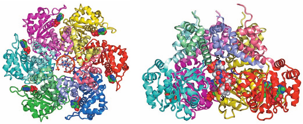
The E1 structure reveals that this helicase, which was
crystallized with ADP and a 13-nt poly(dT) (although
only 6 nt are visible in the X-ray structure), forms a two-
layered hexagonal ring in which the oligomerization
domains form a rigid collar with nearly perfect 6-fold
symmetry. In contrast, the AAA⫹ domains deviate signif-
icantly from this symmetry (Fig. 30-16a). An ADP is
bound at a radially peripheral site between each neigh-
boring pair of AAA⫹ domains. The poly(dT) forms a
right-handed helix that binds in the minimally ⬃13-Å-
diameter central channel of the AAA⫹ domain hexamer
(which is too narrow to admit dsDNA) with its 5¿ end to-
ward the top of the hexamer in Fig. 30-16. The DNA’s
phosphate groups each interact with a positively charged
loop (residues 505–508) that extends radially inward from
each AAA⫹ domain and hence these loops form an
arrangement that resembles a right-handed spiral stair-
case that tracks the ssDNA’s sugar–phosphate backbone.
Apparently, the protein steps through a series of ATP-
driven conformational changes that, via interactions with
the loops, pushes the ssDNA through the channel from
bottom to top in Fig. 30-16b. During this process, each
loop maintains its grip on the same phosphate group.ATP
hydrolysis occurs toward the bottom of the spiral stair-
case and ADP release occurs between subunits located
toward its top. A new ATP then binds to this site, which
causes the topmost loop to drop to the bottom of the
staircase, where it binds the next available phosphate
group and repeats the catalytic cycle.Thus the E1 helicase
mechanically separates the strands of dsDNA by pulling
itself along the groove of one strand in its 3¿S5¿ direc-
tion but without turning relative to the DNA.
b. Rep Helicase Dimers Separate the Strands
of dsDNA via an “Active, Rolling” Mechanism
Two other helicases, Rep helicase and PriA protein,
have been implicated in the replication of various E. coli
phage DNAs (Section 30-3B) and also participate in cer-
tain aspects of E. coli DNA replication (Section 30-3C).
Both proteins translocate along DNA in the 3¿S5¿ di-
rection (and hence along the opposite strand from
DnaB) while hydrolyzing ATP. Rep helicase is not es-
sential for E. coli DNA replication but the rate at which
E. coli replication forks propagate is reduced ⬃2-fold in
rep
⫺
mutants.
Rep helicase is a 673-residue monomer in solution but
dimerizes on binding to DNA. Both subunits of the Rep
dimer bind to ssDNA or dsDNA such that DNA binding to
one subunit strongly inhibits DNA binding to the other
(negative cooperativity). This observation led Timothy
Lohman to propose the “active, rolling” mechanism for
Section 30-2. Enzymes of Replication 1185
Figure 30-16 X-ray structure of bovine papillomavirus E1
helicase in complex with poly(dT) and ADP. (a) The protein is
drawn in ribbon form viewed along the homohexamer’s pseudo-
6-fold axis with each protein subunit differently colored and with
the oligomerization domain of each subunit more lightly shaded
than its AAA⫹ domain.The protein loops extending radially
inward from each subunit to interact with the DNA’s phosphate
groups are purple. The poly(dT), 6 nt of which are visible, is
drawn in stick form with C gray, N blue, O red, and P orange and
(a)
(b)
with successive P atoms joined by orange rods. Its 5¿ end is
closest to the viewer.The ADP is shown in space-filling form
with C green, N blue, O red, and P orange. (b) Side view of the
protein related to that in Part a by a 90° rotation about the
horizontal axis.The blue and green AAA⫹ domains in Part a,
except for their DNA-interacting loops, have been deleted to
expose the DNA, which is drawn in space-filling form. [Based on
an X-ray structure by Leemor Joshua-Tor, Cold Spring Harbor
Laboratory, New York. PDBid 2GXA.]
JWCL281_c30_1173-1259.qxd 8/10/10 9:11 PM Page 1185
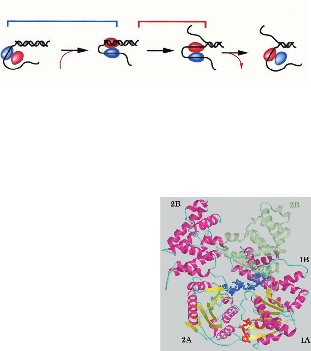
Rep-mediated DNA unwinding in which the two subunits
of the dimer alternate in binding dsDNA and the 3¿ end of
the ssDNA at the ssDNA/dsDNA junction (Fig. 30-17).
The two subunits then “walk” up the DNA while unwind-
ing it in an ATP-dependent manner via a subunit switching
mechanism in which the helicase subunit that is bound to
the dsDNA displaces its 5¿-starting strand while remaining
bound to its 3¿-starting strand. Release of the other subunit
from the 3¿-starting ssDNA then permits this subunit to
bind to and unwind the new end of the dsDNA, thereby
continuing the cycle.
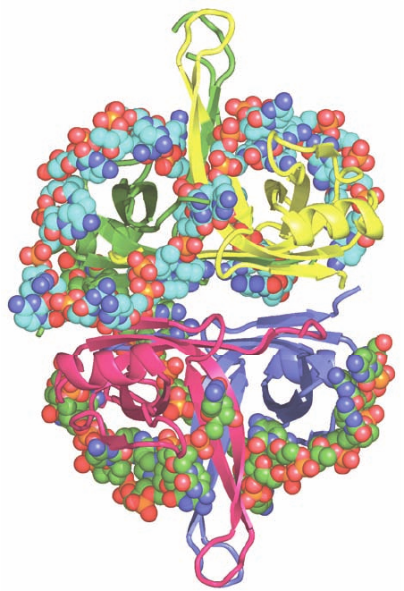
The X-ray structure of E. coli Rep helicase in complex
with the short ssDNA dT(pT)
15
and ADP (Fig. 30-18), de-
termined by Lohman and Waksman, reveals that the rela-
tively straight ssDNA molecule binds two contacting Rep
monomers.A Rep monomer consists of two domains, 1 and
2, each of which is formed by two subdomains, A and B,
with the two N-terminal subdomains (1A and 2A) homol-
ogous to each other. In the two Rep monomers that are
bound to the same ssDNA, subdomain 2B exhibits strik-
ingly different orientations with respect to the other three
subdomains (Fig. 30-18). The Rep monomer that is bound
to the 5¿ end of the ssDNA (which it contacts between
bases 1 and 8) assumes the “open” conformation in which
the four subdomains form an assembly that is reminiscent
of a crab claw with one pincer (subdomain 2B) larger than
the other (subdomain 1B). The DNA is bound at the bot-
tom of the resulting cleft, whose floor is formed by subdo-
mains 1A and 2A. In the Rep monomer that binds to the 3¿
end of the ssDNA (which it contacts between bases 9 and
16), subdomain 2B has reoriented relative to the other sub-
domains via a 130° rotation about a hinge region between
subdomains 2A and 2B, thereby closing the cleft about the
DNA to form the “closed” conformation. This conforma-
tion change is consistent with the active, rolling mechanism
even though the way in which two Rep monomers form the
dimer observed in solution remains unknown. The ADP
binds to Rep between its subdomains 1A and 2A in close
proximity to the DNA, suggesting that conformation
changes at the ATP-binding site arising from ATP hydroly-
sis are transmitted to the DNA-binding site via the second-
ary structural elements that contact both sites. The way in
which Rep separates the two strands of dsDNA is, as yet,
unknown.
1186 Chapter 30. DNA Replication, Repair, and Recombination
Figure 30-17 Active, rolling mechanism for DNA unwinding
by Rep helicase. (1) The subunit of dimeric Rep helicase that is
not bound to ssDNA binds to dsDNA accompanied by ATP
binding. (2) The subunit bound to dsDNA unwinds the double
strand and remains bound to the 3¿-ending strand. (3) In a
Figure 30-18 Superposition of the X-ray structures of Rep
helicase in its open and closed forms in complex with dT(pT)
15
and ADP. The monomer in the open conformation is drawn in
ribbon form colored according to secondary structure (helices
magenta,  sheets yellow, and coil cyan) with its bound ssDNA
segment and ADP drawn in stick form in blue and in red. In the
closed conformation, subdomain 2B (transparent green ribbon)
has rotated via a 130° hinge motion relative to subdomains 1A,
1B, and 2A so as to close over the ssDNA. [Courtesy of Gabriel
Waksman, Washington University School of Medicine. PDBid
1UAA.]
translocation
ATP
1
3'
3'
2
active
unwinding
3'
3
ADP + P
i
3'
process that is accompanied by the release of the ATP hydrolysis
products, the subunit closer to the 3¿ end of the bound ssDNA
releases it preparatory for a new cycle of dsDNA unwinding.
[Courtesy of Gabriel Waksman, Washington University School of
Medicine.]
JWCL281_c30_1173-1259.qxd 8/27/10 7:27 PM Page 1186
c. Single-Strand Binding Protein Prevents ssDNA
from Reannealing
If left to their own devices, the separated DNA strands
behind an advancing helicase would rapidly reanneal to re-
form dsDNA. What prevents them from doing so is the
binding of single-strand binding protein (SSB). It also pre-
vents ssDNA from forming fortuitous intramolecular sec-
ondary structures (helical stems) and protects it from nu-
cleases. Numerous copies of SSB cooperatively coat
ssDNA, thereby maintaining it in an unpaired state. Note,
however,that ssDNA must be stripped of SSB before it can
be replicated by Pol III holoenzyme.
E. coli SSB is a homotetramer of 177-residue subunits.
SSB binds ssDNA in several distinct modes referred to as
(SSB)
n
, which differ by the number of nucleotides (n)
bound to each tetramer.The two major modes are (SSB)
35
,
in which only two of the tetramer’s subunits strongly inter-
act with the ssDNA, and (SSB)
65
, in which all four subunits
interact with the ssDNA.The (SSB)
35
mode displays unlim-
ited cooperativity in that it forms extended strings of con-
tacting tetramers along the length of a bound ssDNA,
whereas the (SSB)
65
mode has limited cooperativity in that
it forms beaded clusters on ssDNA that consist of only a
few contacting tetramers.
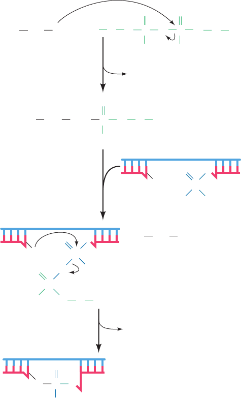
Proteolysis studies have shown that SSB’s ssDNA-
binding site is contained within its 115 N-terminal
residues.The X-ray structure of E. coli SSB’s chymotryptic
fragment (residues 1–135) in complex with dC(pC)
34
,de-
termined by Lohman and Waksman, reveals that the
tetrameric protein has D
2
symmetry and binds two mole-
cules of dC(pC)
34
(Fig. 30-19). For one of these 35-mers, 28
nucleotides (residues 3–30) were visible and these as-
sumed the shape of an elongated horseshoe that wrapped
around two SSB subunits with approximate 2-fold symme-
try and with its apex contacting a third subunit. The other
bound ssDNA was partially disordered such that only two
segments were visible, one with 14 nt (residues 3–16) and
the other with 9 nt (residues 19–27). The paths of the ss-
DNA segments along the surface of the SSB suggested
models that rationalize the different properties of (SSB)
35
and (SSB)
65
. In the (SSB)
65
model, the two ends of a 65-nt
segment emerge from the same side of the tetramer,which
would limit the number of SSB tetramers that can bind to
contiguous 65-nt segments of ssDNA. However, in the
(SSB)
35
model, the two ends of a 35-nt segment emerge
from opposite ends of the tetramer, thereby permitting an
unlimited series of SSB tetramers to interact end-to-end
along the length of a ssDNA.
D. DNA Ligase
Pol I, as we saw in Section 30-2A, replaces the Okazaki
fragments’ RNA primers with DNA through nick trans-
lation. The resulting single-strand nicks between adjacent
Okazaki fragments, as well as the nick on circular DNA
after leading strand synthesis, are sealed in a reaction cat-
alyzed by DNA ligase. The free energy required by this
reaction is obtained, in a species-dependent manner,
through the coupled hydrolysis of either NAD
⫹
to
NMN
⫹
⫹ AMP or ATP to PP
i
⫹ AMP. The E. coli en-
zyme, which is also known as LigA, is a 671-residue
monomer that utilizes NAD
⫹
and catalyzes a three-step
reaction (Fig. 30-20):
1. The adenylyl group of NAD
⫹
is transferred to the
ε-amino group of an enzyme Lys residue to form an un-
usual phosphoamide adduct that is, nevertheless, readily
isolated.
2. The adenylyl group of this activated enzyme is
transferred to the 5¿-phosphoryl terminus of the nick to
form an adenylylated DNA. Here, AMP is linked to the
5¿-nucleotide via a pyrophosphate rather than the usual
phosphodiester bond.
Section 30-2. Enzymes of Replication 1187
Figure 30-19 X-ray structure of E. coli SSB in complex with
dC(pC)
34
. The homotetramer, which has D
2
symmetry, is viewed
along one of its 2-fold axes with its other 2-fold axes horizontal
and vertical. Each of its subunits (which include the N-terminal
134 residues of the 177-residue polypeptide) are differently
colored. Its two bound ssDNA molecules are drawn in space-
filling form colored according to atom type with the upper strand
C cyan, lower strand C green, N blue, O red, and P orange. (The
lower strand is partially disordered and hence appears to consist
of two fragments.) [Based on an X-ray structure by Timothy
Lohman and Gabriel Waksman, Washington University School
of Medicine. PDBid 1EYG.]
JWCL281_c30_1173-1259.qxd 8/10/10 9:11 PM Page 1187
3. DNA ligase catalyzes the formation of a phosphodi-
ester bond by attack of the 3¿-OH on the 5¿-phosphoryl
group, thereby sealing the nick and releasing AMP.
ATP-requiring DNA ligases, such as those of all eukaryotes
and bacteriophage T4, release PP
i
in the first step of the re-
action rather than NMN
⫹
. T4 ligase is also noteworthy in
that, at high DNA concentrations, it can link together two
duplex DNAs (blunt end ligation) in a reaction that is a
boon to genetic engineering (Section 5-5C).
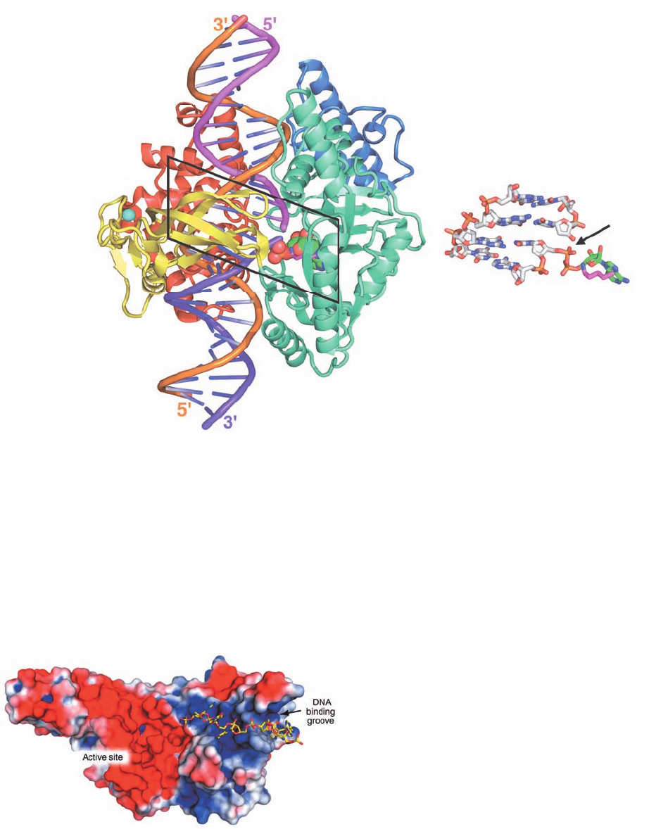
The X-ray structure of E. coli DNA ligase in complex
with a singly nicked 26-bp dsDNA and AMP was deter-
mined by Stewart Shuman.The complex was formed by re-
acting the protein with NAD
⫹
in the presence of Mg
2⫹
(thus forming the phosphoamide product of step 1 of the
DNA ligase reaction; Fig. 30-20), removing the Mg
2⫹
, and
then adding the nicked dsDNA. The X-ray structure of
crystals of this complex revealed that the protein forms a
C-shaped clamp that encircles a 19-bp segment of the
DNA centered on the nick (Fig. 30-21). Moreover, the com-
plex had progressed through step 2 of the reaction, that is,
the adenylyl group had formed a pyrophosphate linkage
with the 5¿-phosphate group at the nick. The reason that
the enzyme did not complete its catalytic cycle by sealing
the nick is presumably due to the absence of Mg
2⫹
.
Residues 587 to 671 form a domain that is not visible in
this X-ray structure although it is poorly resolved in the
X-ray structure of DNA ligase from Thermus filiformis.
Apparently, this domain has high mobility, which suggests
that it folds out to allow the enzyme’s nicked dsDNA sub-
strate to bind to the active site and then folds back to help
immobilize the DNA.
E. Primase
The primases from bacteria and several bacteriophages
track the moving replication fork in close association with
its DNA helicase. Thus, the N-terminal domain of T7 gene
4 helicase/primase forms its primase function (Fig. 30-15),
whereas E.coli primase (DnaG) forms a noncovalent com-
plex with DnaB. Since these DNA helicases translocate
along the lagging strand template DNA in its 5¿S3¿ direc-
tion (Fig. 30-14), the primase must reverse its direction of
travel in order to synthesize an RNA primer in its 5¿S3¿
direction. DnaG, which is held to the RNA primed site by
its association with SSB, can synthesize up to 60-nt primers
in vitro, although in vivo, primers have the length of 11 ⫾ 1 nt.
Since a replication fork in E. coli moves at ⬃1000 nt per
second and Okazaki fragments are ⬃1000 nt in length,
about one RNA primer must be synthesized per second at
each replication fork. Primases tend to initiate synthesis at
specific 3-nt sequences on the template. In E. coli this
sequence is GTA, which is overrepresented in templates
for lagging strand synthesis.
DnaG is a 581-residue monomeric protein. Proteolysis
studies have shown that it consists of three domains: an N-
terminal Zn
2⫹
-binding domain (residues 1–110), which
tetrahedrally ligands a Zn
2⫹
ion via three Cys residues and
a His residue and is implicated in recognizing ssDNA; a
central RNA polymerase domain (residues 111–433) that
carries out primer synthesis; and a C-terminal helicase
binding domain (residues 434–581) that interacts with
DnaB. Isolated DnaG is only weakly active in vitro; it syn-
thesizes primers at a maximum rate of three per hour.
However,in the presence of DnaB,it synthesizes primers at
the rate observed in vivo. Since E. coli have 50 to 100
DnaG molecules per cell, this presumably limits primer
synthesis to the replication fork. The importance of this
function is underscored by T7 gp4, whose helicase and pri-
mase functions reside on the same polypeptide (Fig. 30-15).
The X-ray structure of the DnaG catalytic domain in
complex with a 15-nt ssDNA (Fig. 30-22), determined by
James Berger, reveals a cashew-shaped protein whose fold
is unrelated to those of any other DNA or RNA poly-
merases. It contains an ⬃100-residue segment that is simi-
lar in both sequence and structure to segments in types IA
1188 Chapter 30. DNA Replication, Repair, and Recombination
Figure 30-20 The reactions catalyzed by E. coli DNA ligase.
In eukaryotic and T4 DNA ligases, NAD
⫹
is replaced by ATP so
that PP
i
rather than NMN
⫹
is eliminated in the first reaction
step. The numbered steps are described in the text.
1
NMN
+
O
–
O
–
OH
+
P
–
O
OO
O
–
OH
P
O
O O
O
P
–
O
O
R A
AMP
O
P
O
O
O
–
P
R A
O
O
LysE
2
3
NH
2
LysE+
NAD
+
R ON
–
O
P
O
R AO P
O
O
NH
2
:
Lys
E
+
O
–
NH
2
JWCL281_c30_1173-1259.qxd 8/10/10 9:11 PM Page 1188
and IIA topoisomerases (Section 29-3C) and has therefore
been named the Toprim fold (for topoisomerase and pri-
mase). The Toprim fold consists of a 4-stranded parallel
 sheet flanked by three helices that resembles the
nucleotide-binding (Rossmann) fold (Section 8-3Bi). The
active site is marked by several residues that are highly
conserved in DnaG-type primases, and in particular, a Glu
and two Asp residues, which are invariant in all known
Toprim folds and which, in the X-ray structure of a type
IIA topoisomerase, coordinate an Mg
2⫹
ion.
The ssDNA in the structure, only 5 nt of which are visi-
ble, occupies a positively charged groove on the surface of
the catalytic subunit that feeds into its active site.The pro-
tein makes only a few hydrogen bonding and van der
Waals interactions with the DNA’s sugar–phosphate back-
bone and no specific interactions with its bases. Appar-
ently, this DNA binding groove functions to nonspecifi-
cally capture a DNA template strand and direct it to the
enzyme’s active site.
Section 30-2. Enzymes of Replication 1189
Figure 30-22 X-ray structure of E. coli primase in complex
with ssDNA. The protein is represented by its molecular surface
colored according to its electrostatic potential with red negative,
white nearly neutral, and blue positive.The DNA is drawn in
stick form with C and P yellow, N blue, and O red. Note the
strongly basic character of the DNA binding groove and the
highly acidic nature of the active site region. [Courtesy of James
Berger, University of California at Berkeley. PDBid 3B39.]
(a) (b)
Figure 30-21 X-ray structure of E. coli DNA ligase in
complex with a singly nicked 26-bp dsDNA and AMP. (a) The
enzyme is drawn in ribbon form with its four domains colored,
from N- to C-terminus, blue, aquamarine, yellow, and red.The
dsDNA is shown in ladder form with the sugar–phosphate
backbone of its 26-nt strand orange and those of its
complementary two 13-nt strands magenta and purple. The AMP,
which is covalently bound in pyrophosphate linkage to the
phosphate group at the 5¿-end of the purple strand, is drawn in
space-filling form with C green, N blue, O red, and P orange. The
side chain of Lys 115, which forms a phosphamide adduct in step 1
of the DNA ligase reaction (Fig. 30-20) is shown in stick form
with C magenta and N blue. A Zn
2⫹
ion, represented by a cyan
sphere, is tetrahedrally liganded by four Cys residues. It is distant
from the active site and therefore appears to have structural
rather than catalytic function. (b) The 4 bp of nicked DNA in the
boxed area of Part a in pyrophosphate linkage with the AMP
together with the side chain of Lys 115 are all shown in stick
form.The structure is viewed and colored as in Part a but with
DNA C gray. The arrow points to the DNA’s single-strand nick.
[Based on an X-ray structure by Stewart Shuman, Sloan-
Kettering Institute, New York, New York. PDBid 2OWO.]
JWCL281_c30_1173-1259.qxd 8/10/10 9:11 PM Page 1189
3 PROKARYOTIC REPLICATION
Bacteriophages are among the simplest biological entities
and their DNA replication mechanisms reflect this fact.
Much of what we know about how DNA is replicated
therefore stems from the study of this process in various
phages. In this section we examine DNA replication in the
coliphages (bacteriophages that infect E. coli) M13 and
X174 and then consider DNA replication in E. coli itself.
Eukaryotic DNA replication is discussed in Section 30-4.
A. Bacteriophage M13
Bacteriophage M13 carries a 6408-nt single-stranded circu-
lar DNA known as its viral or (⫹) strand. On infecting an
E. coli cell, this strand directs the synthesis of its comple-
mentary or (⫺) strand to form the circular duplex replica-
tive form (RF), which may be either nicked (RF II) or
supercoiled (RF I). This replication process (Fig. 30-23)
may be taken as a paradigm for leading strand synthesis in
duplex DNA.
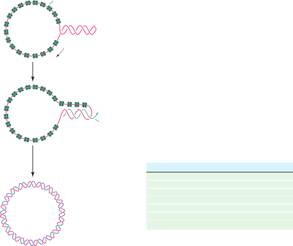
As the M13 (⫹) strand enters the E. coli cell, it becomes
coated with SSB except at a palindromic 57-nt segment
that forms a hairpin. RNA polymerase commences primer
synthesis 6 nt before the start of the hairpin and extends
the RNA 20 to 30 residues to form a segment of
RNA–DNA hybrid duplex. The DNA that is displaced
from the hairpin becomes coated with SSB so that when
RNA polymerase reaches it, primer synthesis stops. Pol III
holoenzyme then extends the RNA primer around the cir-
cle to form the (–) strand. The primer is removed by Pol I-
catalyzed nick translation, thereby forming RF II, which is
converted to RF I by the sequential actions of DNA ligase
and DNA gyrase.
B. Bacteriophage X174
Bacteriophage X174, as does M13, carries a small (5386
nt) single-stranded circular DNA. Curiously, the in vivo
conversion of the X174 viral DNA to its replicative form
is a much more complex process than that for M13 DNA in
that X174 replication requires the participation of a
nearly 600-kD protein assembly known as a primosome
(Table 30-4).
a. X174 (ⴚ) Strand Replication Is a Paradigm
for Lagging Strand Synthesis
X174 (⫺) strand synthesis occurs in a six-step process
(Fig. 30-24):
1. The reaction sequence begins in the same way as that
for M13: The (⫹) strand is coated with SSB except for a
44-nt hairpin. A 70-nt sequence containing this hairpin,
known as pas (for primosome assembly site), is then recog-
nized and bound by the PriA, PriB, and PriC proteins.
2. DnaB and DnaC proteins in the form of a DnaB
6
ⴢ
DnaC
6
complex add to the DNA with the help of DnaT
protein in an ATP-requiring process. DnaC protein is then
released yielding the preprimosome. The preprimosome, in
turn, binds primase yielding the primosome.
1190 Chapter 30. DNA Replication, Repair, and Recombination
Figure 30-23 The synthesis of the M13 (⫺) strand DNA on a
(⫹) strand template to form M13 RF I DNA.
Table 30-4 Proteins of the Primosome
a
Protein Subunit Structure Subunit Mass (kD)
PriA Monomer 76
PriB Dimer 11.5
PriC Monomer 23
DnaT Trimer 22
DnaB Hexamer 50
DnaC
b
Monomer 29
Primase (DnaG) Monomer 60
a
The complex of all primosome proteins but primase is known as the
preprimosome.
b
Not part of the preprimosome or the primosome.
Source: Kornberg,A. and Baker, T.A., DNA Replication (2nd ed.),
pp. 286–288, Freeman (1992).
RNA prime
r
Hairpin
SSB
RNA polymerase
1. DNA replication by
Pol III
2. RNA excision and gap filling by Pol I
3. Gap sealing by DNA ligase
4. Supercoiling by DNA gyrase
M13
RF
I
DNA
M13 (+)
strand DNA
5'
5'
3'
3'
JWCL281_c30_1173-1259.qxd 8/10/10 9:11 PM Page 1190
