Voet D., Voet Ju.G. Biochemistry
Подождите немного. Документ загружается.

X-ray structure). A monomeric unit of Z␣ binds to each
strand of the Z-DNA, out of contact with the Z␣ that binds
to the opposite strand. The protein primarily interacts with
Z-DNA via hydrogen bonds and salt bridges between polar
and basic protein side chains and the Z-DNA’s sugar–phos-
phate backbone. Note that none of the DNA’s bases partic-
ipate in these associations. The protein’s DNA-binding sur-
face, which is complementary in shape to the Z-DNA, is
positively charged, as is expected for a protein that interacts
with several closely spaced, anionic phosphate groups. It
is postulated that ADAR1’s Z␣ domain targets it to the
Z-DNA upstream of actively transcribing genes (for rea-
sons discussed in Section 31-4As).
In an effort to visualize the structure of the junction be-
tween B-DNA and Z-DNA, Rich and Kyeong Kyu Kim
cocrystallized Z␣ with a duplex DNA that has two over-
hanging nucleotides at each end and whose two 17-nu-
cleotide strands have the sequences d(GTCGCGCGC-
CATAAACC) and d(ACGGTTTATGGCGCGCG). The
X-ray structure of this complex reveals that 8 nucleotide
pairs at one end of the double helix are Z-DNA, 6 nu-
cleotide pairs at the other end of the helix are B-DNA, and
the nucleotides that would otherwise form an A ⴢ T base
pair at the junction between these segments have been ex-
pelled from the double helix (Fig. 29-3b).The base pairs of
the B- and Z-DNA segments form a continuous stack,
which stabilizes the structure (Section 29-2C). Four Z␣’s
are bound to the Z-DNA segment, two per polynucleotide
strand, in a manner closely similar to that in the foregoing
Z␣–d(TCGCGCG) complex (for clarity, the Z␣’s are not
shown in Fig. 29-3b) and the B-and Z-DNA segments
adopt their standard conformations. Evidently, under the
proper conditions, the handedness of duplex DNA can be
reversed by breaking one base pair and ejecting its nu-
cleotides from the duplex.
c. RNA-11 and RNA–DNA Hybrids Have
an A-DNA-Like Conformation
Double helical RNA is unable to assume a B-DNA-like
conformation because of steric clashes involving its 2¿-OH
groups. Rather, it usually assumes a conformation resem-
bling A-DNA (Fig. 29-1, left panels) known as A-RNA or
RNA-11, which ideally has 11.0 bp per helical turn, a pitch
of 30.9 Å, and its base pairs inclined to the helix axis by
16.7°. Many RNAs, for example, transfer and ribosomal
RNAs (whose structures are detailed in Sections 32-2B and
32-3A), contain complementary sequences that form dou-
ble helical stems.
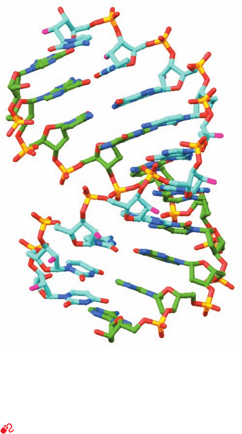
Hybrid double helices, which consist of one strand each
of DNA and RNA, are also predicted to have A-RNA-like
conformations. In fact, the X-ray structure, by Nancy Hor-
ton and Barry Finzel, of a 10-bp complex of the DNA
oligonucleotide d(GGCGCCCGAA) with the complemen-
tary RNA oligonucleotide r(UUCGGGCGCC) reveals
(Fig. 29-4) that it forms a double helix with A-RNA-like
character (Table 29-1) in that it has 10.9 bp per turn, a pitch
of 31.3 Å, and its base pairs are, on average, inclined to the
helix axis by 13.9°. Nevertheless, this hybrid helix also has
B-DNA-like qualities in that the width of its minor groove
(9.5 Å) is intermediate between those for canonical B-DNA
(7.4 Å) and A-DNA (11 Å) and in that some of the ribose
rings of its DNA strand have conformations characteristic
of B-DNA (Section 29-2A), whereas others have conforma-
tions characteristic of A-RNA. Note that this structure is of
biological significance because short segments of RNA ⴢ
DNA hybrid helices occur in both the transcription of RNA
on DNA templates (Section 31-2Ba) and in the initiation of
DNA replication by short lengths of RNA (Section 30-1D).
The RNA component of this helix is a substrate for RNase
H, which specifically hydrolyzes the RNA strands of RNA ⴢ
DNA hybrid helices in vivo (Section 30-4C).
2 FORCES STABILIZING NUCLEIC
ACID STRUCTURES
Double-stranded DNA does not exhibit the structural
complexity of proteins because it has only a limited reper-
toire of secondary structures and no comparable tertiary or
Section 29-2. Forces Stabilizing Nucleic Acid Structures 1151
Figure 29-4 X-ray structure of a 10-bp RNA–DNA hybrid
helix consisting of d(GGCGCCCGAA) in complex with
r(UUCGGGCGCC). The structure is shown in stick form with
RNA C atoms cyan, DNA C atoms green, N blue, O red except
for RNA O2¿ atoms, which are magenta, and P yellow. [Based on
an X-ray structure by Nancy Horton and Barry Finzel,
Pharmacia & Upjohn, Inc., Kalamazoo, Michigan. PDBid 1FIX.]
See Interactive Exercise 31
JWCL281_c29_1143-1172.qxd 10/19/10 10:23 AM Page 1151
HOCH
N
N
N
N
2
HH
HH
OH
O
NH
2
H
H
OH
χ
-Adenosinesyn
HOCH
N
N
N
N
2
HH
HH
OH
O
NH
2
H
H
OH
χ
-Adenosineanti
HOCH
N
N
2
HH
HH
OH
O
NH
2
O
OH
χ
anti-Cytidine
quaternary structures (although see Section 29-3). This is
perhaps to be expected since there is a far greater range of
chemical and physical properties among the 20 amino acid
residues of proteins than there is among the four DNA
bases. However, many RNAs have well-defined tertiary
structures (Sections 31-4A, 32-4Ca, 32-2B, and 32-3A).
In this section we examine the forces that give rise to the
structures of nucleic acids. These forces are, of course,
much the same as those that are responsible for the struc-
tures of proteins (Section 8-4) but, as we shall see, the way
they combine gives nucleic acids properties that are quite
different from those of proteins.
A. Sugar–Phosphate Chain Conformations
The conformation of a nucleotide unit, as Fig. 29-5 indi-
cates, is specified by the six torsion angles of the
sugar–phosphate backbone and the torsion angle describ-
ing the orientation of the base about the glycosidic bond. It
would seem that these seven degrees of freedom per nu-
cleotide would render polynucleotides highly flexible. Yet,
as we shall see, these torsion angles are subject to a variety
of internal constraints that greatly restrict their conforma-
tional freedom.
a. Torsion Angles About Glycosidic Bonds Have Only
One or Two Stable Positions
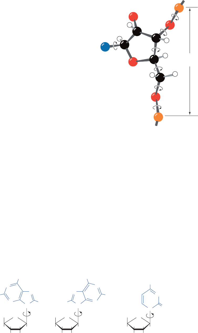
The rotation of a base about its glycosidic bond is
greatly hindered, as is best seen by the manipulation of a
space-filling molecular model. Purine residues have two
sterically permissible orientations relative to the sugar
known as the syn (Greek: with) and anti (Greek: against)
conformations (Fig. 29-6). For pyrimidines, only the
anti conformation is easily formed because, in the syn
conformation, the sugar residue sterically interferes with the
pyrimidine’s C2 substituent. In most double helical nucleic
acids, all bases are in the anti conformation (Fig. 29-1a,c,
left and middle panels). The exception is Z-DNA (Section
29-1Bb), in which the alternating pyrimidine and purine
residues are anti and syn (Fig. 29-1a,c, right panels). This
explains Z-DNA’s pyrimidine–purine alternation. Indeed,the
base pair flips that convert B-DNA to Z-DNA (Fig. 29-2)
are brought about by rotating each purine base about its gly-
cosidic bond from the anti to syn conformation, whereas it
is the sugars that rotate in the pyrimidine nucleotides,
thereby maintaining them in their anti conformations.
b. Sugar Ring Pucker Is Largely Limited to Only
a Few of Its Possible Arrangements
The ribose ring has a certain amount of flexibility that
significantly affects the conformation of the sugar–phos-
phate backbone. The vertex angles of a regular pentagon
are 108°, a value quite close to the tetrahedral angle
(109.5°), so that one might expect the ribofuranose ring to
be nearly flat. However, the ring substituents are eclipsed
when the ring is planar. To relieve the resultant crowding,
which even occurs between hydrogen atoms, the ring puck-
ers; that is, it becomes slightly nonplanar, so as to reorient
the ring substituents (Fig. 29-7; this is readily observed by
the manipulation of a skeletal molecular model).
1152 Chapter 29. Nucleic Acid Structures
To base
Nucleotide
unit
C
2
′
C
1
′
C
4
′
C
3
′
C
5
′
O
5
′
O
4
′
O
2
′
O
3
′
γ
δ
ε
β
α
ζ
χ
P
N
P′
H′5′
H5′
Figure 29-5 The conformation of a nucleotide unit is
determined by the seven indicated torsion angles.
Figure 29-6 The sterically allowed orientations of purine and pyrimidine bases with respect to their attached ribose units.
JWCL281_c29_1143-1172.qxd 7/9/10 12:46 AM Page 1152
C3′- endo
(b)
(a)
C2′- endo
7.0 Å
5.9 Å
P
P
P
P
5′
3′
2′
5′
3′
2′
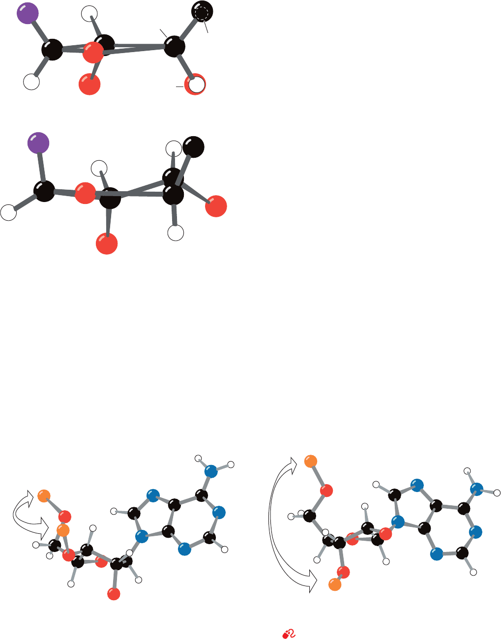
One would, in general, expect only three of a ribose
ring’s five atoms to be coplanar since three points define a
plane. Nevertheless, in the great majority of the 50 nucle-
oside and nucleotide crystal structures that have been
reported, four of the ring atoms are coplanar to within a
few hundredths of an angstrom and the remaining atom is
out of this plane by several tenths of an angstrom (the half-
chair conformation). If the out-of-plane atom is displaced
to the same side of the ring as atom C5¿, it is said to have
the endo conformation (Greek: endon, within), whereas
displacement to the opposite side of the ring from C5¿ is
known as the exo conformation (Greek: exo, out of). In the
great majority of known nucleoside and nucleotide struc-
tures (molecules that are subject to few of the conforma-
tional constraints of double helices), the out-of-plane atom
is either C2¿ or C3¿ (Fig. 29-8). C2¿-endo is the most fre-
quently occurring ribose pucker with C3¿-endo and C3¿-exo
also being common. Other ribose conformations are rare.
The ribose pucker is conformationally important in nu-
cleic acids because it governs the relative orientations of the
phosphate substituents to each ribose residue (Fig. 29-8). For
instance, it is difficult to build a regularly repeating model
of a double helical nucleic acid unless the sugars are either
C2¿-endo or C3¿-endo. In fact, canonical B-DNA has the
C2¿-endo conformation, whereas canonical A-DNA and
RNA-11 are C3¿-endo. In canonical Z-DNA, the purine nu-
cleotides are all C3¿-endo and the pyrimidine nucleotides
are C2¿-endo, which is another reason that the repeating
unit of Z-DNA is a dinucleotide. The sugar puckers ob-
served in the X-ray structures of A-DNA are, in fact, almost
entirely C3¿-endo. However, those of B-DNAs, although
predominantly C2¿-endo, exhibit significant variation in-
cluding C4¿-exo, O4¿-endo, C1¿-exo, and C3¿-exo. This varia-
tion in B-DNA’s sugar pucker is probably indicative of its
greater flexibility relative to other types of DNA helices.
c. The Sugar–Phosphate Backbone
Is Conformationally Constrained
If the torsion angles of the sugar–phosphate chain (Fig.
29-5) were completely free to rotate, there could probably
be no stable nucleic acid structure. However, the compari-
son, by Muttaiya Sundaralingam, of some 40 nucleoside
and nucleotide crystal structures has revealed that these
angles are really quite restricted. For example, the torsion
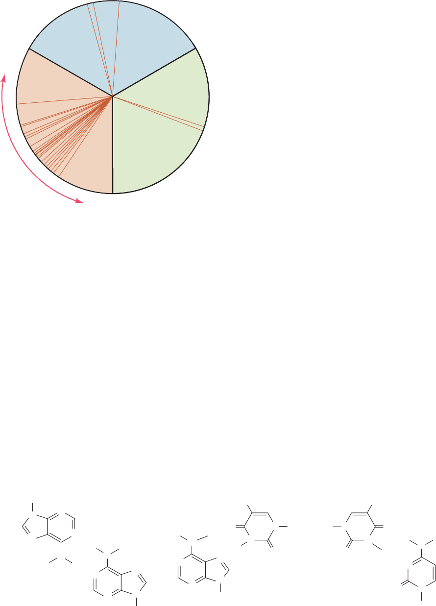
angle about the C4¿¬C5¿ bond ( in Fig. 29-5) is rather
narrowly distributed such that O4¿ usually has a gauche
Section 29-2. Forces Stabilizing Nucleic Acid Structures 1153
Figure 29-8 Nucleotide sugar conformations. (a) The C3¿-endo
conformation (on the same side of the sugar ring as C5¿), which
occurs in A-DNA and RNA-11. (b) The C2¿-endo conformation,
which occurs in B-DNA. The distances between adjacent P atoms
(a)
(b)
C
3
behind
H (eclipsed)
O (eclipsed)
C
1'
C
2'
C
3'
C
4'
C
1'
C
2'
C
4'
O
2'
O
3'
O
4'
Base
Base
O
4'
O
2'
3'
C
5'
C
5'
in the sugar–phosphate backbone are indicated. [After Saenger,
W. , Principles of Nucleic Acid Structure, p. 237, Springer-Verlag
(1983).]
See Kinemage Exercise 17-3
Figure 29-7 Ribose ring pucker. The substituents to (a) a
planar ribose ring (here viewed down the C3¿
¬C4¿ bond) are all
eclipsed.The resulting steric strain is partially relieved by ring
puckering such as in (b), a half-chair conformation in which C3¿
is the out-of-plane atom.
JWCL281_c29_1143-1172.qxd 8/2/10 8:06 PM Page 1153
N
N
N
N
N
N
N
N
H
H
H
H
H
N
N
CH
3
CH
3
...
...
H
...
...
N
N
N
N
N
N
N
R
H
H
O
O
CH
3
CH
3
...
...
H
N
N
N
R
H
O
N
N
O
O
CH
3
CH
3
(b) (c)
(a)
conformation (having a torsion angle of ⬃60°) with respect
to O5¿ (Fig. 29-9).This is because the presence of the ribose
ring together with certain noncovalent interactions of the
phosphate group stiffens the sugar–phosphate chain by
restricting its range of torsion angles. These restrictions are
even greater in polynucleotides because of steric interfer-
ence between residues.
The sugar–phosphate conformational angles of the var-
ious double helices are all reasonably strain free. Double
helices are therefore conformationally relaxed arrange-
ments of the sugar–phosphate backbone. Nevertheless, the
sugar–phosphate backbone is by no means a rigid struc-
ture, so, on strand separation, it assumes a random coil
conformation.
B. Base Pairing
Base pairing is apparently a “glue” that holds together
double-stranded nucleic acids. Only Watson–Crick pairs
occur in the crystal structures of self-complementary
oligonucleotides. It is therefore important to understand
how Watson–Crick base pairs differ from other doubly hy-
drogen bonded arrangements of the bases that have rea-
sonable geometries (e.g., Fig. 29-10).
a. Unconstrained A ⴢ T Base Pairs Assume
Hoogsteen Geometry
When monomeric adenine and thymine derivatives are
cocrystallized, the A ⴢ T base pairs that form invariably
have adenine N7 as the hydrogen bonding acceptor
(Hoogsteen geometry; Fig. 29-10b) rather than N1 (Watson–
Crick geometry; Fig. 5-12). This suggests that Hoogsteen
geometry is inherently more stable for A ⴢ T pairs than is
Watson–Crick geometry. Apparently steric and other envi-
ronmental influences make Watson–Crick geometry the
preferred mode of base pairing in double helices. A ⴢ T
pairs with Hoogsteen geometry are nevertheless of biolog-
ical importance; for example, they help stabilize the terti-
ary structures of tRNAs (Section 32-2Ba). In contrast,
monomeric G ⴢ C pairs always cocrystallize with Watson–
Crick geometry as a consequence of their triply hydrogen
bonded structures.
b. Non-Watson–Crick Base Pairs Are of Low Stability
The bases of a double helix, as we have seen (Section 5-
3A), associate such that any base pair position may
interchangeably be A ⴢ T, T ⴢ A, G ⴢ C, or C ⴢ G without af-
fecting the conformations of the sugar–phosphate chains.
One might reasonably suppose that this requirement of
geometric complementarity of the Watson–Crick base
pairs, A with T and G with C, is the only reason that other
1154 Chapter 29. Nucleic Acid Structures
Figure 29-9 Conformational wheel showing the distribution
of the torsion angle about the C4¿
¬C5¿ bond. The torsion angle
( in Fig. 29-5) was measured in 33 X-ray structures of nucleosides,
nucleotides, and polynucleotides. Each radial line represents the
position of the C4¿
¬O4¿ bond in a single structure relative to the
substituents of C5¿ as viewed from C5¿ to C4¿. Note that most of
the observed torsion angles fall within a relatively narrow range.
[After Sundaralingam, M., Biopolymers 7, 838 (1969).]
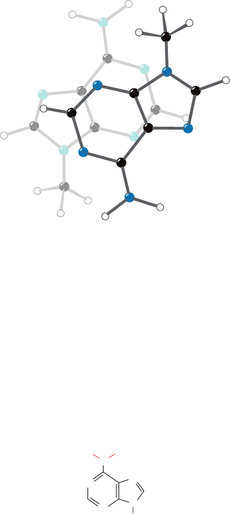
Figure 29-10 Some non-Watson–Crick base pairs. (a) The
pairing of adenine residues in the crystal structure of
9-methyladenine. (b) Hoogsteen pairing between adenine and
thymine residues in the crystal structure of 9-methyladenine ⴢ
H5'
H'5'
O5'
O4'
position
1-methylthymine. (c) A hypothetical pairing between cytosine
and thymine residues. Compare these base pairs with the
Watson–Crick base pairs in Fig. 5-12.
JWCL281_c29_1143-1172.qxd 7/8/10 8:39 PM Page 1154
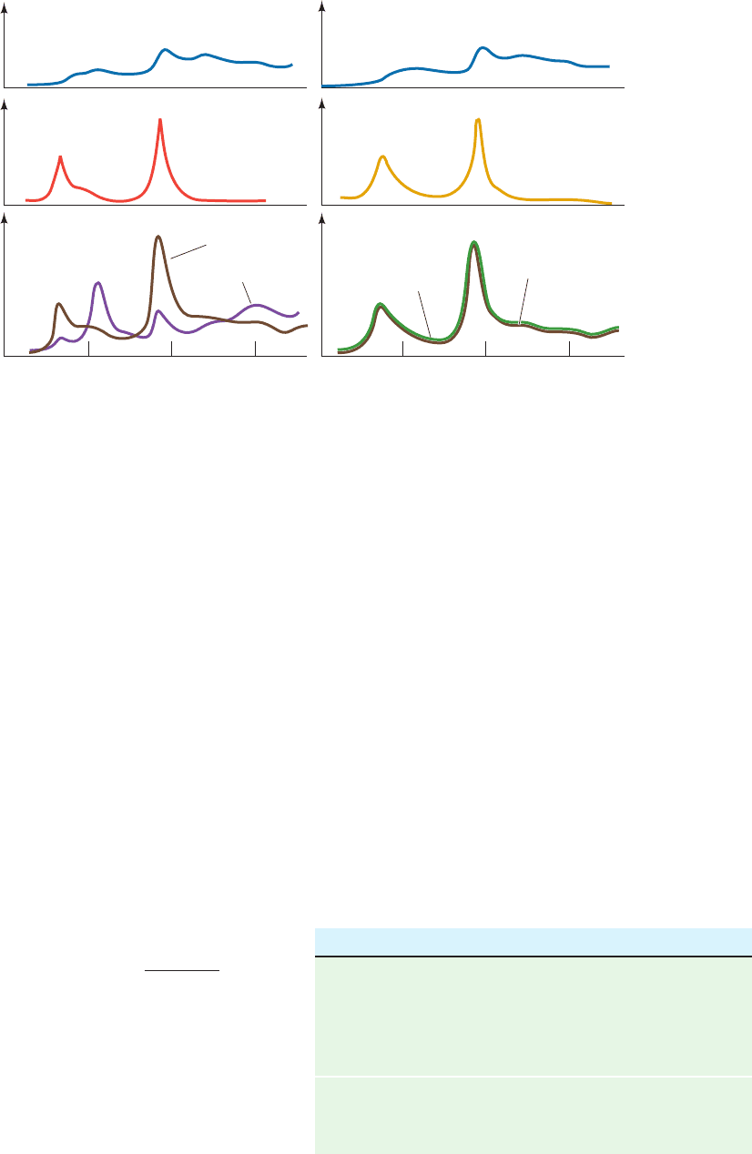
base pairs do not occur in a double helical environment. In
fact, this was precisely what was believed for many years
after the DNA double helix was discovered.
Eventually, the failure to detect pairs of different bases
in nonhelical environments other than A with T (or U) and
G with C led Richard Lord and Rich to demonstrate,
through spectroscopic studies, that only the bases of
Watson–Crick pairs have a high mutual affinity. Figure 29-11a
shows the infrared (IR) spectrum in the N¬H stretch region
of guanine and cytosine derivatives, both separately and in a
mixture.The band in the spectrum of the G C mixture that
is not present in the spectra of either of its components is in-
dicative of a specific hydrogen bonding interaction be-
tween G and C. Such an association, which can occur
between like as well as unlike molecules, may be described
by ordinary mass action equations.
[29.1]
From analyses of IR spectra such as Fig. 29-11, the val-
ues of K for the various base pairs have been determined.
The self-association constants of the Watson–Crick bases
are given in the top of Table 29-2 (the hydrogen bonded as-
sociation of like molecules is indicated by the appearance
of new IR bands as the concentration of the molecule is in-
creased).The bottom of Table 29-2 lists the association con-
stants of the Watson–Crick pairs. Note that each of these
latter quantities is larger than the self-association constants
of either of their component bases, so that Watson–Crick
B
1
B
2
Δ B
1
ⴢ B
2
K
[B
1
ⴢ B
2
]
[B
1
][B
2
]
base pairs preferentially form from their constituents. In
contrast, the non-Watson–Crick base pairs,A ⴢ C, A ⴢ G, C ⴢ
U, and G ⴢ U, whatever their geometries, have association
constants that are negligible compared with the self-pair-
ing association constants of their constituents (e.g., Fig. 29-
11b). Evidently, a second reason that non-Watson–Crick
base pairs do not occur in DNA double helices is that they
have relatively little stability. Conversely, the exclusive pres-
ence of Watson–Crick base pairs in DNA results, in part,
from an electronic complementarity matching A to T and
G to C.The theoretical basis of this electronic complemen-
tarity, which is an experimental observation, is obscure.
Section 29-2. Forces Stabilizing Nucleic Acid Structures 1155
Figure 29-11 The infrared spectra, in the N¬H stretch region,
of guanine, cytosine, and adenine derivatives. The derivatives
were analyzed both separately and in the indicated mixtures.The
solvent, CDCl
3
, does not hydrogen bond with the bases and is
relatively transparent in the frequency range of interest. (a) G
C.The brown curve in the lower panel, which is the sum of the
spectra in the two upper panels, is the calculated spectrum of
Relative absorbance
3500
(a) (b)
3400 3300 3500 3400 3300
Wave number (cm
–1
)
Observed
Calculated sum
Observed
GG0.0008
CA
G + C
0.0016 G + A 0.0016MM
M0.0008M
0.0008M 0.0008M
Calculated sum
a
Data measured in deuterochloroform at 25°C.
Source: Kyogoku, Y., Lord, R.C., and Rich,A., Biochim. Biophys. Acta
179, 10 (1969).
Table 29-2 Association Constants for Base Pair Formation
Base Pair K (M
1
)
a
Self-Association
A ⴢ A 3.1
U ⴢ U 6.1
C ⴢ C28
G ⴢ G10
3
–10
4
Watson–Crick Base Pairs
A ⴢ U 100
G ⴢ C10
4
–10
5
G C for noninteracting molecules.The band near 3500 cm
1
in
the observed G C spectrum (purple) is indicative of a specific
hydrogen bonding association between G and C. (b) G A.The
close match between the calculated and observed spectra of the
G A mixture indicates that G and A do not significantly
interact. [After Kyogoku, Y., Lord, R.C., and Rich, A., Science
154, 5109 (1966).]
JWCL281_c29_1143-1172.qxd 8/2/10 8:06 PM Page 1155
This is because the approximations inherent in theoretical
treatments make them unable to accurately account for the
minor (few kJ ⴢ mol
1
) energy differences between specific
and nonspecific hydrogen bonding associations. The dou-
ble helical segments of many RNAs, however, contain oc-
casional non-Watson–Crick base pairs, most often G ⴢ U,
which have functional as well as structural significance
(e.g., Sections 32-2Ba and 32-2Db).
c. Hydrogen Bonds Only Weakly Stabilize DNA
It is clear that hydrogen bonding is required for the
specificity of base pairing in DNA that is ultimately re-
sponsible for the enormous fidelity required to replicate
DNA with almost no error (Section 30-3D). Yet, as is also
true for proteins (Section 8-4Ba), hydrogen bonding con-
tributes little to the stability of the double helix. For instance,
adding the relatively nonpolar ethanol to an aqueous DNA
solution, which strengthens hydrogen bonds, destabilizes
the double helix, as is indicated by its decreased melting
temperature (T
m
; Section 5-3Ca). This is because hy-
drophobic forces, which are largely responsible for DNA’s
stability (Section 29-2C), are disrupted by nonpolar sol-
vents. In contrast, the hydrogen bonds between the base
pairs of native DNA are replaced in denatured DNA by en-
ergetically nearly equivalent hydrogen bonds between the
bases and water. This accounts for the thermodynamic
observation that hydrogen bonding contributes only 2 to
8 kJ/mol to base pairing stability.
C. Base Stacking and Hydrophobic Interactions
Purines and pyrimidines tend to form extended stacks of
planar parallel molecules. This has been observed in the
structures of nucleic acids (e.g., Fig. 29-1) and in the several
hundred reported X-ray crystal structures that contain nu-
cleic acid bases. The bases in these structures are usually
partially overlapped (e.g., Fig. 29-12). In fact, crystal struc-
tures of chemically related bases often exhibit similar
stacking patterns. Apparently stacking interactions, which
in the solid state are a form of van der Waals interaction
(Section 8-4Ab), have some specificity, although certainly
not as much as base pairing.
a. Nucleic Acid Bases Stack in Aqueous Solution
Bases aggregate in aqueous solution, as has been demon-
strated by the variation of osmotic pressure with concen-
tration.The van’t Hoff law of osmotic pressure is
[29.2]
where is the osmotic pressure, m is the molality of the
solute (mol solute/kg solvent), R is the gas constant, and T
is the temperature. The molecular mass, M, of an ideal
solute can be determined from its osmotic pressure since
M c/m, where c g solute/kg solvent.
If the species under investigation is of known molecular
mass but aggregates in solution, Eq. [29.2] must be rewrit-
ten:
[29.3]RTm
RTm
where , the osmotic coefficient, indicates the solute’s de-
gree of association. varies from 1 (no association) to 0
(infinite association).The variation of with m for nucleic
acid bases in aqueous solution (e.g., Fig. 29-13) is consistent
with a model in which the bases aggregate in successive
steps:
where n is at least 5 (if the reaction goes to completion,
1/n). This association cannot be a result of hydrogen
bonding since N
6
, N
6
-dimethyladenosine,
which cannot form interbase hydrogen bonds, has a greater
degree of association than does adenosine (Fig. 29-13).Ap-
parently the aggregation arises from the formation of stacks
of planar molecules. This model is corroborated by proton
NMR studies: The directions of the aggregates’ chemical
shifts are compatible with a stacked but not a hydrogen
bonded model. The stacking associations of monomeric
bases are not observed in nonaqueous solutions.
Single-stranded polynucleotides also exhibit stacking in-
teractions. For example, poly(A) shows a broad increase of
CH
3
N
N
N
N
N
H
3
C
Ribose
N
6
, N
6
-Dimethyladenosine
A A Δ A
2
A Δ A
3
A Δ
# # #
Δ A
n
1156 Chapter 29. Nucleic Acid Structures
Figure 29-12 Stacking of adenine rings in the crystal structure
of 9-methyladenine. The partial overlap of the rings is typical of
the association between bases in crystal structures and in double
helical nucleic acids. [After Stewart, R.F. and Jensen, L.H., J.
Chem. Phys. 40, 2071 (1964).]
JWCL281_c29_1143-1172.qxd 8/2/10 8:06 PM Page 1156
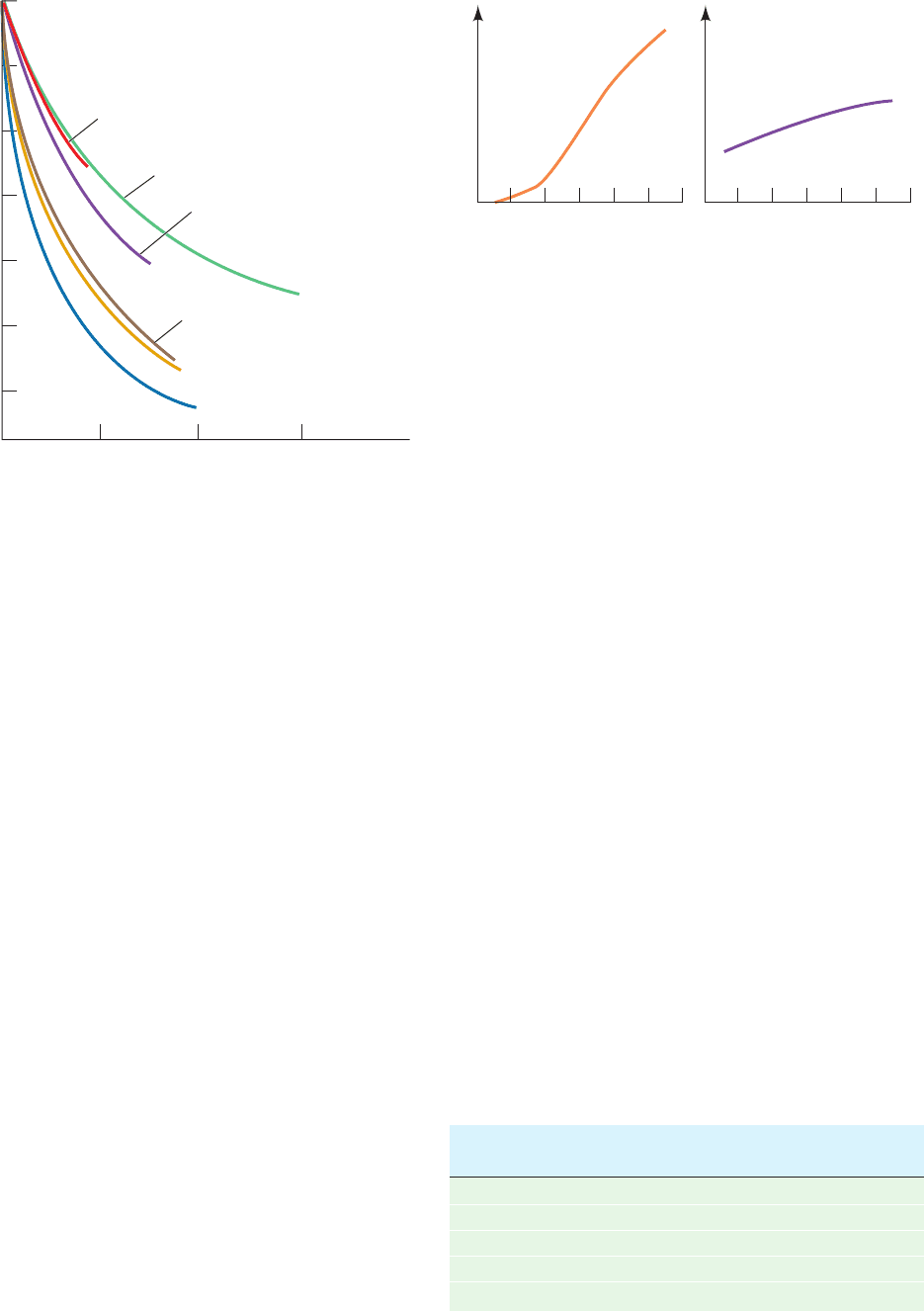
UV absorbance with temperature (Fig. 29-14a).This hyper-
chromism (which is indicative of nucleic acid denaturation;
Section 5-3Ca) is independent of poly(A) concentration, so
that it cannot be a consequence of intermolecular disaggre-
gation. Likewise, it is not due to a reduction in intramolec-
ular hydrogen bonding because poly(N
6
,N
6
-dimethyl-
adenosine) exhibits a greater degree of hyperchromism
than does poly(A). The hyperchromism must therefore
arise from some sort of stacking associations within a single
strand that melt out with increasing temperature.This is not
a very cooperative process, as is indicated by the broadness
of the melting curve and the observation that short polynu-
cleotides, including dinucleoside phosphates such as ApA,
exhibit similar melting curves (Fig. 29-14b).
b. Nucleic Acid Structures Are Stabilized
by Hydrophobic Forces
Stacking associations in aqueous solutions are largely
stabilized by hydrophobic forces. One might reasonably
suppose that hydrophobic interactions in nucleic acids are
similar in character to those that stabilize protein struc-
tures. However, closer examination reveals that these two
types of interactions are qualitatively different in charac-
ter. Thermodynamic analysis of dinucleoside phosphate
melting curves in terms of the reaction
(Table 29-3) indicates that base stacking is enthalpically
Δ dinucleoside phosphate(stacked)
Dinucleoside phosphate(unstacked)
driven and entropically opposed. Thus the hydrophobic in-
teractions responsible for the stability of base stacking asso-
ciations in nucleic acids are diametrically opposite in char-
acter to those that stabilize protein structures (which are
enthalpically opposed and entropically driven; Section 8-
4C). This is reflected in the differing structural properties
of these interactions. For example, the aromatic side chains
of proteins are almost never stacked and the crystal struc-
tures of aromatic hydrocarbons such as benzene, which re-
semble these side chains, are characteristically devoid of
stacking interactions.
Hydrophobic forces in nucleic acids are poorly under-
stood. The observation that they are different in character
from the hydrophobic forces that stabilize proteins is nev-
ertheless not surprising because the nitrogenous bases are
considerably more polar than the hydrocarbon residues of
proteins that participate in hydrophobic interactions.
There is, however, no theory available that adequately ex-
plains the nature of hydrophobic forces in nucleic acids
(our understanding of hydrophobic forces in proteins, it
will be recalled, is similarly incomplete).They are complex
interactions of which base stacking is probably a signifi-
cant component. Whatever their origins, hydrophobic
forces are of central importance in determining nucleic
acid structures.
Section 29-2. Forces Stabilizing Nucleic Acid Structures 1157
Figure 29-13 Variation of the osmotic coefficient with the
molal concentrations m of adenosine derivatives in H
2
O. The
decrease of with increasing m indicates that these derivatives
aggregate in solution. [After Broom, A.D., Schweizer, M.P., and
Ts’o, P.O.P., J. Am. Chem. Soc. 89, 3613 (1967).]
Figure 29-14 Melting curves for poly(A) and ApA. The broad
temperature range of hyperchromic shifts at 258 nm of (a) poly(A)
and (b) ApA is indicative of noncooperative conformational
changes in these substances. Compare this figure with Fig.
5-16. [After Leng, M. and Felsenfeld, G., J. Mol. Biol. 15, 457 (1966).]
Adenosine
2'-
O -Methyladenosine
2'-Deoxyadenosine
N
6
-Methyladenosine
N
6
-Methyl-2'-deoxyadenosine
N
6
,N
6
-Dimethyladenosine
Osmotic coefficient, φ
1.0
0.9
0.8
0.7
0.6
0.5
0.4
0.1 0.2 0.3
m
Relative absorbance at 258 nm
Temperature (°C)
0 20406080100 0 20406080100
(a) Poly(A) (b) ApA
Source: Davis, R.C. and Tinoco, I., Jr., Biopolymers 6, 230 (1968).
Table 29-3 Thermodynamic Parameters for the Reaction
Dinucleoside phosphate 34 dinucleoside phosphate
(unstacked)(stacked)
Dinucleoside H
stacking
T S
stacking
Phosphate (kJ ⴢ mol
1
) (kJ ⴢ mol
1
at 25°C)
ApA 22.2 24.9
ApU 35.1 39.9
GpC 32.6 34.9
CpG 20.1 21.2
UpU 32.6 36.2
JWCL281_c29_1143-1172.qxd 8/2/10 8:06 PM Page 1157
D. Ionic Interactions
Any theory of the stability of nucleic acid structures must
take into account the electrostatic interactions of their
charged phosphate groups. Polyelectrolyte theory approxi-
mates the electrostatic interactions of DNA by considering
the anionic double helix to be a homogeneously charged
line or cylinder. We shall not discuss the details of this the-
ory here, but note that it is often in reasonable agreement
with experimental observations.
The melting temperature of duplex DNA increases with
the cation concentration because these ions bind more
tightly to duplex DNA than to single-stranded DNA due to
the duplex DNA’s higher anionic charge density. An in-
creased salt concentration therefore shifts the equilibrium
toward the duplex form, thus increasing the DNA’s T
m
.The
observed relationship for Na
is
[29.4]
where X
GC
is the mole fraction of G ⴢ C base pairs (recall
that T
m
increases with the G C content; Fig. 5-17);
the equation is valid in the ranges 0.3 X
GC
0.7 and
10
3
M [Na
] 1.0M. Other monovalent cations such as
Li
and K
have similar nonspecific interactions with phos-
phate groups. Divalent cations, such as Mg
2
,Mn
2
, and
Co
2
, in contrast, specifically bind to phosphate groups, so
that divalent cations are far more effective shielding agents
for nucleic acids than are monovalent cations. For example, an
Mg
2
ion has an influence on the DNA double helix compa-
rable to that of 100 to 1000 Na
ions. Indeed, enzymes that
mediate reactions with nucleic acids or just nucleotides (e.g.,
ATP) usually require Mg
2
for activity. Moreover, Mg
2
ions
play an essential role in stabilizing the complex structures
assumed by many RNAs such as transfer RNAs (tRNAs;
Section 32-2B) and ribosomal RNAs (rRNAs; Section 32-3A).
3 SUPERCOILED DNA
See Guided Exploration 24: DNA supercoiling Genetic analyses
indicate that numerous viruses and bacteria have circular
genetic maps, which implies that their chromosomes are
T
m
41.1X
GC
16.6 log[Na
] 81.5
likewise circular. This conclusion has been confirmed by
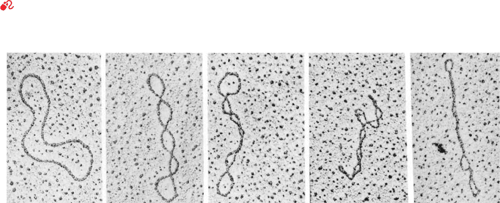
electron micrographs in which circular DNAs are seen
(Fig. 29-15). Some of these circular DNAs have a peculiar
twisted appearance, a phenomenon that is known equiva-
lently as supercoiling, supertwisting, and superhelicity. Su-
percoiling arises from a biologically important topological
property of covalently closed circular duplex DNA that is
the subject of this section. It is occasionally referred to as
DNA’s tertiary structure.
A. Superhelix Topology
Consider a double helical DNA molecule in which both
strands are covalently joined to form a circular duplex mol-
ecule as is diagrammed in Fig. 29-16 (each strand can be
joined only to itself because the strands are antiparallel). A
geometric property of such an assembly is that the number
of times one strand wraps about the other cannot be altered
without first cleaving at least one of its polynucleotide
strands. You can easily demonstrate this to yourself with a
buckled belt in which each edge of the belt represents a
strand of DNA.The number of times the belt is twisted be-
fore it is buckled cannot be changed without unbuckling or
cutting the belt (cutting a polynucleotide strand).
This phenomenon, as James White proved mathemati-
cally in 1969, is expressed
[29.5]
in which:
1. L, the linking number (also symbolized Lk), is the
number of times that one DNA strand winds about the other.
This integer quantity is most easily counted when the mole-
cule’s duplex axis is constrained to lie in a plane (see below).
However, the linking number is invariant no matter how the
circular molecule is twisted or distorted so long as both its
polynucleotide strands remain covalently intact; the linking
number is therefore a topological property of the molecule.
2. T, the twist (also symbolized Tw ), is the number of
complete revolutions that one polynucleotide strand
makes about the duplex axis in the particular conformation
L T W
1158 Chapter 29. Nucleic Acid Structures
Figure 29-15 Electron micrographs of circular duplex DNAs.
Their conformations vary from no supercoiling (left) to tightly
supercoiled (right). [Electron micrographs by Laurien Polder.
From Kornberg, A. and Baker,T.A., DNA Replication (2nd ed.),
p. 36, W.H. Freeman (1992). Used with permission.]
JWCL281_c29_1143-1172.qxd 8/2/10 8:06 PM Page 1158
Large writhing
number,
small twist
Small writhing
number,
large twist
under consideration. By convention, T is positive for right-
handed duplex turns, so that, for B-DNA in solution, the
twist is normally the number of base pairs divided by 10.5
(the number of base pairs per turn of the B-DNA double
helix under physiological conditions; see Section 29-3Bc).
3. W, the writhing number (also symbolized Wr), is the
number of turns that the duplex axis makes about the su-
perhelix axis in the conformation of interest. Its value is
readily determined by projecting the DNA onto a plane
and counting the number of times the duplex axis crosses
itself (Fig. 29-17). The writhing number is a measure of the
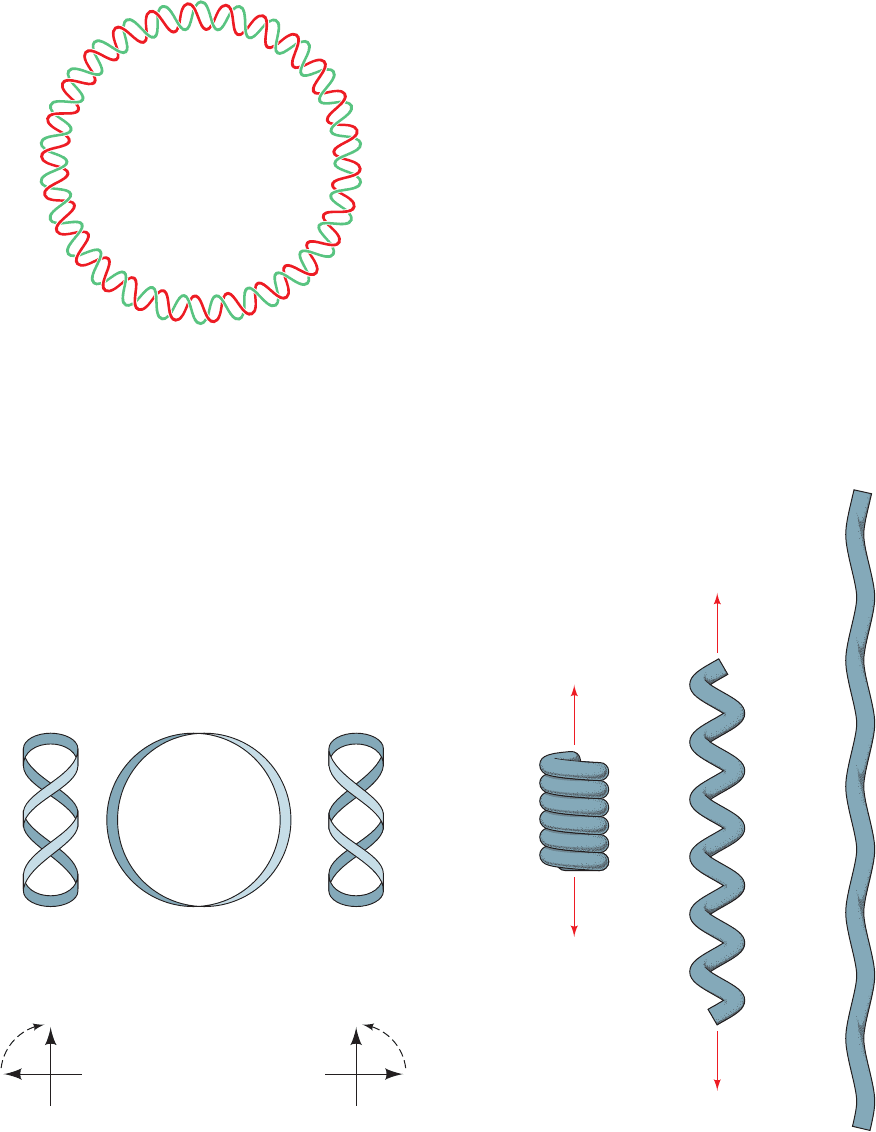
DNA’s superhelicity. The difference between writhing and
twisting is illustrated by the familiar example in Fig. 29-18.
W 0 when the DNA’s duplex axis is constrained to lie in
a plane (e.g., Fig. 29-16); then L T, so L may be evaluated
by counting the DNA’s duplex turns.
The two DNA conformations diagrammed on the right of
Fig. 29-19 are topologically equivalent; that is, they have
the same linking number, L, but differ in their twists and
writhing numbers. Note that T and W need not be integers
(at least mathematically), only L. Although, strictly speak-
ing, superhelicity is only defined for covalently closed cir-
cular duplex DNA, a linear segment of duplex DNA that is
mechanically constrained from rotating at both ends (e.g.,
by protein anchors) has identical topological properties.
Section 29-3. Supercoiled DNA 1159
Figure 29-16 Schematic diagram of covalently closed circular
duplex DNA that has 26 double helical turns. Its two
polynucleotide strands are said to be topologically bonded to
each other because, although they are not covalently linked, they
cannot be separated without breaking covalent bonds.
Figure 29-17 Topological relationships in covalently closed
duplex DNA. (a) DNA molecules are represented by circular
ribbons. DNA with no torsional strain is said to be relaxed.
Underwinding or overwinding produces negative (1) or
positive (1) supercoils. (b) A negative writhe has a crossover in
which a clockwise rotation of the front segment of 180° aligns
it over the back segment, whereas a positive writhe has the
corresponding counterclockwise rotation. [After Deweese, J.E.,
Osheroff, M.A., and Osheroff, N., Biochem. Mol. Biol. Educ. 37, 2
(2009).]
Figure 29-18 The difference between writhing and twist as
demonstrated by a coiled telephone cord. Here the cord
represents a double helical DNA molecule. In its relaxed state
(left), the cord is in a helical form that has a large writhing
number and a small twist.As the coil is pulled out (middle) until
it is nearly straight (right), its writhing number becomes small as
its twist becomes large.
L = 26
Negative
supercoiling
Positive
supercoiling
Relaxed
Node convention
(
1)
(
1)
(
1)(1)
(
1)
(
1)
(a)
(b)
JWCL281_c29_1143-1172.qxd 7/8/10 8:39 PM Page 1159
(a) Toroidal
(b) Interwound
Since L is constant in an intact duplex DNA circle, for
every new double helical twist, T, there must be an equal and
opposite superhelical twist, that is, W T. For example,
a closed circular DNA without supercoils (Fig. 29-19, upper
right) can be converted to a negatively supercoiled confor-
mation (Fig. 29-18, lower right) by winding the duplex helix
the same number of positive (right-handed) turns.
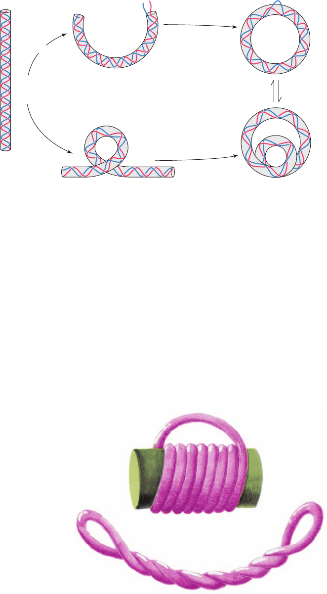
a. Supercoils May Be Toroidal or Interwound
A supercoiled duplex may assume two topologically
equivalent forms:
1. A toroidal helix in which the duplex axis is wound as
if about a cylinder (Fig. 29-20a).
2. An interwound helix in which the duplex axis is
twisted around itself (Fig. 29-20b).
Note that these two interconvertible superhelical forms
have opposite handedness. Since left-handed toroidal turns
may be converted to left-handed duplex turns (e.g., Fig. 29-
19), left-handed toroidal turns and right-handed inter-
wound turns both have negative writhing numbers.Thus an
underwound duplex (T number of bp/10.5), for example,
will tend to develop right-handed interwound or left-
handed toroidal superhelical turns when the constraints
causing it to be underwound are released (the molecular
forces in a DNA double helix promote its winding to its
normal number of helical turns).
b. Supercoiled DNA Is Relaxed by Nicking
One Strand
Supercoiled DNA may be converted to relaxed circles
(as appears in the leftmost panel of Fig. 29-15) by treatment
with pancreatic DNase I, an endonuclease (an enzyme that
cleaves phosphodiester bonds within a polynucleotide
strand) that cleaves only one strand of a duplex DNA. One
single-strand nick is sufficient to relax a supercoiled DNA.
This is because the sugar–phosphate chain opposite the
nick is free to swivel about its backbone bonds (Fig. 29-5) so
as to change the molecule’s linking number and thereby al-
ter its superhelicity. Supercoiling builds up elastic strain in a
DNA circle, much as it does in a rubber band. This is why
the relaxed state of a DNA circle is not supercoiled.
B. Measurements of Supercoiling
Supercoiled DNA, far from being just a mathematical cu-
riosity, has been widely observed in nature. In fact, its dis-
covery in polyomavirus DNA by Jerome Vinograd stimu-
lated the elucidation of the topological properties of
superhelices rather than vice versa.
a. Intercalating Agents Control Supercoiling by
Unwinding DNA
All naturally occurring DNA circles are underwound;
that is, their linking numbers are less than those of their
1160 Chapter 29. Nucleic Acid Structures
Figure 29-19 Two ways of introducing one
supercoil into a DNA with 10 duplex turns.
The two closed circular forms shown (right)
are topologically equivalent; that is, they are
interconvertible without breaking any
covalent bonds.The linking number L, twist T,
and writhing number W are indicated for each
form. Strictly speaking, the linking number is
only defined for a covalently closed circle.
Figure 29-20 Toroidal and interwound supercoils. A rubber
tube that has been (a) toroidally coiled in a left-handed helix
around a cylinder with its ends joined such that it has no twist
jumps to (b) an interwound helix with the opposite handedness
when the cylinder is removed. Neither the linking number, the
twist, nor the writhing number are changed in this
transformation.
L = 10
T = 10
W = 0
L = 9
T = 9
W = 0
L = 9
T = 9
W = 0
L = 9
T = 10
W = –1
L = 9
T = 10
W = –1
Unwind one turn
Turn once
Close circle
Close circle
Equivalent
topologically
JWCL281_c29_1143-1172.qxd 7/9/10 12:46 AM Page 1160
