Seuront L. Fractals and Multifractals in Ecology and Aquatic Science
Подождите немного. Документ загружается.

174 Fractals and Multifractals in Ecology and Aquatic Science
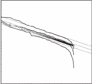
a log-log plot, exhibits the different characteristic features previously identied: a power-law behav-
ior not signicantly different from the original one (that is, a′ ≈ a = 0.24) followed by a roll-off
toward low Y
8r
values (Figure 5.20). As stated above, successive positive random uctuations might
lead to local increasing trends slightly diverging from a power law (gray arrow) but should not be
associated with a step function.
Next, consider a situation where the positive and negative uctuations are purely randomly
driven as:
Y
9r
= X
r
± kX
r
(5.33)
and
Y
10r
= X
r
± eX
r
(5.34)
where k is a constant (0 ≤ k ≤ 1) and e is a random-noise process, that is, e ∈ [0, 1], whose amplitude
is dened as being a given percentage of the maximum value of X
r
; k and e are randomly chosen as
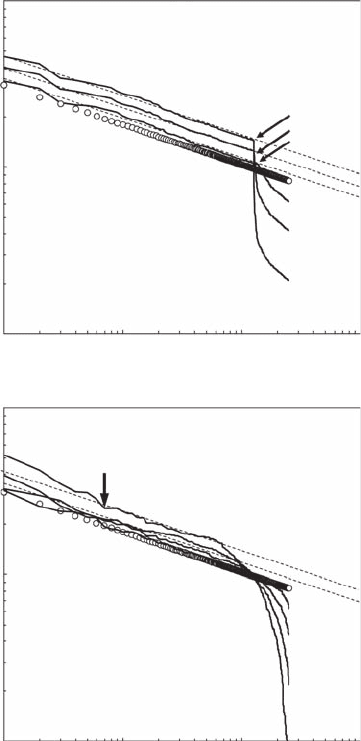
being positive or negative. The resulting Zipf signatures of the functions Y
9r
and Y
10r
are shown in
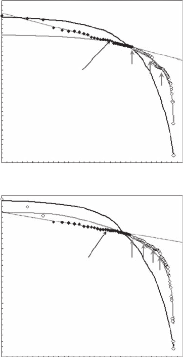
Figure 5.21 as log-log plots. The positive and negative components of Equation (5.33) clearly appear
as separated by a step function (Figure 5.21A). The positive components lead to power laws that are
not signicantly different from the original. A separate analysis of the range of values separated
from the power laws by step functions (arrows; Figure 5.21A) did not show any power-law behavior.
Alternatively, the effects of Equation (5.34) on the initial power-law behavior are the characteristic
downward roll-off signature related to randomness and the uctuations around a power-law behav-
ior that are not signicantly different from the original (Figure 5.21B). To ensure the relevance of
Zipf analysis, as introduced by Equations (5.17) and (5.22), the few data points diverging upward,
or attening, toward the rst-rank values must not be included in the regression analysis aimed at
estimating a. Indeed, the former case describes a distribution marked by the dominance of a few
(ultimately one) “hotspots” that is likely to be chronically undersampled (Seuront et al. 1999), while
the latter case refers to a distribution that has been systematically oversampled. These issues have
100
10
1
1 10010 1000
r
Y
8r
Figure 5.20 Log-log plot signature of the Zipf behavior expected in case of a power law X
r
(open dots)
competing with random growth and mortality components (Y
8r
= X
r
+ (e
1
− e
2
) X
r
), where the random processes
e
1
and e
2
have been chosen as e
1
= 0.75 and e
1
= 0.25, e
1
= 0.50 and e
1
= 0.25, and and e
2
= 0.75 (from bottom
to top). The dashed lines indicate the power-law behavior of the initial values X
r
. (Modied from Seuront and
Mitchell, 2008.)
2782.indb 174 9/11/09 12:11:38 PM
Frequency Distribution Dimensions 175
been specically detailed elsewhere in the framework of information theory (Mandelbrot 1953) and
led to the modied version of the generalized Zipf law presented above; see Equation (5.19).
It is now known that the distributions of nutrients, phytoplankton, and zooplankton exhibit differ-
ent levels of persistence (Tsuda 1995; Seuront et al. 1996a, 1996b, 1999, 2002; Seuront and Lagadeuc
2001). In addition, the interplay between the biotic properties of individuals and populations and
abiotic processes produce space-time structures characterized by long-range correlation (that is,
persistence) (Kendall et al. 2000). We consider, nally, a situation where the local concentration of
a phytoplankton population initially driven by a power law (X
r
∝ r
−0.24
) could be inuenced by a
fractional Brownian motion resulting from the combined effects of local biological (nutrient uptake,
100
10
A
1
1 10010 1000
r
Y
9r
100
10
B
1
1 10010 1000
r
Y
10r
Figure 5.21 Log-log plot signature of the Zipf behavior expected in case of a power law X
r
(open dots)
competing with combined constant random and growth components (Y
9r
= X
r
± kX
r
; (A) and combined random
growth and mortality components (Y
9r
= X
r
± eX
r
; (B), where the constant k and the noise e have been chosen
as k = 0.25, 0.50, and 0.75, and e = 0.25, 0.50, 0.75, and 100 (from bottom to top). The arrows indicate a step
function (A) and the beginning of a local departure from a pure power law due to successively increasing
random increments (B). The dashed lines indicate the power-law behavior of the initial values X
r
. (Modied
from Seuront and Mitchell, 2008.)
2782.indb 175 9/11/09 12:11:40 PM
176 Fractals and Multifractals in Ecology and Aquatic Science
inter- and intraspecic competition, grazing pressure, infection) and physical (advection, diffusion,
turbulence) processes following
Y
11r
= X
r
± fBmX
r
(5.35)
where fBm is a persistent fractional Brownian motion (see Figure 5.9b) whose amplitude is dened
as being a given percentage of the maximum value of X
r
, and randomly chosen as being positive
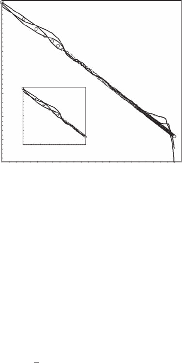
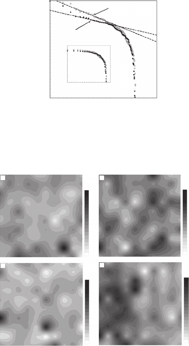
or negative. The resulting Zipf signature (Figure 5.22) exhibits the downward roll-off characteris-
tic of randomness for high rank values, and long-range correlations around a power-law behavior
(Y
11r
∝ r
−a
′, with a′ = 0.240 ± 0.005;
x ±SD
) that is signicantly different from the original power
law (X
r
∝ r
−0.24
, p > 0.05). These long-range correlations exist whatever the values of r but are more
clearly visible for the low values of r, that is, high values of X
r
in Equation (5.35).
5.5.5.1.6 On the Relevance of Zipf’s Law to Diagnose Ecosystem Complexity
The previous sections illustrate the potential for the seldom-used Zipf’s law to be a powerful tool in
the analysis and the classication of marine and terrestrial ecosystems in the presence of random-
ness, monotonic and periodic trends, internal and external noise, and both biotic and abiotic forc-
ings. Specically, Zipf analysis can be directly and easily applied to any data set without intensive
computational, mathematical, or statistical analysis, and with a minimum amount of calculation.
It can be conducted in a few minutes with most standard software packages, even for a data set
of several thousand data points. The results of Zipf analysis should not, however, be used without
a preliminary visual inspection of the data (an absolute prerequisite in data analysis that is often
neglected, especially by undergraduate students), as they could erroneously be used as a direct index
of patchiness. For instance, a distribution characterized by a patch of 10 high-density data points,
10 randomly or regularly spaced hotspots, or 10 ranked hotspots will return exactly the same Zipf
shape. This issue has also been raised in the framework of power spectrum analysis in marine ecol-
ogy (Franks 2005).
The one-to-one correspondence between Zipf and Pareto distributions analytically derived here
(see Box 5.1) could be regarded as a way to reconcile previous and future works using one or
1.5
1.2
1.3
1.4
1.1
1.0
0.9
0.8
0.0 0.5
0.0 0.2 0.4 0.6 0.8 1.0 1.2 1.4
1.5
1.1
1.2
1.3
1.4
2.01.0 1.5 2.5
Log r
Log Y
11r
Log Y
11r
Log r
Figure 5.22 Log-log plot signature of the Zipf behavior expected in case of a power law X
r
(open dots) com-
peting with a persistent fractional Brownian motion. (Modied from Seuront and Mitchell, 2008.)
2782.indb 176 9/11/09 12:11:44 PM
Frequency Distribution Dimensions 177
the other technique. The strength of the Zipf framework also has the desirable properties of not
requiring any assumptions about the statistical distribution, regularity of sampling intervals, and
stationarity of the data set that are sometimes absolute prerequisites to some statistical data analy-
sis techniques.
Finally, the Zipf framework can be conveniently used as a tool to identify and classify structures
in marine ecosystems and also to infer the underlying processes that generate the observed pat-
terns. The characteristic shapes introduced above, and most importantly their potential changes in
time and space, make it possible to hypothesize the origin and the ecological implications of such
modications, as well as providing useful insights on what further analysis to conduct and how to
design sampling schemes. For instance, a transect study providing a step function in a Zipf plot (see
Figure 5.9d) will indicate different levels of organization within the same populations, and maybe
different subpopulations, that would require separate analysis or additional sampling. In the specic
case of phytoplankton distribution, it is easy to imagine that mixing or changing nutrient and/or
zooplankton concentrations will alter distribution and intensity to the extent that the characteristic
exponents a and b for a set of data will vary due to natural processes. Thus, a phytoplankton popu-
lation exhibiting a single power-law behavior before wind stratication and investigated temporally
from a xed point might exhibit successive changes (see, for example, Figure 5.17 and Figure 5.18),
and the identication and the classication of the Zipf shapes will then allow one to infer the nature
of the observed changes. In turn, a study mainly focusing on phytoplankton distributions that results
in transitions such as those shown in Figure 5.13 and Figure 5.15 in spring and autumn respectively
may well be modied and adapted to investigate the potential differences in the grazer community.
More generally, if phytoplankton properties such as growth or distribution follow a power law, then
mortality processes such as grazing and lysis may well follow a similar but competing power law, as
hypothesized and illustrated above. Thus, if such power-law behavior can be shown in phytoplank-
ton, the removal of the rst ranks (large values) could be interpreted as an indication of predation.
However, the ubiquity of power laws is not an absolute requirement as many non-power-law pro-
cesses could be involved in the modication of the pure power-law behavior as well as the removal
of the last ranks (low values).
5.5.5.2 case study: zipf laws of two-dimensional Patterns
5.5.5.2.1 Ecological Framework
An extensive amount of work has still to be done to cover spatial gaps between remote sensing,
which provides low-resolution data over large areas, and experimental approaches, which give infor-
mation about local processes but are unable to provide continuous, spatially explicit data over large
areas. This is a particularly salient issue considering the increasing awareness of the heterogeneous
nature of the microscale distribution of nutrient, bacteria, phytoplankton, and microphytobenthos
(Seymour et al. 2000, 2004, 2007, 2008; Seuront and Spilmont 2002; Seuront and Leterme 2006;
Seuront et al. 2002; Waters and Mitchell 2002; Waters et al. 2003). Large-scale and small-scale pat-
terns and processes could thus be reconciled by achieving a full understanding of how the effects
of small-scale and microscale processes on the biology and the ecology of individual organisms
propagate toward larger scales, for example, at the population level.
In this framework, the objective of the present case study is to demonstrate the applicability of
the Zipf method described above (Section 5.5.5.1) to characterize two-dimensional patterns. This
method, investigated theoretically by Seuront and Mitchell (2008) and applied to one-dimensional
data sets (Mitchell and Seuront 2008), does not require any assumptions about the distribution of
the data set or regular sampling intervals, and presents the desirable feature of being extremely
easy to implement. To ensure the generality and the relevance of this work, original data sets of
centimeter-scale spatial distributions of bacterioplankton, phytoplankton, and microphytobenthos
are considered.
2782.indb 177 9/11/09 12:11:44 PM
178 Fractals and Multifractals in Ecology and Aquatic Science
5.5.5.2.2 Experimental Procedures and Data Analysis
5.5.5.2.2.1 Centimeter-Scale (1.2 cm) Bacterioplankton Distribution
Sampling sites. The distribution of bacterial marine populations has been investigated from two
coastal sites, Port Noarlunga and Port River, in the metropolitan area of Adelaide, South Australia.
These sites have been chosen, rst, because their bacterial concentrations have been shown to dif-
fer by one order of magnitude and exhibit different degrees of variability in their total abundance
(Seymour et al. 2000, 2004) and, second, because they are characterized by two different hydrody-
namic and hydrological regimes. Port Noarlunga is an oligotrophic environment, exposed to turbu-
lence induced by waves breaking over a reef, while Port River is located in a eutrophic, sheltered
estuary inuenced by high levels of urban and industrial waste and intermittent ows of freshwa-
ter. Samples were taken from subsurface waters from the end of a coastal pier at Port Noarlunga
(35°09′S, 138°28′E) on March 21, 2002, and from a oating pontoon platform at Port River (34°49′S,
138°30′E) on May 9, 2002.
Microscale sampling. The two-dimensional distribution of bacterial distributions has been
investigated using a sampling device conceptually similar to the millimeter-scale resolution system
extensively described elsewhere (Seymour et al. 2000). The system consists of a 10×10 array of 1 ml
syringes, each separated by a distance of 1.2 cm and set to sample volumes of 100 µl. A messenger
weight would release the sampling mechanism and 100 subsamples would simultaneously be taken
across an area of 116 cm
2
(Seymour et al. 2004). Subsamples were subsequently transferred to 1
ml cryovials and immediately incubated with 2.5% paraformaldehyde for 20 minutes, before being
quick-frozen in liquid nitrogen and subsequently stored at –80°C.
Enumeration of bacterioplankton. Prior to ow cytometric analysis, frozen samples were quick-
thawed and transferred to 5 ml cytometry tubes. Samples were then stained with SYBR-I Green
solution (1:10000 dilution; Molecular Probes), and incubated in the dark for 15 minutes (Marie et al.
1997, 1999). Fluorescent beads of 1 µm diameter (Molecular Probes) were added to samples in a
nal concentration of ca. 10
5
beads ml
-1
(Gasol and del Giorgio 2000), and all measured cytometry
parameters were normalized to bead concentration and uorescence. After each cytometry session,
working bead solutions were enumerated using epiuorescent microscopy to ensure consistency
of the bead concentration (Gasol and del Giorgio 2000). Samples were analyzed using a Becton
Dickinson FACScan ow cytometer, with phosphate buffered saline (PBS) solution employed as a
sheath uid. For each sample, forward-angle light scatter (FALS), right-angle light scatter (RALS),
green (SYBR-I) uorescence, red uorescence, and orange uorescence were acquired. Acquisition
was run until at least 50 to 100 µl of the sample was analyzed at an approximate rate of 40 µl mn
−1
.
To avoid coincidence of particles, it was ensured that the rate of analysis was kept below 1000
events sec
−1
by diluting samples with 0.2 µm ltered seawater collected from the study site at time
of sampling when necessary (Gasol and del Giorgio 2000). Data were analyzed and bacterial popu-
lations were identied and enumerated using WinMDI (Scripps Research Institute) and CYTOWIN
(Vaulot 1989) ow cytometry analysis software.
5.5.5.2.2.2 Centimeter-Scale (1.2 cm) Phytoplankton Distribution
Sampling sites. Sampling was conducted on 9 December 2003, from a oating pontoon platform, in
the above described Port River estuary, Adelaide, South Australia (34°49′S, 138°30′E).
Microscale sampling. Two-dimensional samples were collected using the above-mentioned spring-
loaded 10×10 syringe array sampler, set up to simultaneously collect 100 samples of 200 µl each.
During sample collection, the array sampler was oriented vertically, 10 cm below the water surface
with the syringe inlets facing upstream. Sampling consisted of four sets of 100 samples, collected in
succession, at a time interval of 10 minutes, and referred to as P1, P2, P3, and P4 hereafter. At the
completion of sampling, syringe contents were subsampled (150 µl), transferred to cryovials with 2%
nal concentration paraformaldehyde, frozen in liquid nitrogen, and subsequently stored at −80°C.
2782.indb 178 9/11/09 12:11:45 PM
Frequency Distribution Dimensions 179
Enumeration of phytoplankton. Total phytoplankton cell concentrations were estimated using
a FACScan ow cytometer (Becton Dickinson) at the Flinders Medical Centre of South Australia.
The nozzle diameter of the ow cytometer was 70 µm, which was taken to be the maximum size of
cells enumerated. Samples were quick-thawed and analyzed at a rate of approximately 20 µl min
−1
,
employing PBS as sheath uid. For each sample, natural orange uorescence (from phycoerythrin)
and red uorescence (from chlorophyll), together with FALS and RALS parameters, were recorded
on three decade logarithmic scales, sorted in list mode, and analyzed using CYTOWIN custom-
designed software (Vaulot 1989). All parameters were normalized to a known concentration of 1
µm uorescent marker beads (Molecular Probes), which were added to the sample prior to analysis
at a nal concentration of ca. 10
5
beads ml
−1
.
5.5.5.2.2.3 Centimeter-Scale (6.6 cm) Microphytobenthos Distribution
Sampling sites. The two study sites, located on the French coast of the eastern English Channel, were
chosen because of their intrinsic sharp differences in terms of hydrodynamic exposure, sediment
nature, and biotic properties.
The rst study site, an intertidal at of sand in Wimereux (50°45′896 N, 1°36′364 E), is typi-
cal of the hydrodynamically sandy beach habitats that dominate the littoral zone along the French
coast of the eastern English Channel. Measurements were performed on a at area located in the
upper intertidal zone, without sharp topographical features such as ripple marks, high pinnacles, or
deep surge channels. The substrate was homogeneous, medium-size sand (200 to 250 µm, modal
size), typical of the surrounding sandy habitat. Because of the substrate homogeneity and the weak
biomass, productivity and production of phyto- and zoobenthic organisms, the microphytobenthos
biomass distribution is a priori expected to be rather homogeneous (Seuront and Spilmont 2002). In
addition, due to the highly dynamic environment, microphytobenthos is resuspended, and surface
concentrations at low tide are low.
The second study site is located in the Bay of Somme, at Le Crotoy (50°13′524 N, 1°36′506 E),
which is the second-largest estuarine system in France, after the Seine estuary, and the largest
sandy-muddy (72 km²) intertidal area on the French coasts of the eastern English Channel. The
sampling site was chosen in a topographically homogeneous area, where the substrate grain size
typically varied between 125 and 250 µm (modal size), and is characterized by higher phyto-
and zoobenthos biomass, activity, and spatial heterogeneity when compared to the Wimereux site
(Seuront and Leterme 2006). Because of the weak hydrodynamic conditions, the microphytoben-
thos biomass is only weakly inuenced by resuspension processes, and surface concentrations at
low tide are high.
Microscale sampling. All measurements were performed at low tide, on October 9 and 10, 2003,
at the Wimereux and Bay of Somme study sites, respectively. The two-dimensional spatial distri-
bution of microphytobenthos was investigated for scales smaller than 1 m
2
, which is usually the
nest grain considered in both landscape ecology (He et al. 1994) and intertidal benthic ecology
(MacIntyre et al. 1996; Blanchard and Bourget 1999). A rigid 1 m
2
aluminum quadrant was used,
and 225 sediment samples were collected every 6.67 cm using 1.9 cm
2
plastic cores. The cores were
pushed into the sediment down to a depth of 1 cm, where the most of the active cells are concen-
trated, carefully removed, mixed with 5 ml of methanol, and then stored in an insulated, cool box,
brought back to the laboratory, and stored in the dark at –20°C.
Chlorophyll content analysis. Five ml of methanol were added directly to the sampled sediment
sections, and the chlorophyll content was assayed in a Turner 450 uorometer previously calibrated
with a pure chlorophyll a solution (Anacystis nidulans extract, Sigma Chemicals) after an extraction
time of 4 hours. Chlorophyll a concentrations in the sediment sections were then converted in terms
of Chl.a m
−2
, taking into account the surface (1.9 cm
2
) of the sampling units.
2782.indb 179 9/11/09 12:11:46 PM
180 Fractals and Multifractals in Ecology and Aquatic Science
5.5.5.2.3 Results
5.5.5.2.3.1 Centimeter-Scale Bacterioplankton Distribution
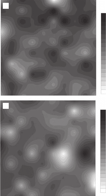
Zipf analyses clearly show that the two-dimensional bacterial distributions (Figure 5.23) are not
uniformly distributed (Figure 5.24). The Zipf plots estimated for bacterial populations sampled at
Port Noarlunga and Port River exhibit a linear behavior with a = 0.05 (r
2
= 0.98) for bacterial
concentrations ranging from 3.1 × 10
5
to 3.7 × 10
5
cell ml
−1
(Figure 5.24A), and with a = 0.03
(r
2
= 0.98) for bacterial concentrations ranging from 37.7 × 10
5
to 39.8 × 10
5
cell ml
−1
(Figure 5.24B).
Respectively, 33% and 28% of the values observed in Port Noarlunga and Port River are included
in the identied power-law behaviors. While the Port Noarlunga Zipf plot exhibits a power-law
behavior up to the highest bacterial concentrations (Figure 5.24A), note that the three highest con-
centrations (that is, the three rst ranks) observed in Port River were not included in the regression
analysis (Figure 5.24B). As discussed on the basis of Zipf analyses of simulated data (Seuront and
Mitchell 2008), such a local increasing trend is intrinsically caused by random uctuations and
should be regarded as a source of contamination of the observed power law resulting in a distribu-
tion dominated by a few hotspots rather than a breakpoint indicative of structural discontinuities
(see Section 5.5.5.1). A proper normalization further shows that for concentrations lower than 0.37
× 10
6
cell ml
−1
and 3.98 × 10
6
cell ml
–1
respectively at Port Noarlunga and Port River, the Zipf plots
are extremely similar (Figure 5.25A), continuously diverging from a power-law behavior as a step
function toward the lowest concentrations (Figure 5.24A,B and Figure 5.25A). Because a step func-
tion might be indicative of the presence of structural discontinuities within the distributions, we
18
19
20
21
22
23
24
25
26
27
28
29
30
31
32
33
34
35
36
23
24
25
26
27
28
29
30
31
32
33
34
35
36
37
38
39
40
41
42
43
44
A
B
Figure 5.23 Two-dimensional distributions of the bacterioplankton abundance sampled in (A) Port
Noarlunga (×10
4
cell ml
−
1
) and (B) Port River (×10
5
cell ml
−
1
) with a spatial resolution of 1.2 cm.
2782.indb 180 9/11/09 12:11:49 PM
Frequency Distribution Dimensions 181
performed separate Zipf analyses for the ranges of concentrations separated by identied break-
points (gray arrows in Figure 5.24A,B). The resulting Zipf plots (Figure 5.25B), shown here for con-
centrations lower than 0.37 × 10
−6
cell ml
−1
at Port Noarlunga and 3.98 × 10
−6
cell ml
−1
at Port River,
do not exhibit any power-law behavior, but instead produce a continuous roll-off from a horizontal
line (that is, a → 0) to a vertical line (that is, a → ∞). This is representative of the fact that no value
is more likely to be more common than any other one, a characteristic of uniformity.
5.5.5.2.3.2 Centimeter-Scale Phytoplankton Distribution
The 1.2-cm resolution, two-dimensional phytoplankton distributions obtained from the four “replicate”
experiments conducted in this study (hereafter referred to as P1, P2, P3, and P4) exhibit specic
5.25
0.0 0.3 0.6
α = 0.027; (r
2
= 0.982)
α = 0.049; (r
2
= 0.983)
0.9
Log r
1.2 1.5 1.8 2.1
5.30
5.35
5.40
5.45
Log X
r
5.50
5.55
5.60
A
B
6.35
0.0 0.3 0.6 0.9
Log r
1.2 1.5 1.8 2.1
6.40
6.45
6.50
Log X
r
6.55
6.60
6.65
Figure 5.24 Zipf plots of the bacterioplankton abundance in Port Noarlunga (A) and Port River (B), shown
in log-log plots. The black diamonds correspond to the range of abundance values exhibiting a power-law
behavior, and used to estimate the exponent a as the slope of the linear t maximizing the coefcient of
determination and minimizing the total sum of the residuals in the regression (dotted lines). The continuous
black and gray lines correspond to the Zipf plots obtained from 100 simulated uniform distributions with the
same minimum and maximum values as the empirical ones and from 100 simulated normal distributions with
the same mean and variance as the empirical ones, respectively. The gray arrows indicate the structural break
points in the Zipf distributions.
2782.indb 181 9/11/09 12:11:52 PM
182 Fractals and Multifractals in Ecology and Aquatic Science
–0.6
Log r
Log X
r
0.0
–0.1
–0.2
–0.3
–0.4
–0.5
–0.6
0.51.0 1.52.0 2.0
0.0 0.5 1.0
α = 0.027; (r
2
= 0.982)
α = 0.049; (r
2
= 0.983)
1.5
Log r
2.0 2.5
–0.5
–0.4
–0.3
Log X
r
–0.2
–0.1
0.0
A
B
Figure 5.25 Zipf plots of (A) the normalized bacterioplankton abundance in Port Noarlunga (open rhombs)
and Port River (black rhombs) showing the similarity of their overall shape and (B) the bacterioplankton
abundance that did not exhibit a power-law behavior in (A).
55
52
50
47
45
42
40
37
35
32
30
27
25
22
20
17
15
12
10
43
42
41
40
38
36
34
32
30
28
26
24
22
20
18
16
14
12
27
28
29
26
25
24
23
22
21
20
19
18
17
16
15
14
13
12
11
10
350
340
330
320
300
280
260
240
220
200
180
160
140
120
100
90
80
70
60
AB
C
D
Figure 5.26 Replicate two-dimensional distributions of the phytoplankton abundance (×10
3
cell ml
−
1
) sam-
pled in Port River with a spatial resolution of 1.2 cm.
2782.indb 182 9/11/09 12:11:56 PM
Frequency Distribution Dimensions 183
features such as localized gradients, hotspots, and “coldspots” (Figure 5.26) that are not compatible
with a homogeneous or a normal distribution. As previously observed for bacterioplankton distribu-
tions, the Zipf analysis of two-dimensional phytoplankton patterns shows that phytoplankton cells
are not uniformly distributed (p < 0.01) and exhibit two different types of organization (Figure 5.27).
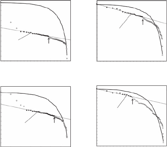
P2 and P4 thus exhibit a linear behavior starting from the highest values, while distributions P1
and P3 present local increasing trends characterizing a distribution dominated by a few hotspots.
Phytoplankton patterns thus exhibit a power-law behavior for cell concentrations ranging from 38.2
× 10
3
to 31.8 × 10
3
cell ml
−1
with a = 0.12 (r
2
= 0.99) for P1 (Figure 5.27A), 35.9 × 10
3
to 24.3 × 10
3
cell ml
–1
with a = 0.13 (r
2
= 0.99) for P2 (Figure 5.27B), 26.7 × 10
3
to 22.8 × 10
3
cell ml
−1
with a
= 0.09 (r
2
= 0.97) for P3 (Figure 5.27C), and 29.8 × 10
3
to 25.6 × 10
3
cell ml
−1
with a = 0.06 (r
2
=
0.97) for P4 (Figure 5.27D). The percentage of values contributing to the power laws are 20% for
P1, 23% for P2, 36% for P3, and 11% for P4. As stated above, separate analyses were performed for
the ranges of concentrations separated by breakpoints (arrows in Figure 5.27). Except in the case of
the distribution P4 that shows a power law for concentrations ranging from 21.4 × 10
3
to 25.1 × 10
3
cell
ml
−1
with a = 0.05 (r
2
= 0.97), no power laws were observed. The corresponding Zipf plots instead
exhibit a continuous roll-off from a horizontal line (that is, a → 0) to a vertical line (that is, a → ∞),
representative of uniformity (Seuront and Mitchell 2008).
4.0
0.0 0.3 0.6
α = 0.119 (r
2
= 0.988)
1.2 1.5
0.9
Log r
1.8 2.1
4.2
4.4
4.6
Log X
r
4.8
5.0
5.2
A
4.0
3.9
3.8
0.0 0.3 0.6
α = 0.125 (r
2
= 0.991)
1.2 1.5
0.9
Log r
1.8 2.1
4.1
4.2
4.3
Log X
r
4.4
4.5
4.6
B
4.0
0.0 0.3 0.6
α = 0.089 (r
2
= 0.969)
1.2 1.5
0.9
Log r
1.8 2.1
4.1
4.2
4.3
Log X
r
4.4
4.5
4.6
4.7C
4.0
0.0 0.3 0.6
α = 0.059 (r
2
= 0.968)
1.2 1.5
0.9
Log r
1.8 2.1
4.1
4.2
4.3
Log X
r
4.4
4.5D
Figure 5.27 Zipf plots of the phytoplankton abundance sampled in Port River, shown in log-log plots. The
black diamonds correspond to the range of abundance values exhibiting a power-law behavior, and used to
estimate the exponent a as the slope of the linear t maximizing the coefcient of determination and minimiz-
ing the total sum of the residuals in the regression (dotted lines). The continuous black lines correspond to the
Zipf plots obtained from 100 simulated uniform distributions with the same minimum and maximum values
as the empirical ones. The gray arrows indicate the structural breakpoints in the Zipf distributions.
2782.indb 183 9/11/09 12:11:59 PM