Schlick T. Molecular Modeling and Simulation: An Interdisciplinary Guide
Подождите немного. Документ загружается.


150 5. Nucleic Acids Structure Minitutorial
the {P, χ} combinations of C2
–endo/anti,C3
–endo/anti,C2
–endo/syn,and
C3
–endo/ syn; two maxima are also apparent. See also [414] for a recent
quantum-mechanical study of the glycosyl torsion energetics in DNA.
5.3.5 Basic Helical Descriptors
Besides the sugar, backbone, and glycosyl conformational variables introduced
above, additional helical parameters are defined to describe the global arrange-
ment of a base pair (bp) in a double helix [307, 1080]. See Dickerson [306]and
the text [139] for complete definitions of all translational and rotational variables.
Names, symbols, and sign conventions were decided at an international work-
shop [307]. Before we introduce some of these (as well as other) conformational
variables, we define basic features of helix descriptors that are relevant for model
helices, whose geometries can be described by characteristic values, as shown in
Tables 5.3 and 5.4.
• Helix sense refers to the handedness of the double helix.
8
• Helix pitch (per turn), P
h
, measures the distance along the helix axis for
one complete turn (see Figure 5.1).
• Number of residues per turn, n
b
, is the number of bps for every complete
helix turn.
• Axial rise, h, is the characteristic vertical distance along the double helix
axis between adjacent bps.
• Unit twist or rotation per residue for a repetitious helix is Ω = 360
o
/n
b
and
describes the characteristic rotation about the global helix axis between two
neighboring base pairs.
• Helix diameter refers to the geometric diameter of the helical cylinder
(around 20
˚
A for DNA).
• Major and minor grooves (Mgr and mgr in Figure 5.1) refer to the spaces
generated by the asymmetry of the DNA. The two different-sized grooves
are most apparent from a side view of the double helix (Fig. 5.13). The
minor groove side is defined as the space generated along the edge closer
to the two glycosyl linkages of a bp, and the major groove side is generated
by the other edge, farther from those links (see also Figure 5.11).
In the classic DNA described by Watson and Crick, the minor groove is
narrower (around 6
˚
A wide) compared to the major groove (which is dou-
bly wide) and slightly deeper (8.5 vs. 7.5
˚
A). For the A-model of DNA
8
In a right-handed form, a right hand held with the thumb pointing upward in the direction of
the helix axis will wrap right (counterclockwise) and around the axis to follow the chain; a left hand
with an upward-pointing thumb will wrap to the left (clockwise) to follow the chain direction of a
left-handed helix.

5.3. Nucleic Acid Conformational Flexibility 151
discussed below, the ‘minor’ groove is as large, or larger, and also more
shallow, than the ‘major’ groove according to the above definition.
Characterizing helical grooves is important for describing interactions of
nucleic acids with solvent molecules and with proteins. A larger accessible
area of a groove can facilitate nonspecific, as well as sequence-directed,
protein recognition and binding. The edges of the bps, which form the
bottom of the grooves, contain nitrogen and oxygen atoms available for
contacting protein side chains via hydrogen bonds. The hydrogen bonds
form the basis of sequence-specific recognition of DNA by proteins.
Table 5.3. Mean properties of representative DNA forms.
Property A-DNA B-DNA Z-DNA
Handedness Right Right Left
Representative GGCCGGCC CGCGAATTCGCG CGCGCG
Structures CGTATACC
Bps/turn 11 10 12 (6 dimers)
Rise/base pair 2.6
˚
A 3.4
˚
A 3.8
˚
A(ave.)
Helix diameter ≈26
˚
A ≈20
˚
A ≈18
˚
A
Helix pitch ≈28
˚
A ≈34
˚
A ≈45
˚
A
Twist/residue 33
o
36
o
−60
o
/dinuc.
Bp inclination 20
o
0
o
−7
o
Sugar pucker C3
-endo C2
-endo C2
-endo (C)/
C3
-endo (G)
Glycosyl anti anti anti (C)/
rotation (higher) syn (G)
Major groove narrow & deep wide & deep convex
Minor groove very wide & narrow & very narrow &
shallow deep deep
5.3.6 Base-Pair Parameters
The geometric variables above and others are necessary to describe the global
and local arrangements of base pairs in nucleic acid helices. The global parame-
ters describe the overall arrangement of the bps in double-stranded helices, while
the local variables specify the orientation between successive bps. The global he-
lical parameters are thus measured for a particular bp with respect to the overall
(global) helix axis, while the local variables are defined in the local framework of
two successive bps. The global and local helical parameters can be entirely dif-
ferent quantities. See [678] for related transformations between global and local
variables.

152 5. Nucleic Acids Structure Minitutorial
Table 5.4. Selected parameters for constructing model DNA from nucleotide geometric
variables (dinucleotide for Z-DNA) according to Figure 5.13, as developed in [1102]and
used to generate the structures in Figure 5.13. See also Figure 5.11 for definitions of the
translational variables dx and dy and the rotational variables tip (θ), inclination (η), and
twist (Ω). The parameter h is the helical rise.
Helix αβγδP/τ
max
χ
A-DNA −62 173 52 88 3/38 −160
B-DNA −63 171 54 123 131/36 −117
Z-DNA (dG) 47 179 − 165 9 −1/23 68
Z-DNA (dC) −137 −139 56 138 152/35 −159
Helix dx dy h θ η Ω
A-DNA 4.0 0 2.87 0 13.5 32.2
B-DNA 0 0 3.33 0 0 37.3
Z-DNA (dG) −3.0 2.5 −3.72 0 −7 52 (G→C)
Z-DNA (dC) −3.0 −2.5 −3.72 0 −7 8 (C→G)
In addition to these two groups of conformational variables, other parameters
describe the orientation of the two bases in a hydrogen-bonded bp.
Below, we define some of the global parameters for bp orientations (like tip
and inclination), local variables (associated with successive bps) like roll, tilt,
and twist, and a variable that describes orientations within a bp (propeller twist).
See Dickerson [306] for complete definitions of all rotational and translational
variables.
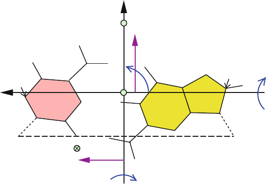
The description of these quantities requires definition of a reference coordinate
frame, which we introduce next.
Reference Frame
The commonly used reference frame shown in Figure 5.11 [307] defines a co-
ordinate system with unit vectors {e1, e2, e3} so that e1 represents the short bp
axis, e2 represents the long bp axis, and e3 is the normal to e1 and e2 which
completes the right-handed coordinate system (i.e., defined as the cross product
e3 = e1 × e2). The direction of the long-bp axis can be defined by connecting
the two C1
atoms of the pyrimidine and purine atoms. The short-bp axis (also
called the dyad) can be constructed by passing a perpendicular vector from the
midpoint of this C1
(purine) to C1
(pyrimidine) line. The intersection of the
dyad with the C8 (purine) to C6 (pyrimidine) line is considered the origin of this
plane.
An alternative standard reference frame describing nucleic-acid bp geom-
etry was proposed at an international workshop held in 1999 in Tsukuba,
Japan [941].
5.3. Nucleic Acid Conformational Flexibility 153
.
e1
C6
e2
C1´ C1´
C8
B−DNA
Pyrimidine
(C)
Purine
(G)
Mgr
mgr
INCLINATION (η)
TIP (θ)
dx
dy
TWIST (Ω)
A−DNA
.
Z−DNA
Figure 5.11. The local base-pair coordinate system {e1, e2, e3} representing the short
base-pair axis, the long base-pair axis, and the normal to both which completes a
right-handed coordinate system. Associated translational and rotational parameters are in-
dicated as detailed in the text. The symbols Mgr and mgr define major and minor groove
sides of the base pair, respectively. The positions of A-DNA and Z-DNA helix centers are
only illustrated for perspective relative to B-DNA. The global helix direction for Z-DNA
would point in the opposite direction (down from the paper plane) according to standard
definitions [307].
Global Variables (Base Pair Orientations With Respect to Helical Axis)
The reference frame defined above can be used to define deviations of the bp
as a whole with respect to the overall helical axis. These include the rotational
variables tip and inclination, and translational variables like dx and dy.
• Tip (θ in standard conventions) measures the rotational deformation of
the bp as a whole about the long bp axis e2. It is considered positive when
the rotation is clockwise as shown in Figure 5.11, moving the far side of the
bases, or the major groove side, as viewed along e1, below the paper plane.
• Inclination (η in standard conventions) measures the rotational deformation
of the bp as a whole about the short bp axis e1. This angle is considered
positive when it is clockwise as shown, moving the far base when viewed
along e2 (G in Figure 5.11) down below the paper plane.
• The displacement parameters dx and dy denote the translations of the mean
bp plane from the global helical axis, along e1 and e2, respectively (see
Figure 5.11). They indicate the shift of the bp origin (the point through
which e3 passes for a particular helical model). A positive dx indicates
154 5. Nucleic Acids Structure Minitutorial
translation towards the major groove direction, and a positive dy denotes
displacement toward the first nucleic acid strand of the duplex (see Strand
I in Figure 5.12).
For A and B-DNA, the helix axis lies approximately on the dyad, but for
Z-DNA the helix axis is displaced from the dyad toward a pyrimidine atom
and points down rather than up.
The A-DNA double helix lies on the major groove side (dx > 0 as shown in
Figure 5.11 with respect to the B-DNA helical axis), while the Z-DNA helix
lies on the minor groove side (dx < 0, as shown in Figure 5.11). Note that
the signs of dx and dy should be reversed when meaning the displacements
of the mean A-DNA and Z-DNA bp planes from their respective global
helical axes.
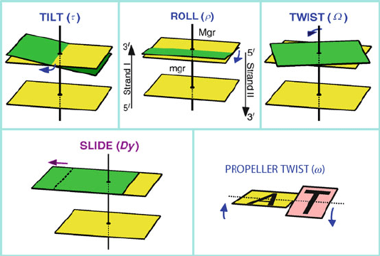
Local Variables (Base-Pair Step Orientations)
The roll, tilt,andtwist angles (see Figure 5.12) define the rotational deforma-
tions that relate the local coordinate frames of two successive bps. The three local
translational variables are slide, shift,andrise.
• Roll (ρ) defines the deformation along the long axis of the bp plane
and describes DNA groove bending: a positive roll angle opens up a bp-
step towards the minor groove, while a negative roll angle opens up a
bp-step towards the major groove.
• Tilt (τ) is the deformation defined with respect to the short axis of the bp
plane. A positive tilt angle opens the bps on the side of the sequence strand
(Strand I in Figure 5.12).
• Twist (Ω) is the helical rotation between successive bps, as measured by the
change in orientation of the C1
–C1
vectors between two successive bps,
projected down the helix axis, as shown in Figure 5.12.
• The translational slide (Dy) motion describes the relative displacement of
successive bps along their long axes as measured between the midpoint
of each pyrimidine–purine long-bp axis. It is considered positive when the
direction is toward the first nucleic acid strand (i.e., positive dy direction),
as shown in Figure 5.12. The other local translational variables are the shift
(Dx)andrise(Dz).
Deviations Within a Base Pair
• The propeller twist (ω in standard conventions) measures the angle be-
tween the normal vectors associated with the planes of the two bases in
a hydrogen-bonded pair (from the torsion angle between the individual
base planes). Imagine the motion of a helicopter propeller where the two
bases twist in opposite directions about the long bp axis e2 (one up and one
down), as shown in Figure 5.12.
5.4. Canonical DNA Forms 155
Figure 5.12. Base-pair step (tilt τ ,rollρ,twistΩ, slide Dy) and base pair (propeller
twist sω) orientation parameters, with positive displacements shown; see text for de-
tails. Mgr and mgr denote the major and minor-groove sides of the bps, respectively. The
sequence strand (I) and the complementary strand (II) are indicated in the roll illustration.
According to nucleic-acid conventions [307], when the angle is viewed
from the minor groove edge of a bp, it is positive when the base on the
left moves its minor groove edge up while the base on the right moves
down. Alternatively, we define a positive angle when each base rotates in a
counterclockwise manner when viewed from its attached sugar/phosphate
backbone. The propeller twist has a negative sign under normal conditions.
• The two other rotational variables that describe deformations within a
hydrogen-bonded bp are buckle (κ), about the short bp axis e1,andopening
(σ), about the e3 [307].
5.4 Canonical DNA Forms
Small changes in the global and local parameters introduced above can lead to
large overall changes in helix geometries. The double helix described by Watson
and Crick in 1953 — now known as B-DNA — was deduced by adjusting wire
models so as to fit the X-ray diffraction patterns recorded in the 1950s from calf
thymus DNA fibers, first manually and later by various model-building and re-
finement analyses (summarized in [215]). The fiber diffraction patterns provide an
excellent reference for describing features of canonical B-DNA forms since they
are generally devoid of the end effects evident in analysis of crystal structures of
DNA oligomers. They also represent average structures over all sequences in the
fiber.
156 5. Nucleic Acids Structure Minitutorial
The right-handed B-form is the dominant form under physiological condi-
tions. One possibility for its prevalence is that the B-DNA helix can be smoothly
bent about itself to form a (left-handed) superhelical (plectoneme or toroid-like
form; see next chapter) with minimal changes in the local structure. This was
first suggested by Levitt by early molecular simulations [745]. This deformability
property facilitates the packaging of long stretches of the hereditary material in
the cell (especially of circular and topologically-constrained DNA) by promoting
volume condensation as well as protein wrapping.
Yet we now recognize numerous variations in polynucleotide structures —
both helical and nonhelical forms — that depend profoundly on the nucleotide
sequence composition and the environment (counterions, relative humidity, and
bound ligands or other biomolecules).
The canonical B-DNA was deduced from X-ray diffraction analyses of the
sodium salt of DNA fibers at 92% relative humidity. Another form of DNA —
now termed A-DNA — emerged from early X-ray diffraction studies of various
forms of nucleic acid fibers at the much lower value of 75% relative humidity. This
alternative helical geometry is prevalent in double-helical RNA structures and in
duplex DNA under extreme solvation conditions in certain sequences (such as
runs of guanines).
Though both these early diffraction-based models were inherently low in reso-
lution and contained several incorrect structural details, later analyses of single
DNA crystals concurred with these basic fiber diffraction findings. The DNA
fiber structure analyses also served as a reference by which to analyze the
sequence-dependent trends that emerged from oligomer crystallography [215].
Both the A and B-DNA forms are right handed. A rather surprising finding,
first discovered by single crystal X-ray diffraction and rediscovered 25 years after
Watson and Crick’s description of DNA, was a peculiar left-handed helix with a
zigzag pattern. Andrew Wang, Alexander Rich, and their collaborators observed
this form in crystals of cytosine/guanine polymers (dCGCGCG) at high salt con-
centrations and dubbed it Z-DNA (for its zigzag pattern) [1327]. This high ionic
environment stabilizes Z-DNA relative to B-DNA by shielding the closer phos-
phate groups on opposite strands and hence minimizing the otherwise increased
repulsive interactions.
The biological function of Z-DNA remains in the forefront of research, but
recent evidence suggests that the conversion of helical segments from B to Z-like
acts as a genetic regulator.
Below, these three families of DNA helices are detailed; see Figures 5.13, 5.14,
and 5.15 for comparative illustrations.
5.4.1 B-DNA
B-DNA can be distinguished by the following characteristics:
• The helix axis runs through the center of each bp (dx, dy ≈ 0).
5.4. Canonical DNA Forms 157
• The bps stack nearly perpendicular to the helix axis (small inclination of the
bps and very small roll and tilt values). This implies that bases in adjacent
steps of the same strand overlap vertically (stack), and bases on the opposite
strands do not stack.
• The mean helical twist (Ω) is about 34–36
o
.
• There are about 10–10.5 bps per turn.
• Deoxyriboses favor a C2
-endo sugar conformations (S region of pseudoro-
tation cycle); see Figure 5.8.
• The glycosyl bond orientation is typically higher anti; see Figure 5.9.
• The major groove is wider (12
˚
A) and deeper (8.5
˚
A) than the minor groove,
which is 6
˚
A wide and only slightly less deep.
Overall, these features produce a model helix of the form shown in
Figures 5.13, 5.14,and5.15. Note that the top view in the space-filling stereo
figure (5.15) reveals no hole in the helix cylinder since the global helix axis inter-
sects the bps. Ordered water spines in DNA structures have been reported along
the minor groove and around the phosphate groups (see next chapter) [125].
5.4.2 A-DNA
The A-DNA helix is very different in overall appearance than B-DNA.
Specifically:
• The bp center is shifted from the global helix axis (dx ≈ 4
˚
A, dy ≈ 0 in
Figure 5.11).
• A prominent inclination is noted for the bp planes, as large as 20
o
on av-
erage. This implies a combination of both intrastrand and interstrand base
stacking for most sequences (the exception being pyrimidine/purine steps,
which overlap in an interstrand manner only).
• The mean helical twist is less than for B-DNA, about 33
o
.
• There are thus 11 bps per turn, producing a shorter helix length than B-DNA
for the same number of bps.
• The sugars favor a C3
-endo pucker (N region of the pseudorotation cycle)
rather than C2
-endo as in B-DNA; see Figure 5.8.
• The glycosyl bond orientation is typically anti,asinB-DNA;see
Figure 5.9.
• The minor groove is not as deep as in B-DNA, but the major groove is
narrower and deeper than the minor groove.
158 5. Nucleic Acids Structure Minitutorial
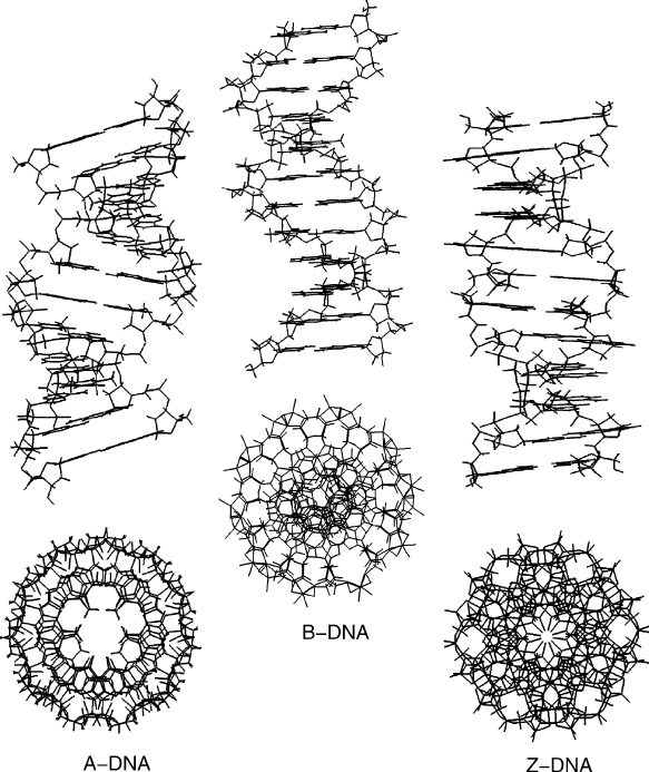
Figure 5.13. Model A, B, and Z-DNA (top and side views) as generated by the polynu-
cleotide building program developed in [1102] based on parameters listed in Table 5.4.
From Figure 5.13, note the dramatic inclination of the bps and the hollow
top view of the helix, a consequence of the bps being pulled closer to the
sugar/phosphate backbone.
A-DNA regions might exist within a generally B-DNA helix (e.g., in runs of
poly(dG)·poly(dC)) under extremes conditions only. Certain RNA molecules that
adopt partially double-helical forms, such as tRNA, rRNA, and parts of mRNA,
tend to be A-like in the duplex regions, with characteristic C3
-endo sugar puck-
ers, because the B-DNA conformation leads to steric clashes between the two
sugar hydroxyl groups.
5.4. Canonical DNA Forms 159
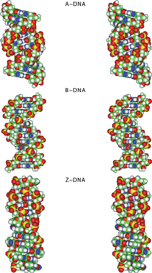
Figure 5.14. Space-filling stereo figures of model A, B, and Z-DNA, as generated by
the polynucleotide building program developed in [1102] based on parameters listed in
Table 5.4. The stereo side-view images are rotated about 8
o
relative to one another, with
the center of images separated by about 6.5 cm, the average distance between two human
eyes. Images are prepared for cross-eyed viewing from about 46–51 cm. See Figure 5.15
for corresponding top views.