Schlick T. Molecular Modeling and Simulation: An Interdisciplinary Guide
Подождите немного. Документ загружается.

120 4. Protein Structure Hierarchy
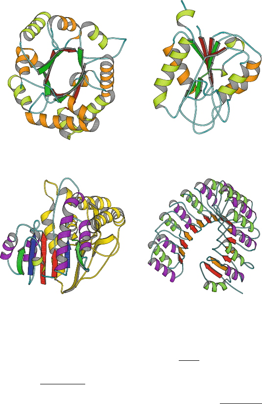
Flavodoxin (1AG9,
175 residues, open twisted)
Maltate Dehydrogenase
(2CMD, 312 residues)
Rossmann fold in
res. 1−145, rest − yellow
Ribonuclease Inhibitor
(1A4Y, 460 residues, horseshoe)
Triose Phosphate Isomerase
(1TIM, 247 residues, TIM barrel)
Figure 4.6. Examples of α/β-proteins. TIM (triosephosphate isomerase) displays an
architecture of 8 twisted parallel β-strands which form a barrel
surrounded by α-helices.
Flavodoxin, an electron transport protein that binds to a flavin mononucleotide prosthetic
group, displays an open twisted
α/β fold made of three layers (2 helices at left, 5 β-strands
in the middle, and 2 helices at right). Maltate dehydrogenase contains the (βαβαβ)
2
Rossmann fold in the subunit shown. Ribonuclease inhibitor, in the leucine-rich class of
α/β folds, displays a horseshoe structure.
consists of infectious nucleic acids. For example, the poliovirus — a spherical
complex of 310
˚
A in diameter — has a shell of 60 copies of each of four pro-
teins. The coat of tobacco mosaic virus combines 2130 identical protein units,
each of 158 residues, arranged in a helix around a coiled RNA structure of 6400
nucleotides. This results in a rod-shaped complex 3000
˚
A long and 18
˚
Ain
diameter.
4.10. Quaternary Structure 121
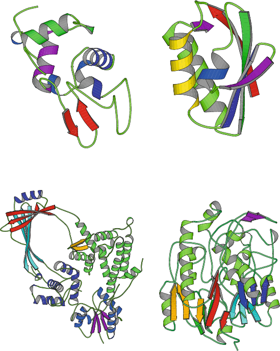
Lysozyme
(1REY, 130 residues)
Phosphocarrier protein
(1SPH, 176 residues)
E. Coli TopoisomeraseI
(1ECL, 597 residues)
L−Arginine: Glycine
Amidinotransferase
(1JDW, 423 residues)
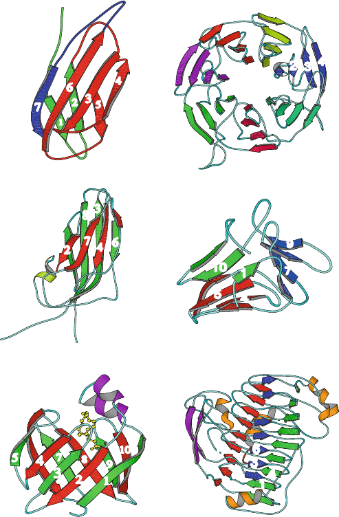
Figure 4.7. Examples of α + β-proteins: lysozyme, phosphocarrier protein, DNA
topoisomerase I,andglycine amidinotransferase.
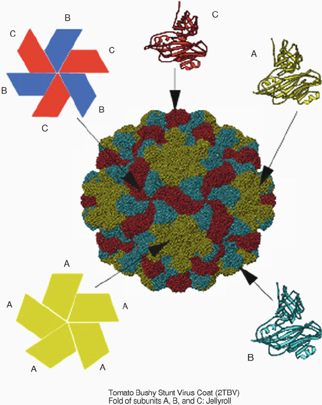
Figure 4.11 illustrates the structure of the 180-chain tomato bushy stunt virus
that infects many plants, including tomatoes and cherry trees. Interestingly, virus
coats are assemblies of similar proteins rather than one huge protein or combi-
nations of different proteins, because the relatively small amount of viral nucleic
acids must encode this protein coat; at the same time, the nucleic acids must be
covered completely. Hence a large protein shell consisting of repetitive motifs
satisfies both of these criteria.
122 4. Protein Structure Hierarchy
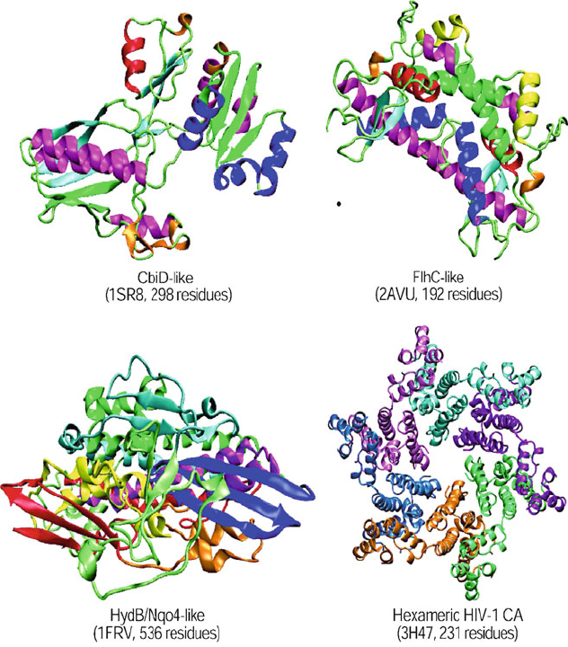
Figure 4.8. Examples of multidomain proteins: CbiD-like protein with two domains;
FlhC-like protein with three domains; HydB/Nqo4-like with four domains; and
Hexameric HIV-1 CA with two domains.
Among the larger molecular structures determined by X-ray crystallography at
moderate resolution (i.e., approaching 3.5
˚
A) is the core particle of bluetongue
virus, an agent of disease in both plants and mammals. Its transcriptionally ac-
tive compartment measures 700
˚
A in diameter and is composed of two principal
structural proteins that assemble in two layers, a core and a subcore, together en-
capsulating the RNA genome (10 segments of doubled-stranded RNA, ∼19,000
base pairs total). The crystal structure revealed how these approximately 1000
protein components self-assemble through a complex mixture of packing mech-
anisms involved in each of the two layers, using triangulation and geometrical
quasi-equivalence packing motifs [484].
4.10. Quaternary Structure 123
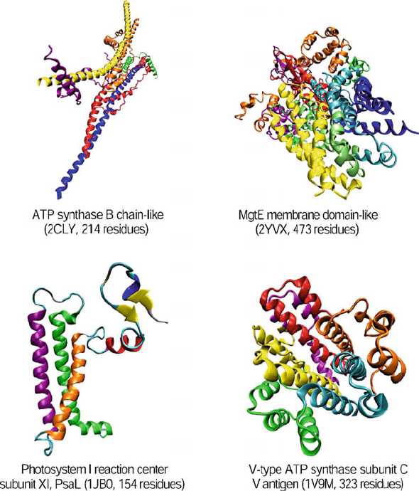
Figure 4.9. Examples of membrane and cell surface proteins and peptides: ATP synthase
B chain-like protein, with a long helix; MgtE membrane domain-like protein, with five
transmembrane helices; Photosystem I reaction center subunit protein, with three trans-
membrane helices; and V-type ATP synthase subunit C protein, with nine transmembrane
helices.
4.10.2 From Ribosomes to Dynamic Networks
Other examples of quaternary structure are noted for the ribosome, muscle-
fiber complexes, bacterial flagellar filaments, and photosynthetic assemblies of
membrane proteins.
The E. Coli ribosome is a ribonucleoprotein complex with a diameter of
about 200
˚
A constructed from 3 RNA molecules and 55 protein chains [419].
The Nobel Prize in Chemistry was awarded in 2009 to three scientists who
independently obtained atomic-level crystallographic views of this magnificent
124 4. Protein Structure Hierarchy
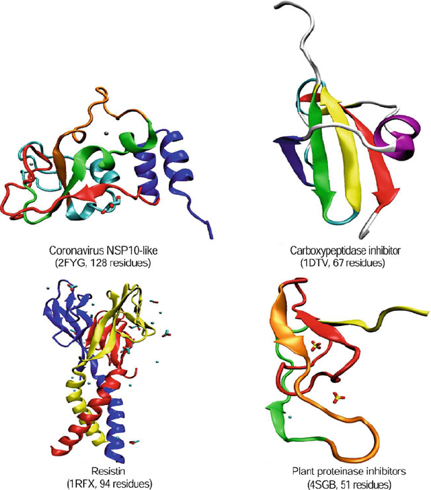
Figure 4.10. Examples of small proteins: Coronavirus NSP10-like, binds two zinc ion
per subunit; Carboxypeptidase inhibitor, disulfide-rich, α+β; Resistin, disulfide-rich
six-stranded β-sandwich; and Plant proteinase inhibitor complexed with calcium
and SO
4
.
RNA/protein machine: Ada Yonath, Venkatraman Ramakrishnan, and Thomas
Steitz. For example, the Yonath lab solved the large ribosomal subunit from
Deinococcus radiodurans [516] and the small ribosomal subunit from Thermus
thermophilus [1135](seeFig.1.1). The Steitz lab reported the structure of the
large ribosomal subunit from Haloarcula marismortui (2833 of the subunit’s 3045
nucleotides and 27 of its 31 proteins) [85], and Ramakrishnan’s group reported the
structure of the small subunit of T. thermophilus [1379]. These eagerly awaited
structures of the bacterial ribosome were aided by cryo-electron microscopy re-
constructions — first reported in 1995 for the ribosome from E. Coli (see recent
4.10. Quaternary Structure 125
Figure 4.11. The structure of tomato bushy stunt virus, a spherical arrangement of 180
polypeptide chains, each of 387 amino acids, with every 3 chains making up an asymmetric
unit (the subunits are colored blue, green, and red).
views in Figure 1.2 [710]) — which helped crystallographers estimate the initial
phasing of their X-ray data (see [171] for a perspective). The combined structural
characterizations of the ribosome provided clear evidence that the ribosome is a
ribozyme — that is, that the ribosome RNA’s component likely catalyzes peptide
bond formation (see Chapter 7, RNA sections).
Muscle cells contain parallel myofibrils composed of two kinds of filaments,
each with the following proteins: myosin (thick filament), and actin,
126 4. Protein Structure Hierarchy
tropomyosin,andtroponin (thin filament); around these filaments, titin —
itself two extremely long proteins — plus nebulin form a flexible mesh. Muscle
contraction is produced by the interaction of actin and myosin.
The bacterial flagellar motor of the protein flagellin [1085] represents an-
other challenging motor complex solved recently. Filaments of flagellin are
formed by an arrangement of stacked flagellin proteins (‘protofilaments’) lined
up side by side; an arrangement like loosely rolled sheets of paper results.
The remarkable cooperativity among the different filaments leads to conversions
between a macroscopic left-handed form — used for swimming — and a right-
handed form — used for reorientation of motion. The high-resolution flagellin
crystal suggests how this possible structural switch (between left and right-handed
supercoiled forms) might occur to direct function.
Insights into the solar energy converters in the membranes of bacteria and
plants were provided by the crystal structure of photosystem I, a large photosyn-
thetic assembly of membrane proteins and other cofactors from the thermophilic
cyanobacterium S. elogatus [616]. The detailed atomic picture (at 2.5
˚
Areso-
lution) of the network of 12 proteins subunits and 127 cofactors (chlorophylls,
lipids, ions, waters, others) shows the beautiful coordination of all components
for efficient absorption and conversion of solar energy into chemical energy.
4.11 Protein Structure Classification
Many groups worldwide are working on classifying known protein structures;
see [47, 48, 952, 1259] for a perspective of protein structure and function
evolution. Several classification schemes and associated software products ex-
ist.A popular program is SCOP: “Structural Classification of Proteins” [887].
(See scop.mrc-lmb.cam.ac.uk/scop/ or connect to SCOP through links avail-
able in many mirror sites such as PDB) [262]. These classifications are currently
assigned manually, by visual inspection, but some automated tools are being used
for assistance.
Also noteworthy is the PROSITE (www.expasy.ch/prosite/) database of pro-
tein families and domains intended to help researchers associate new sequences
with known protein families. Other databases of patterns and sequences of protein
families are PFAM and PRODOM;see[881] for a comprehensive list.
The SCOP levels (top-to-bottom) are: class, fold, superfamily, family, and do-
main. The sequence, or reference PDB structure, can be considered at the very
bottom of this tree.
The top level of the SCOP hierarchy is the class (all-α, all-β, α/β, α + β,
multi-domain, membrane and cell-surface, and small proteins). Each class
denotes common, global topologies of secondary structure.
Next comes the fold, which clusters proteins that have the same global struc-
ture, that is, similar packing and connectivity schemes for the secondary structural
elements. Folds are often also called supersecondary structure. From 50 to several
4.11. Protein Structure Classification 127
hundred folds are currently known for each class, with the repertoire increasing
steadily. An example mentioned above, the α/β barrel fold, groups TIM with
other proteins like RuBisCo(C), Trp biosynthesis,andglycosyltransferase into
a superfamily, the next level of the classification hierarchy.
The superfamily groups proteins with low sequence identity but likely evo-
lutionary similarity, as judged by similar overall folds and/or related functions.
Members of the same superfamily are thus thought to evolve from a common
ancestor. Another superfamily, for example, contains actin, the ATPase domain
of the heat shock protein,andhexokinase. Superfamilies often pose the greatest
challenge in the task of protein classification.
Superfamilies are further divided into families, which cluster proteins with sub-
stantial sequence, structure, and function similarity. Generally, this requirement
implies a sequence identity of at least 30%, but there are instances of low se-
quence identity (e.g., 15%) but definitive structural and functional similarities, as
in the case of globin proteins. For example, families of glycosyltransferase include
β-galactosidases, β-glucanase, α-amylase, and β-amylase.
Finally, at the bottom of the tree of the SCOP classification lies the domain cat-
egory, to distinguish further structurally-independent regions that may be found
in larger proteins.
For updated information on the number of identified folds, superfamilies, and
domains, check scop.mrc-lmb.cam.ac.uk/scop/count.html.
As our knowledge of protein structure increases, our classification schemes and
software tools will evolve quickly. Automation of the classification is important
for rapid structural analysis and ultimately for relating the sequence and structure
to biological function.
The reader is encouraged to re-read at this point the sections in Chapter 2 on
protein folding/misfolding (Sections 2.2 and 2.3).
128 4. Protein Structure Hierarchy
7
8
10
5
9
6
4
3
1
2
1
3
6
8
5
4
7
2
1
5
2
4
7
6
3
4
3
2
1
4
1
5
2
6
3
9
6
5
8
4
7
11
2
3
10
1
5
Nucleic Acids Structure Minitutorial
Chapter 5 Notation
S
YMBOL DEFINITION
Vectors
{e
1
, e
2
, e
3
} local base-pair coordinate system
Scalars & Terms
endo/exo sugar labeling (e.g., C3
-endo)
dx, dy
x and y-displacements (helical parameters)
h
helical rise
q
wave amplitude (pseudorotation description)
n
number of DNA base pairs
n
b
number of base pairs per turn
z
0
–z
4
perpendicular displacements of sugar ring atoms
Dx,Dy, Dz
shift, slide, and rise translations (between successive
base pairs)
P
phase angle of sugar pseudorotation
P
h
helix pitch
α, β, γ, δ, , ζ
polynucleotide backbone torsion angles
η
inclination angle (helical parameter)
θ
tip angle (helical parameter)
ρ
roll angle (base-pair step parameter)
τ
tilt angle (base-pair step parameter)
τ
0
–τ
4
internal sugar ring torsion angles
τ
max
puckering amplitude of sugar pseudorotation
φ
phase-shift angle (pseudorotation description)
χ
glycosyl (sugar/base) torsion angle
ω
propeller twist (base pair parameter)
Ω
twist angle
T. Schlick, Molecular Modeling and Simulation: An Interdisciplinary Guide, 129
Interdisciplinary Applied Mathematics 21, DOI 10.1007/978-1-4419-6351-2
5,
c
Springer Science+Business Media, LLC 2010