Reed S.J.B. Electron microprobe analysis and scanning electron microscopy in geology
Подождите немного. Документ загружается.

used formats, preferably uncompressed TIFF, which retains all the original
information content, in preference to a compressed format such as JPEG.
The simplest way of representing an image is to make the brightness, or ‘grey
level’, directly proportional to the recorded intensity of the relevant signal.
Output
In
p
ut
1 0.5
γ = 2
Fig. 4.27. A ‘gamma function’ used to modify image contrast.
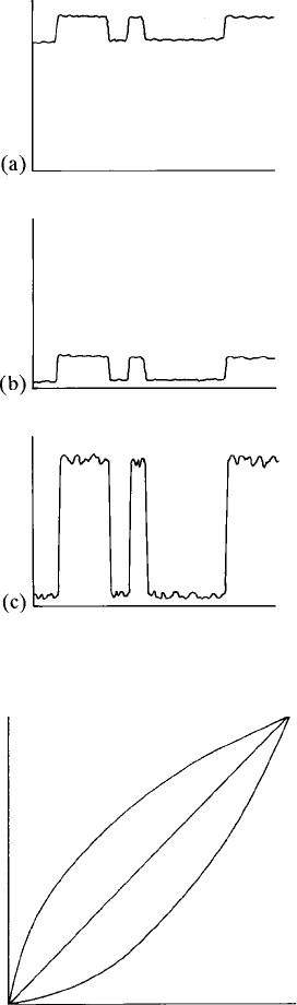
Fig. 4.26. Line-scan profiles in which low contrast in the original image
(a) is enhanced by shifting the black level (b) and increasing the amplification (c).
4.7 Image enhancement 65
Often it is advantageous to modify this relationship by applying an ‘intensity
transform’ relating recorded intensity to grey level by a nonlinear function. In
addition to the gamma function described in the preceding section, more
complex functions may be used to manipulate image contrast in different
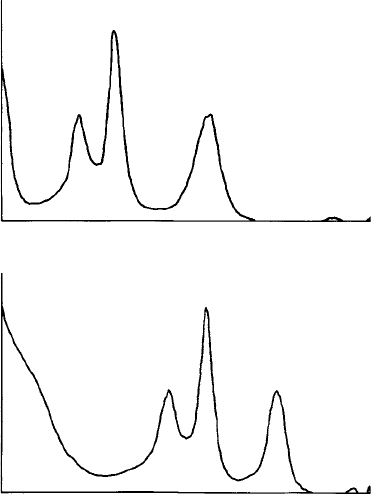
ways. A useful aid is the ‘grey-level histogram’, in which the numbers of pixels
for each grey level are plotted: the distribution of pixels can be modified by
applying an intensity transform, as illustrated in Fig. 4.29.
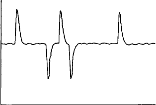
In digital images, differentiation (which in an analogue image affects only
boundaries intersecting the horizontal lines) can be made equally effective for
all boundary orientations, by means of a numerical operator such as a ‘kernel’,
of the following form:
1 1 1
181
1 1 1
For each pixel in turn the sum of the products of the adjacent pixel contents
and the kernel coefficients is calculated. For areas of uniform brightness the
result is zero, but a non-zero result is obtained on moving across a boundary,
which enhances image sharpness (at the expense of increased noise).
Noisy images can be improved by using a smoothing kernel, an example of
which is
111
181
111
This has the effect of averaging the intensity in neighbouring pixels, thereby
reducing statistical variance (but also degrading the sharpness of the image).
Grey level
X deflection
Fig. 4.28. A derivative signal obtaine d by differentiating the original signal in
Fig. 5.8, to emphasise boundaries between areas of uniform brightness.
66 Scanning electron microscop y
Too much smoothing causes undesirable artefacts to appear and it is prefer-
able to obtain a less noisy image in the first instance, with increased beam
current or acquisition time.
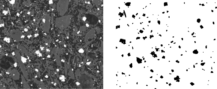
For some purposes it is useful to convert images into binary form in which
each pixel is either black or white, which is done by applying a grey-level
‘threshold’. For BSE images, in which grey level is related to atomic number,
thresholding can be used to discriminate between phases of different compos-
itions (Fig. 4.30), as a preliminary to modal analysis (Section 6.8).
4.7.2 False colours
The visual impact of an image is enhanced if the grey scale is replaced by a
range of colours, represented by a ‘look-up table’ (LUT) relating colour to
grey level. Such ‘false-colour’ images are used mostly for BSE or X-ray images,
for which the signal used has a direct relationship to a quantifiable character-
istic such as mean atomic number or elemental concentration, rather than for
Grey level
(a)
(b)
No. of pixels
Grey level
No. of pixels
Fig. 4.29. Histograms showing numbers of pixels for different grey levels:
(a) for the original image and (b) for the same image with expanded dark
tones.
4.7 Image enhancement 67
topographic images, for which the use of colour is less meaningful. The best
effects are obtained by using a smooth colour scale, usually ranging from dark
and ‘cool’ colours to light and ‘hot’ ones. An example of a false-colour image is
shown in Fig. 4.31.
4.8 Other types of image
Most SEM work involves the use of SE or BSE images showing predominantly
topographic and compositional contrast, respectively. Various other types of
image can be produced, however, as described in the following sections.
4.8.1 Absorbed-current images
The current flowing from the specimen to earth (equal to the incident beam
current minus the current lost owing to backscattering and secondary-electron
emission) can be amplified and used to produce an ‘absorbed-current’ image.
Contrast in such images is reversed compared with normal images, since regions
from which a large number of electrons are emitted appear dark rather than light.
Absorbed current is governed solely by the number of electrons leaving each
point in the image (whereas in other imaging modes direction and energy
influence detection efficiency), therefore shadow effects observed in SE and
BSE images obtained with directional detectors are absent. Topographic
contrast originates from variations in the local angle of the surface (which
(a) (b)
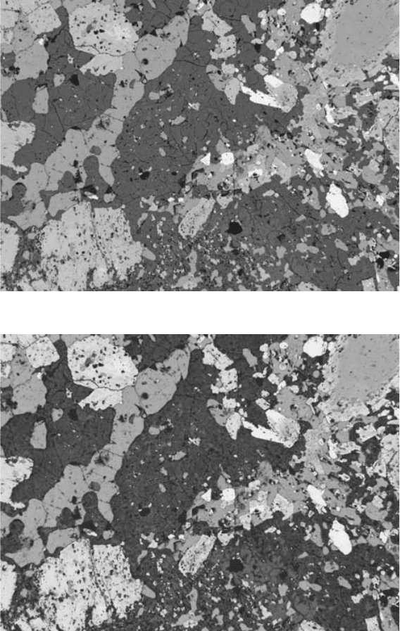
Fig. 4.30. (a) A BSE image of a rock section (500 mm 450 mm); (b) the same
after applying ‘threshold’ and inverting, showing only grains of phases of
high mean atomic number.
68 Scanning electron microscop y
(a)
(b)
Fig. 4.31. BSE images of igneous rock: (a) original monochrome image; and
(b) the same image with the grey scale converted to ‘thermal’ colours. See
plate section for a colour version.
4.8 Other types of image 69
affects both forms of electron emission). Compositional differences also
influence the image through the contribution of backscattered electrons.
4.8.2 Magnetic-contrast images
Magnetic domains can be revealed in SEM images by exploiting variations in
the detection efficiency of secondary electrons resulting from deflection by the
field immediately above the surface of the specimen. The biassed grid of the
E–T detector attracts secondary electrons too strongly, giving only rather poor
magnetic contrast, but this can be improved by placing an aperture in front
of the detector to make it more selective. For further details, see Newbury et al.
(1986).
An alternative way of viewing magnetic domains is the ‘Bitter method’,
whereby a fluid containing suspended colloidal magnetite particles is applied
to the surface and dried; the particles decorate domain walls and can be seen
easily in a SE image (Moskowitz, Halgedahl and Lawson, 1988).
4.8.3 Electron-backscatter diffraction images
The EBSD technique is increasingly commonly being used as an alternative to
electron diffraction in the TEM, having the advantages of faster data acquisi-
tion and simpler specimen preparation. The crystallographic information
obtained can be used for phase identification and texture analysis. A typical
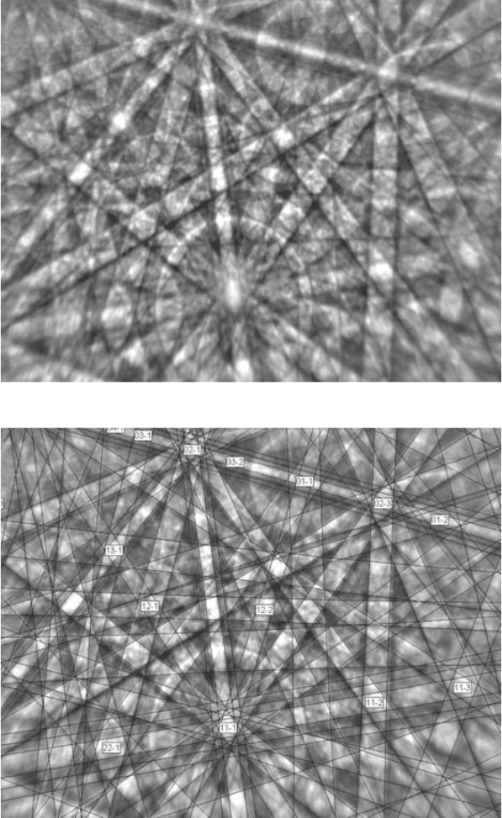
‘Kikuchi’ pattern produced by EBSD is shown in Fig. 4.32. The boundaries of
the bands represent positive and negative Bragg angles (typically of the order
of 18) for a given set of crystallographic planes. The pattern is produced by
electrons backscattered from close to the point of impact of the beam with very
little energy loss (those that have lost more energy merely contribute to the
diffuse background). Image processing can be applied to EBSD patterns to
remove background, reduce noise and enhance contrast. Software for auto-
matic indexing of the patterns and deriving data on lattice parameters and
orientation is applicable to most mineral structures, with some exceptions. See
Schwartz, Kumar and Adams (2000) for further details.
The signal from a ‘forescatter detector’ (Section 3.12.3) that detects elec-
trons scattered over a limited angular range in the forward direction combines
the effects of atomic number and orientation, and can be used to produce
‘orientation-contrast’ (OC) images (Prior et al., 1996, 1999) in which different
grey levels (or false colours) are related to orientation.
‘Orientation maps’ can be generated from patterns recorded at each point
in a grid, which are obtained by moving the specimen. Orientation can be
70 Scanning electron microscop y
(a)
(b)
Fig. 4.32. The EBSD pattern of zircon: (a) as recorded; (b) with indexing.
(By courtesy of P. Trimby.)
4.8 Other types of image 71
displayed by means of a colour code and boundary angles can also be dis-
played (Fig. 4.33). The spatial resolution is typically better than 1 mm (100 nm
is possible in favourable cases using a field-emission electron gun).
Normal specimen preparation results in a damaged surface layer, which
seriously hinders the production of EBSD images, but, by using appropriate
methods, a sufficiently damage-free surface can be obtained (see Section 9.4).
Specimens should preferably be uncoated (approximate charge balance is
obtained at the oblique incidence angle used for EBSD images), or coated
with a very thin carbon layer, though this tends to reduce image contrast.
Contamination with carbon can seriously degrade the quality of EBSD
patterns and should be minimised (see Section 3.10.1).
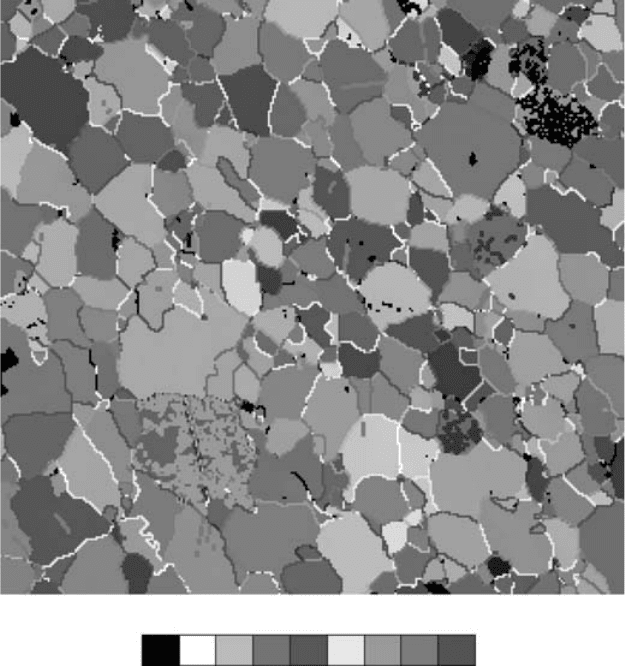
Fig. 4.33. An orientation map of calcite grains in marble de rived from EBSD
patterns; misorientations across boundaries are coloured according to the
scale shown (0–90
o
). (By courtesy of G. Lloyd.) See plate section for a colour
version.
72 Scanning electron microscop y
4.8.4 Cathodoluminescence images
Cathodoluminescence occurs in a range of different minerals and is caused by
either crystal-structure defects or trace elements (see Section 2.8). Despite the
fact that the origins of varying CL intensity and colour are often obscure, CL
images can play a useful petrographic role alongside BSE images and optical
microscopy.
Quartz gives rather low CL intensity, but since neither BSE images nor X-ray
maps generally provide useful information, SEM-CL has a particularly
significant role. The causes of variations in CL emission are thought to be a
combination of crystal defects and substitution of trace elements such as
Al and Ti. In sandstones the difference between detrital and authigenic quartz
(difficult to see in the optical microscope) shows clearly, the former being
brighter, owing to crystallisation at a higher temperature giving a higher
density of defects. Details of cementation, recrystallisation, fracture healing,
etc. are also revealed. These effects are usually visible in panchromatic mode,
but more information is obtainable when colour is taken into account
(see Fig. 4.34; also Laubach et al. (2004)). Demars et al. (1996) noted the
existence of an emission band in the UV region, which is apparently related
to coupled substitution of Al and Li, and occurs with greater intensity in
authigenic quartz. Growth zones that are otherwise invisible can also be
observed in CL images of quartz in volcanic rocks (Watt et al., 1997) and
granite (D’Lemos et al., 1997), and filled microfractures appear bright in CL
images, owing to their high density of defects (Watt, Oliver and Griffin, 2000).
Haloes due to radiation damage can be seen in Fig. 4.34(b). Deformation
lamellae in quartz that has experienced meteorite-impact shock show as black
lines, since the very high defect density suppresses CL emission (Boggs et al.,
2001).
There are greater composi tional va riations amongs t feldspa rs, giv ing
rise to a range of CL intensity and colour. Cathodoluminescence images
reveal ef fects related to cry stal growth and alteration that are somewhat
similar to those of quartz. Differences between plagioclase and orthoclase
provide a convenient means o f identifying these minerals in fine-grained
intergrowths.
Calcite is well known for its intense orange CL emission caused by Mn.
Dolomite also luminesces, but with a redder colour, which is useful as an aid to
identification. The marked banding observed in calcite overgrowths is con-
trolled by varying concentrations of both Mn and Fe, the latter having a
quenching effect. Though this is easily visible with a CL microscope, SEM-
CL enables finer detail to be observed, especially in darker regions (Fig. 4.35).
4.8 Other types of image 73
(a)
(b)
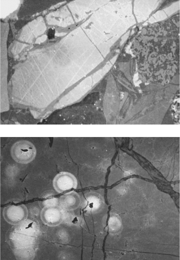
Fig. 4.34. ‘Real-colour’ SEM cathodoluminescence images obtained by
combining images recorded with red, green and blue filters: (a) quartz-
cemented sandstone (700 mm 500 mm) with two episodes of fracturing
revealed by dark blue and red luminescent quartz infilling (Markowitz and
Milliken, 2003); and (b) haloes in quartz (550 mm 350 mm) revealing
radiation damage caused by radioactive elements in small inclusions (Oliveira
et al., 2003). See plate section for a colour version.
74 Scanning electron microscop y