Reed S.J.B. Electron microprobe analysis and scanning electron microscopy in geology
Подождите немного. Документ загружается.

3.11 Electron detectors
The commonest type of SEM image is derived from secondary electrons
ejected from the sample by the incident electrons (Section 2.4). In most cases
a detector for backscattered as well as secondary electrons (Section 2.3.1)is
also provided. Both modes of detection are also available in EMPs.
3.11.1 Secondary-electron detectors
Usually secondary electrons are detected by means of a ‘scintillator’, which
produces light when bombarded with electrons, the light being converted into
an electrical signal by a photomultiplier. However, secondary electrons are
emitted with energies of only a few electron volts and must be accelerated to
produce a reasonable output from the scintillator. A positive potential (e.g.
10 kV) is therefore applied to a thin metal coating on the scintillator.
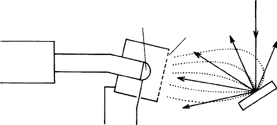
The detector most commonly used in SEMs is the Everhart–Thornley (E–T)
type illustrated in Fig. 3.14. This has a mesh in front of the scintillator, which
can be biassed to control electron collection. With a positive bias (e.g. 200 V),
secondary electrons are attracted and, after passing through the spaces in the
mesh, are accelerated towards the scintillator.
When the specimen is immersed in the magnetic field of the final lens in
order to achieve the highest possible spatial resolution (see Section 3.3),
Photo-
multiplier
Scintillator
Light pipe
Grid
BSE
Electron
beam
SE
Specimen
+10
kV+200 V
Fig. 3.14. An Everhart–Thornley detector, as used in SEMs: low-energy
secondary electrons (SE) are attracted by þ200 V on the grid and
accelerated onto the scintillator by þ10 kV; light produced by the
scintillator passes along a transparent ‘light pipe’ to an external
photomultiplier, which converts light into an electrical signal; backscattered
electrons (BSE) are also detected, but less efficiently because they have higher
energy and are not significantly deflected by the grid potential.
3.11 Electron detectors 35
secondary electrons are trapped in the field and are unable to reach a detector
of the conventional type. To overcome this problem a detector is mounted
inside the column.
A special type of detector is also required in the low-vacuum or environ-
mental SEM (Section 3.10.2), since secondary electrons cannot move freely in
the relatively high gas pressure prevailing in the specimen chamber. For this
purpose an insulated plate is mounted on the final lens polepiece and held at a
positive potential of around 1 kV. Secondary electrons are detected by means
of the signal appearing at the cathode owing to ionisation in the gas.
3.11.2 Backscattered-electron detectors
If a negative voltage is applied to the mesh of an E–T detector, secondary
electrons are repelled and only backscattered electrons, which are unaffected
by the bias owing to their much higher energy, are detected. However, the
efficiency in this mode is low because of the small solid angle subtended by
the detector.
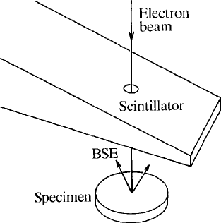
A more efficient alternative is the ‘Robinson’ detector, which consists of a
scintillator located immediately above the specimen, usually mounted on a
retractable arm, with a hole to allow the beam to pass (Fig. 3.15). The large
solid angle enables relatively noise-free BSE images to be obtained.
High efficiency can also be obtained with a solid-state detector mounted
coaxially with the beam directly above the specimen. These are often divided
into sectors (Fig. 3.16), enabling different types of signal to be produced by
combining the outputs of the sectors in different ways, and are commonly
mounted on a retractable arm. An alternative arrangement used in some
Fig. 3.15. A backscattered-electron scintillation (Robinson) detector.
36 Instrumentation

electron microprobes is to dispose a number of individual detectors around the
specimen between the spectrometer ports, the signals from which are com-
bined. Solid-state detectors have a slower response than scintillators and are
less suitable for use in fast scanning mode. Also, they are usually inefficient for
electron energies below about 10 keV, and are sensitive to light from the
microscope lamp (if any).
3.12 Detection of other types of signal
Various other types of signal can be used in SEM and EMP instruments, the
most important being X-rays (see the following chapter). Some others are
covered in the following sections.
3.12.1 Auger electrons
Auger electrons are emitted with energies (mostly in the range 0–3 keV)
characteristic of the element concerned (Section 2.7). In order to detect them,
an electron spectrometer is required. Usually this employs a cylindrical elec-
trostatic mirror: the energy of the electrons reaching the detector via the exit
slit is determined by the potential of the mirror, which is swept in order to
produce a spectrum. Scanning images showing elemental distributions are
obtained by setting the spectrometer on a particular line.
Fig. 3.16. An annular solid-state backscattered-electron detector divided into
sectors allowing discrimination between topographic and compositional
contrast.
3.12 Detection of other types of signal 37
Auger analysis requires a very clean specimen surface, so ultra-high vacuum
(e.g. 10
10
torr) is necessary. Ordinary SEMs do not satisfy this requirement,
hence a purpose-built ‘scanning Auger microprobe’ (SAM) is preferable. High
Auger-electron collection efficiency can be obtained with a cylindrical mirror
spectrometer mounted directly above the specimen.
3.12.2 Cathodoluminescence
Cathodoluminescence (CL) from specimens under electron bombardment
(Section 2.8) can be observed in an electron microprobe through the optical
microscope. By defocussing or scanning the beam, CL emission from a large
area (limited by the field of view) may be observed. Images of larger areas may
be obtained by scanning the specimen stage, with a focussed beam.
In SEMs the light can be detected with a photomultiplier (PM) mounted on
a window in the specimen chamber, and scanning CL images of any magnifi-
cation (within the available range) can be produced by modulating the display
with the PM output. The light may be focussed onto the PM entrance window
with a lens mounted in the specimen chamber. By inserting colour filters
different wavelength bands can be selected; ‘real’ colour images can then be
reconstructed by combining red, green and blue images.
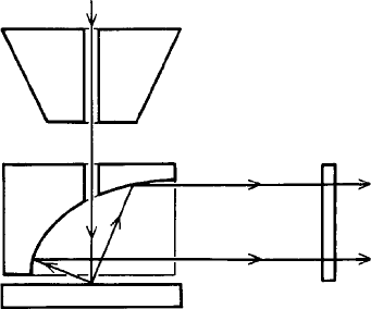
Higher sensitivity can be obtained by using a retractable ellipsoidal or
paraboloidal mirror with a hole to allow the beam to pass (Fig. 3.17). With
the point of impact of the beam at the focus of the mirror the collection
Electron
beam
Pole-
piece
Mirror
Light
Window
Specimen
Fig. 3.17. A cathodoluminescence light-collection system with a high-
efficiency paraboloidal mirror focussing light into a parallel beam for
detection by an externally mounted phot omultiplier.
38 Instrumentation
efficiency is very high for a small area (typically of the order of 100 mm
diameter); larger areas can be covered by defocussing the mirror, at the
expense of reducing its efficiency. This form of light collection is often used
in conjunction with a grating spectrograph, in order to obtain adequate
intensity. Movable mirrors allow operation in either ‘panchromatic’ mode
(detection of light of all wavelengths) or ‘monochromatic’ mode (collection
of light of a narrow band of wavelengths as selected by the spectrograph). By
using different diffraction gratings and detectors, wavelengths from ultra-
violet to infra-red can be recorded. Resolution is controlled by the widths of
the entrance and exit slits (high resolution entails a sacrifice in intensity). By
placing a CCD camera in the focal plane of the spectrograph a range of
wavelengths can be recorded in ‘parallel’ mode, enabling complete spectra to
be recorded at each point in a scanned raster.
Cathodoluminescence emission can also be observed with an optical micro-
scope fitted with a relatively simple device in which the specimen is bombarded
with a broad beam of electrons. However, using an electron microprobe or
SEM equipped with a CL detector as described above enables a lower current
to be used, with less risk of damage to the specimen; also higher resolution and
magnification are available. In addition, weaker CL emission and a wider
range of wavelengths (extending beyond the visible region) can be detected,
though the capabilities of the CL microscope can be enhanced by using a
sensitive CCD camera in place of film.
For a discussion of applications of CL, see Section 4.8.4.
Electron
beam
Pole-
piece
Specimen
BS
electrons
Phosphor
screen
Camera
Forescatter
detector
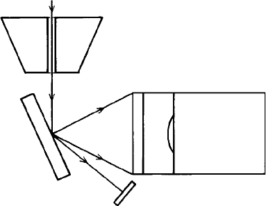
Fig. 3.18. An electron -backscatter diffraction (EBSD) detector.
3.12 Detection of other types of signal 39
3.12.3 Electron-backscatter diffraction
The necessary additional equipment for EBSD is available as an SEM acces-
sory. The specimen is steeply tilted in order to obtain optimum diffraction
conditions and the pattern appears on a phosphor screen that converts elec-
trons to visible photons, and is recorded with a camera (Fig. 3.18). For fast
recording, a normal video camera can be used, but for maximum sensitivity a
slow-scan cooled CCD camera is required. Often a ‘forescatter’ detector is
mounted below the camera, providing an alternative means for recording
images. The detector assembly is normally retractable to allow normal SEM
operation.
Applications of EBSD are discussed in Section 4.8.3.
40 Instrumentation
4
Scanning electron microscopy
4.1 Introduction
The scanning electron microscope consists essentially of the following (as
described in Chapter 3): a source of electrons; lenses for focussing them to
a fine beam; facilities for sweeping the beam in a raster; arrangements for
detecting electrons (and possibly other signals) emitted by the specimen;
and an image-display system. Secondary-electron (SE) images, which show
topographic features of the specimen, are the most commonly used type.
Backscattered-electron (BSE) images are principally used to reveal compos-
itional variations. An X-ray spectrometer (Chapter 5) is an optional extra
enabling the SEM to be used for element mapping and analysis (Chapters 6–8).
Other types of image can also be produced, as described at the end of this
chapter.
4.2 Magnification and resolution
The magnification of a scanning image is equal to the ratio of the size of the
image as viewed by the user to that of the raster scanned by the beam on the
specimen. The minimum magnification is determined by the maximum angle
through which the beam can be deflected, and depends on the working
distance, being least when this is greatest. The typical minimum magnification
is about 10, with a scanned area of the order of 1 cm
2
. Magnification can be
increased by reducing the amplitude of the scanning waveform. There is no
advantage in increasing magnification beyond the point at which the image
starts to appear unsharp. The useful maximum is thus related to resolution,
and for most purposes is in the range 10
4
–10
6
, according to the type of image,
the nature of the specimen and the operating conditions. The ability to ‘zoom’
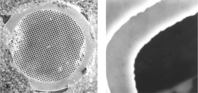
over a wide range is illustrated in Fig. 4.1.
41
Resolution, defined as the size of the smallest detail clearly visible in the
image, is limited not only by the diameter of the electron beam but also by the
interaction between the electrons and the specimen. Beam diameter is deter-
mined by various instrumental factors, as discussed in Section 3.4, and can be
reduced to a few nanometres in principle. In many applications, however, the
ultimate resolution is not needed and a larger beam diameter can be used, with
the advantage that more current is then available. The resolution limit as
determined by beam–specimen interaction ranges from approximately 1 mmin
X-ray images to less than 10 nm for SE images (under favourable circumstances).
In digital images the pixel size limits the maximum attainable resolution.
4.3 Focussing
Correct focus setting is achieved by adjusting the control to obtain the sharpest
possible image of fine detail in the specimen, preferably with the magnification set
to a high value. For this purpose fast (TV rate) scanning is desirable. The beam is
perfectly focussed only in a single plane. However, the depth of focus is large
compared with that of an optical microscope, owing to the small convergence
angle of the beam, so sharpness is often adequate for all parts of the specimen.
For tilted specimens, dynamic focus correction linked to deflection can be used.
4.3.1 Working distance
A short working distance is optimal for high-resolution SE imaging but is
incompatible with some other types of image (for example, an X-ray detector
(a) (b)
Fig. 4.1. SEM images of an electron microscope grid (3 mm diameter);
magnification (a) 10 and (b) 10 000.
42 Scanning electron microscop y

may be unable to ‘see’ the sample when the working distance is less than a
certain value). The working distance can be set by adjusting the final lens focus
to the required setting, then moving the specimen stage vertically until the
image is sharp. The focus setting depends on the accelerating voltage, and must
be adjusted when this is changed.
4.4 Topographic images
The principal function of the SEM is to produce images of three-dimensional
objects (the main advantages over the optical microscope are greater depth of
focus and higher resolution). Usually secondary-electron images are employed
to show topographic contrast, but backscattered-electron images, though
mostly used to show compositional differences, may also contain topographic
information.
4.4.1 Secondary-electron images
Secondary electrons are emitted from very near the surface of the sample,
with energies of a few electron volts (Section 2.4). The SE yield increases
with decreasing angle between beam and specimen surface (Fig. 4.2), giving a
result that is very similar to that produced by illuminating a solid object with
partly directional and partly diffuse light, making the topographic inform-
ation contained in such images easy to assimilate intuitively (Fig. 4.3(a)).
The Everhart–Thornley type of electron detector (Section 3.11.1) attracts
Fig. 4.2. The variation in secondary-electron yield with the angle of tilt of the
specimen surface relative to the horizontal.
4.4 Topographic images 43

secondary electrons, including those emitted from the far side of protruding
features (Fig. 4.4) and from inside cavities, so that the image is relatively free
of shadows. The detector should ideally be positioned adjacent to the top of
the scanned raster (i.e. at the back of the specimen chamber), giving a ‘top-
lighting’ effect.
More secondary electrons can escape from the sample at an edge (Fig. 4.5),
causing it to appear bright (see Fig. 4.3(b)). This ‘edge effect’ is most promi-
nent when a high accelerating voltage is used, owing to the greater penetration
of the electrons.
(a) (b)
Fig. 4.3. Secondary-electron images, showing (a) the three-dimensional
effect resulting from the dependence of SE emission on the angle of the
surface to the beam; and (b) the edge effect (for a razor blade); scale bar ¼
5 mm.
Fig. 4.4. Collection of secondary electrons from a three-dimensional specimen
by a detector with a positively biassed grid.
44 Scanning electron microscop y
