Reed S.J.B. Electron microprobe analysis and scanning electron microscopy in geology
Подождите немного. Документ загружается.

the bore, where it is immersed in the magnetic field of the lens. Such a lens can
be operated with a very short focal length, thereby minimising aberrations (see
the next section). It is harder to collect electron and other signals, but this
drawback is avoided in the ‘snorkel’ lens, in which the focussing field extends
below the polepiece, and short focal length can be combined with larger
specimens and easier access.
Somewhat different design criteria apply in the case of the electron microprobe,
in which an optical microscope and multiple WD spectrometers must be accom-
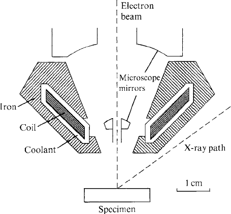
modated, and a very small beam diameter is not necessary. Figure. 3.5 shows
a ‘mini-lens’ used in an EMP: this employs a coil of small dimensions carrying a
relatively high current, allowing maximum space for other components.
3.3.1 Aberrations
The strength of a magnetic electron lens increases with the distance of the
electron trajectory from the axis, giving rise to ‘spherical aberration’ analo-
gous to that of a simple biconvex glass lens (Fig. 3.6). This defect is important
only in the final lens, and can be controlled by means of a limiting aperture.
Also, it decreases with decreasing focal length, and for high-resolution SEM
imaging may therefore be minimised by decreasing the ‘working distance’
Fig. 3.5. The final lens design for an electron microprobe (by courtesy of JEOL
Ltd): a liquid-cooled miniature lens allows space for optical microscope
components and X-ray paths to spectrometers.
3.3 Electron lenses 25

between specimen and lens (this is not applicable to EMP instruments, in
which this distance is fixed).
Another important aberration is astigmatism caused by small lens imperfec-
tions or contamination on apertures, the effect of which is that two elliptical
foci (with perpendicular axes) occur in separate planes. The result is loss of
resolution in SEM images. This can be corrected by means of a ‘stigmator’
consisting of coils that create astigmatism that cancels out that already pres-
ent. In EMP instruments, the stigmator can be adjusted by observing the
change in shape of the spot of light produced by the beam on a cathodolumi-
nescent sample as the lens is swept through focus (Fig. 3.7). Alternatively, the
adjustment can be made while observing a scanning image and varying the
stigmator controls, which is the method used in SEMs.
Fig. 3.6. Spherical aberration: the lens focusses outer rays more strongly than
those close to the axis, resulting in enlargement of the beam diameter at the
focus.
Fig. 3.7. The appearance of the electron beam in a through-focus series:
(a) with and (b) without astigmatism (this behaviour can be observed on
a cathodoluminescent sample).
26 Instrumentation
3.3.2 Apertures
An aperture limiting the beam diameter in the final lens is essential to control
spherical aberration, as noted in the previous section. This consists of a thin
disc (diaphragm) with a central hole allowing the beam to pass through,
usually made of a metal such as platinum or molybdenum (Fig. 3.4). Several
interchangeable apertures of different sizes may be provided: for high spatial
resolution a small aperture should be selected, but, when a larger beam
diameter is acceptable, more current can be obtained with a larger aperture.
Additional ‘spray apertures’ located between the electron gun and the final
lens intercept the outer parts of the beam which might otherwise be scattered
on striking lens bores etc. When astigmatism caused by contamination
becomes too much to be corrected by the stigmator, the relevant aperture
must be either cleaned or replaced.
3.4 Beam diameter and current
As described in the preceding sections, the beam diameter in the specimen
plane is determined principally by the effective source size, demagnification by
the electron lenses and spherical aberration. Demagnification is determined by
the condenser lens settings (sometimes labelled ‘spot size’). On increasing the
condenser strength in order to reduce the beam diameter, the divergence of the
beam increases and the fraction passing through the final aperture decreases.
Also, the reduction in aperture size necessary in order to minimise spherical
aberration entails a further decrease in current. As a result of these factors the
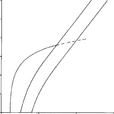
maximum current obtainable varies with beam diameter, d, approximately as
d
8/3
(Fig. 3.8). More current can be obtained in a beam of given diameter by
decreasing the working distance (possible only in SEMs) so that spherical
aberration is reduced (see Section 3.3.1). The minimum beam diameter attain-
able with a high-performance SEM is typically about 2 nm, but is larger
for EMPs.
3.5 Column alignment
The direction of the electron beam emerging from the gun is sensitive to the
position of the filament relative to the wehnelt aperture and not only is it
impossible to set this initially with perfect precision, but also the filament tip is
liable to wander in use. In addition, the components of the electron optical
column (electron gun, lenses etc.) are subject to small misalignments.
Adjustment of beam alignment can be carried out by means of magnetic
3.5 Column alignment 27
coils (in place of mechanical centring adjustments) that ‘steer’ the beam along
the correct path. In a computer-controlled instrument the adjustment can
be done automatically, and optimum settings (which vary with accelerating
voltage) can be saved in a ‘set-up’ file.
Usually the final lens aperture is centred mechanically, by finding the
position for which there is no lateral movement of the beam as the lens focus
is varied. A useful aid is a ‘wobbler’, which oscillates the focus setting while the
operator adjusts the aperture position.
3.6 Beam current monitoring
It is desirable (especially for X-ray analysis) to have means for monitoring
beam current. The current flowing from specimen to earth does not give a true
indication of the current in the incident beam because secondary and back-
scattered electrons leaving the sample reduce the apparent current by a vari-
able amount. It is therefore preferable to collect the current in a ‘Faraday cup’
(or ‘cage’) consisting of a deep recess in a solid conducting block, preferably
made of a material of low atomic number (e.g. carbon) to minimise
1 µA
100
nA
10
nA
1
nA
100
pA
FE
W
LaB
6
10 pA
1
pA
Beam current
Beam diameter (nm)
1 10 100 1000
Fig. 3.8. Beam current versus diameter for a typical instrument, using
different types of electron source: W, tungsten filament; LaB
6
, lanthanum
hexaboride; and FE, field emission.
28 Instrumentation
backscattering. This may be mounted on the specimen holder or, more conven-
iently, on a retractable arm (a standard feature of EMPs, but not SEMs).
As noted in Section 3.4, there is a direct relationship between the strength of
the condenser lenses and the beam current. The required current may therefore
be obtained by adjustment of the condenser lenses, which, in a computer-
controlled instrument with current monitoring, can be done automatically by
means of a software command. (This also has the effect of changing the final
beam diameter.)
Drift in beam current as a function of time is caused mainly by movement of
the tip of the filament, which can be corrected using beam alignment coils (as
discussed in the previous section). Such drift is a potential source of error
in automated quantitative X-ray analysis sessions, which may extend over
several hours. One solution to this problem is to insert the Faraday cup before
each measurement and normalise the intensities. A better alternative, however,
is to use a regulating system that continuously adjusts the condenser lenses to
maintain a constant current. This requires a double aperture, in which part of
the beam passing through the first aperture is intercepted by the second
(smaller) one, and the current collected by this is used as input to a feedback
amplifier. (It is assumed that this current is proportional to that passing
through the second aperture.) This is a normal feature of EMPs but not SEMs.
3.7 Beam scanning
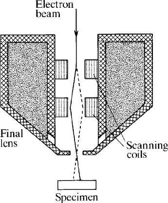
Scanning images are produced by sweeping the beam across the sample in a
television-like ‘raster’ while displaying the output of an electron detector on
the screen of a synchronously scanned visual display unit (VDU), as shown in
Fig. 3.9. The electron beam is deflected by means of coils located above the
final lens, which enable the beam to be ‘pivoted’ about the centre of the lens.
These are supplied with a ‘sawtooth’ current waveform derived from line and
frame scan generators. The ratio of frame and line scan frequencies determines
the number of lines in the image (typically 500–2000).
Instead of scanning a rectangular raster, the beam can be swept along a
single line by using only one set of coils, in order to produce a line plot, which is
useful for some purposes. The deflection system can also be used to move the
beam around in ‘spot’ mode for X-ray or other forms of analysis on selected
points.
‘Analogue’ scanning systems have been superseded by digital systems in
which the beam deflection is computer-controlled via a digital-to-analogue
converter (DAC) and the output from the signal detector is converted into
digital form by an analogue-to-digital converter (ADC), so that the intensity at
3.7 Beam scanning 29
each point (or ‘pixel’) in the image is recorded as a number. Such images are
displayed on a computer monitor and can be saved on the hard disc of the
computer, from which they can be printed out, copied to other media for long-
term storage, or transferred to another computer via a network connection.
For scanning large areas, stage movement rather than beam deflection can
be used. However, owing to the relatively slow speed of the mechanical stage
movements, this entails a long acquisition time, which is appropriate for X-ray
‘maps’ used to show element distributions, but less so for electron images.
3.8 The specimen stage
Specimens for SEM are commonly fixed to a ‘stub’ consisting of a metal disc
with a projecting peg (Fig. 3.10). This is mounted on the stage mechanism,
which incorporates linear movements in the x and y directions (perpendicular
to the column axis), enabling different areas of the specimen to be imaged
and/or analysed, and in the z direction (parallel to the axis), which serves to
locate the surface of the specimen at the required height relative to the final
electron lens (also light microscope and X-ray spectrometers, if fitted). Tilt
and rotation movements allow adjustment of the orientation of the specimen
relative to the beam and electron (or other) detectors in order to optimise the
Fig. 3.9. Electron-beam scanning using double deflection coils in the bore of
the final lens.
30 Instrumentation
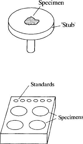
image. It is desirable for these motions to be ‘eucentric’ (centred about the
column axis).
For quantitative X-ray analysis, constant geometry is required, ideally with
normal electron incidence, and the arrangements used in EMPs are therefore
different. A typical EMP specimen holder is illustrated in Fig. 3.11. Several speci-
mens and standards may be mounted together, with their front surfaces lying in a
common plane defined by a ledge at the front of the holder (described as ‘front
referencing’, as distinct from ‘back referencing’ of SEM specimens mounted as
described above). The holder is attached to a mechanism that provides orthogonal
movements, which in modern instruments are computer-controlled. The minimum
step size is typically 1 mm or less, and ‘micro-stepping’ is sometimes available to
give ultra-fine control of position.
Optional specialised facilities include cooling (using either a thermo-electric
device or liquid nitrogen), which enables aqueous samples to be examined in a
frozen state, and heating, enabling the response of mineral samples etc. to be
viewed ‘live’ (for example, see Kloprogge, Bostro
¨
m and Weier (2004)). The
latter is most suitable for use in an environmental SEM (Section 3.10.2), owing
to the emission of gases during heating.
Fig. 3.10. The ‘stub’ used for mounting SEM specimens.
Fig. 3.11. A typical specimen holder for EMPA, holding a number of small
standards and several specimens (round in this case, but they may be
rectangular).
3.8 The specimen stage 31

3.9 The optical microscope
Optical microscopes in current EMP instruments use a reflecting objective lens
mounted coaxially with the electron beam, as shown in Fig. 3.12 (though other
configurations have been used in the past). This enables the sample to be viewed
at normal incidence while under electron bombardment, so that cathodo-
luminescence (Section 2.8) can be used as an aid in determining the location of
the beam, on a suitable target such as benitoite, willemite, periclase, glassy
carbon, or microscope-slide glass.
The focal plane of the microscope is fixed, and the specimen is moved in the
vertical (z) direction to obtain a sharp image, ensuring that the surface is always
in the same plane, which is essential for WD (though not ED) analysis. In some
instruments automatic focussing is available. Usually a CCTV camera is fitted in
place of the eyepiece to provide an image on a video screen or computer monitor.
In instruments with fixed magnification this is fairly high, as required for
precise location of points for analysis. Sometimes there is provision for ‘zoom-
ing’ to low magnification, which is useful when searching specimens for areas
of interest. Illumination by transmitted light, with polariser and analyser, is an
optional feature that is highly desirable for geological applications.
Some SEMs have a low-power microscope using oblique incidence, but
most have none. This is a handicap for geological applications, though back-
scattered electron images serve as a partial substitute.
Fig. 3.12. A reflecti ng microscope mounted coaxially with the electron beam,
as fitted to electron microprobe instruments.
32 Instrumentation
3.10 Vacuum systems
Electron beam instruments must be evacuated sufficiently well to avoid
damage to the electron source and high-voltage breakdown in the gun, as
well as allowing electrons to reach the specimen without being scattered.
Taking these considerations into account, it is desirable for the operating
pressure to be below 10
5
mbar*, though a lower pressure in the electron
gun is required when using a high-brightness electron source (Section 3.2.1).
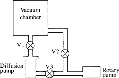
A simple vacuum system employing a mechanical rotary pump and an oil
diffusion pump is shown schematically in Fig. 3.13. To pump the chamber
from atmospheric pressure, V2 is opened (with V1 and V3 closed). When a
pressure of about 0.1 torr is attained with the rotary pump, V2 is closed and
both V1 and V3 are opened, bringing into operation the diffusion pump (which
must be backed by the rotary pump whilst in use). In contemporary instru-
ments the vacuum system is computer-controlled, so that the required opera-
tions are executed automatically and inappropriate actions that could cause
damage are barred. Pump-down time is minimised by venting with dry nitro-
gen when changing specimens, etc., to avoid introducing water vapour. For
some purposes better vacuum than that provided by the oil diffusion pump is
needed (for example when a field emission source is used), in which case a
turbomolecular or ion pump may be used.
The following additional features are sometimes provided (in EMPs
more often than in SEMs): (1) a gun-isolation valve to enable the gun to be
vented and pumped independently for filament replacement; (2) a specimen
airlock to enable specimens to be changed without venting the whole column;
Fig. 3.13. A simple vacuum system with diffusion and rotary pumps (see the
text for details of operation).
* Units of pressure are the torr (1 mm Hg), the millibar (10
3
atm) and the pascal (1 N m
2
), which are
related as follows: 1 torr = 1.3 mbar = 130 Pa.
3.10 Vacuum systems 33
and (3) a separate chamber for WD spectrometers, allowing them to be
evacuated by rotary pump only.
3.10.1 Contamination
Residual hydrocarbon molecules adsorbed on exposed surfaces in the vacuum
chamber are polymerised by electrons, leaving a carbon deposit. This causes
contamination of apertures and other components, which can lead to instabil-
ity due to charging. Carbon is also deposited on the specimen at the point of
impact of the beam. In many applications this is not very important, but
absorption of long-wavelength X-rays passing through the contamination
layer can be significant. The following measures may be applied to minimise
contamination: (1) replacing the diffusion pump by an oil-free type of high-
vacuum pump (see above); (2) fitting a vapour trap in the rotary pump backing
line; (3) replacing the rotary pump by an oil-free type; (4) using a liquid-
nitrogen cold trap above the pump and/or close to the specimen; and (5)
introducing a jet of air or oxygen via a fine capillary tube close to the specimen.
The cold trap should be warmed up before venting, to avoid condensation of
water. In the case of the gas jet, the flow rate must be adjusted so that the
pressure rise in the chamber is within acceptable bounds.
3.10.2 Low-vacuum or environmental SEM
Instead of the SEM specimen chamber being maintained at the same low
pressure as the column, a relatively high pressure is advantageous for some
purposes. This is achieved by a valve leaking gas into the specimen chamber,
with differential pumping between the specimen chamber and the upper sec-
tions of the column. An instrument designed to operate in this mode is known
as a ‘low-vacuum’ or ‘environmental’ SEM (LVSEM or ESEM). The term
‘variable-pressure SEM’ (VPSEM) refers to the ability to function in either
normal or low-vacuum mode.
The main advantage of low vacuum is that specimen charging is neutralised by
positively ionised gas atoms, making coating of insulators unnecessary. For this
purpose, a pressure of 0.1 mbar is sufficient. With a pressure of about 20 mbar,
liquid water is stable and wet samples can be used (this is of interest mainly for
biological applications). Under low-vacuum conditions, normal backscattered-
electron detectors can be employed, but special arrangements are necessary for
SE imaging (see Section 3.11.1). The relatively poor vacuum does not seriously
interfere with X-ray detection, but spatial resolution is degraded by scattering of
the electrons in the beam (for further details, see Danilatos (1994)).
34 Instrumentation
