Reed S.J.B. Electron microprobe analysis and scanning electron microscopy in geology
Подождите немного. Документ загружается.


The long decay time of CL emission in some minerals can be minimised by
increasing the dwell time or, in the case of calcite, by selecting a wavelength in
the violet–UV region (Reed and Milliken, 2003).
Cathodoluminescence images of zircon commonly show considerable detail
related to growth processes. Since the CL emission is at least partly dependent
on compositional variations (typically involving rare earths and yttrium), BSE
images usually show similar general features, though CL images often reveal
finer detail. Cathodoluminescence images are used to characterise different
groups of detrital zircons in sediments and for selecting areas for ion micro-
probe U–Pb dating (see Fig. 4.36).
For further information on applications of CL, see Marshall (1988) and
Pagel et al. (2000).
Fig. 4.35. An SEM-CL image of zoned calcite with a complex growth history
(1 mm 1 mm); finer detail can be resolved than with a CL microscope.
(By courtesy of M. Lee; first published in Microscopy and Analysis, no. 79,
September 2000, p.15.)
4.8 Other types of image 75

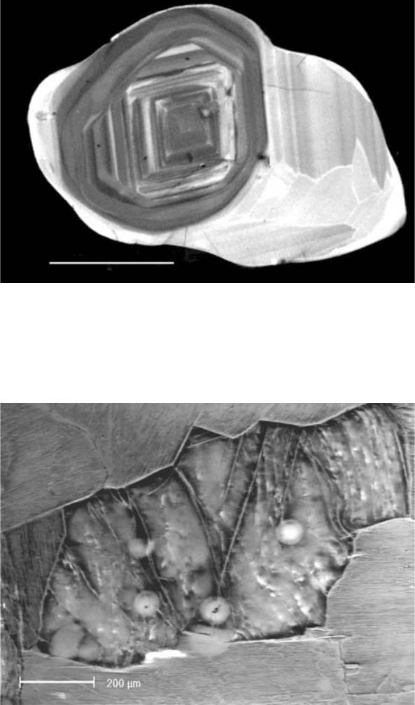
Fig. 4.36. A cathodoluminescence image of multi-generation zircon, in which
brightness is related to composition and defect density, used as a guide for
selection of areas for ion microprobe U–Pb dating. The original zoned core
(2850 Ma) has been overgrown by a weakly luminescent band
(2550–2510 Ma) and a highly luminescent banded and sector-zoned over-
growth (2490 Ma). Scale bar ¼ 100 mm. (By courtesy of N. Kelly.)
Fig. 4.37. A charge-contrast image of deformed biotite in granite (Watt,
Griffin and Kinny, 2000), showing folded cleavage, linear fractures and
radiation haloes caused by small monazite inclusions (for explanation of
the contrast mechanism, see Section 4.8.5). (By courtesy of B. Griffin.)
76 Scanning electron microscop y
4.8.5 Charge-contrast images
Secondary-electron images of uncoated samples of materials with poor elec-
trical conductivity obtained with an ‘environmental’ or low-pressure SEM
(Section 3.10.2) may, under certain conditions, exhibit contrast related to the
local charge density in the surface region (Watt, Griffin and Kinny, 2000). To
achieve this, the gas pressure in the sample chamber should be adjusted to a
level at which specimen charging is only partly neutralised by positive ions.
Variations in the local surface potential then influence secondary-electron
emission, producing contrast dependent on conductivity. Figure 4.37 shows
an example of such a ‘charge-contrast image’ (CCI). This type of contrast is
related to crystal defects connected with either growth processes or trace
elements and is related to contrast of similar origin observed in cathodolumi-
nescence images (see Section 4.8.4). Higher spatial resolution is attainable in
CCI images and they can be obtained for a wider range of sample types,
including phases that do not luminesce.
4.8.6 Scanning Auger images
The combination of an Auger electron spectrometer with an SEM to give a
‘scanning Auger microscope’ (SAM) is described in Section 3.12.1. Elemental
distribution ‘maps’ are formed by the signal obtained with the spectrometer set
to an Auger line of the element of interest. For this surface-sensitive technique
the use of a conductive coating is inappropriate and specimen charging is
therefore a problem in geological applications. It can be avoided, however,
by using a low accelerating voltage and low beam current (e.g. 3 kV and a few
nanoamps) and by tilting the specimen, which increases the number of back-
scattered and secondary electrons leaving the sample.
The spatial resolution is very high with respect to depth, owing to the small
‘escape depth’ of Auger electrons. Lateral resolution is limited by the contri-
bution of Auger electrons produced by backscattered electrons leaving the
specimen, but is typically a fraction of 1 mm (considerably better than for X-ray
images). The technique thus provides a method of surface analysis with high
lateral resolution. Geological applications have been reviewed by Hochella,
Harris and Turner (1986) and Hochella (1988).
4.8 Other types of image 77
5
X-ray spectrometers
5.1 Introduction
X-ray spectrometers are of two kinds. The energy-dispersive (ED) type records
X-rays of all energies effectively simultaneously and produces an output in the
form of a plot of intensity versus X-ray photon energy. The detector consists of
one of several types of device producing output pulses proportional in height
to the photon energy. The wavelength-dispersive (WD) type makes use of
Bragg reflection by a crystal, and operates in ‘serial’ mode, the spectrometer
being ‘tuned’ to only one wavelength at a time. Several crystals of different
interplanar spacings are needed in order to cover the required wavelength
range. Spectral resolution is better than for the ED type, but the latter is faster
and more convenient to use. X-ray spectrometers attached to SEMs are
usually of the ED type, though sometimes a single multi-crystal WD spec-
trometer is fitted. Electron microprobe instruments are fitted with up to five
WD spectrometers, and often have an ED spectrometer as well. These two
types of spectrometer are described in detail in Sections 5.2–5.4 below.
5.2 Energy-dispersive spectrometers
The modes of operation and characteristics of the detectors and associated
electronics used as ED spectrometers are described in the following sections.
5.2.1 Solid-state X-ray detectors
In ED spectrometers the X-ray detection medium is a semiconductor (either
silicon or germanium), in which the valence band is normally fully occupied by
electrons. The valence and conduction bands are separated by an energy gap
(1.1 eV for Si, 0.7 eV for Ge), and at room temperature very few electrons have
sufficient thermal energy to jump this gap: the conductivity is therefore
78
normally very low. When an X-ray photon is absorbed it generates Auger
electrons and photo-electrons (Section 2.4), which dissipate their energy partly
by raising valence electrons to the conduction band. The arrival of each
photon thus creates a brief pulse of current caused by electrons in the former
and ‘holes’ in the latter, moving in opposite directions under the influence of
the bias voltage applied to the detector.
The mean energy used in generating one electron–hole pair is 3.8 eV for
Si (2.9 eV for Ge). The size of the output pulse depends on the number of such
pairs, which is given by the X-ray energy divided by the mean energy. Hence a
1.487-keV Al Ka photon, for example, produces an average of 391 electron– hole
pairs in a Si detector, whereas a Ni Ka photon (7.477 keV ) produces 1970 (the
actual numbers are subject to some statistical fluctuation).
Even highly refined silicon contains impurities, which have undesirable
effects. These are counteracted by introducing lithium using a process
known as ‘drifting’ – hence the name ‘lithium-drifted silicon’, or ‘Si(Li)’,
detector. Germanium detectors are usually made of high-purity material
(‘HPGe’), which does not require the addition of Li. A typical Si(Li) detector
consists of a silicon slice about 3 mm thick, with an area of 10 mm
2
(though
larger sizes are available). The front surface is covered by a thin layer of gold,
which serves as a contact for the bias voltage. The rear is connected to a field-
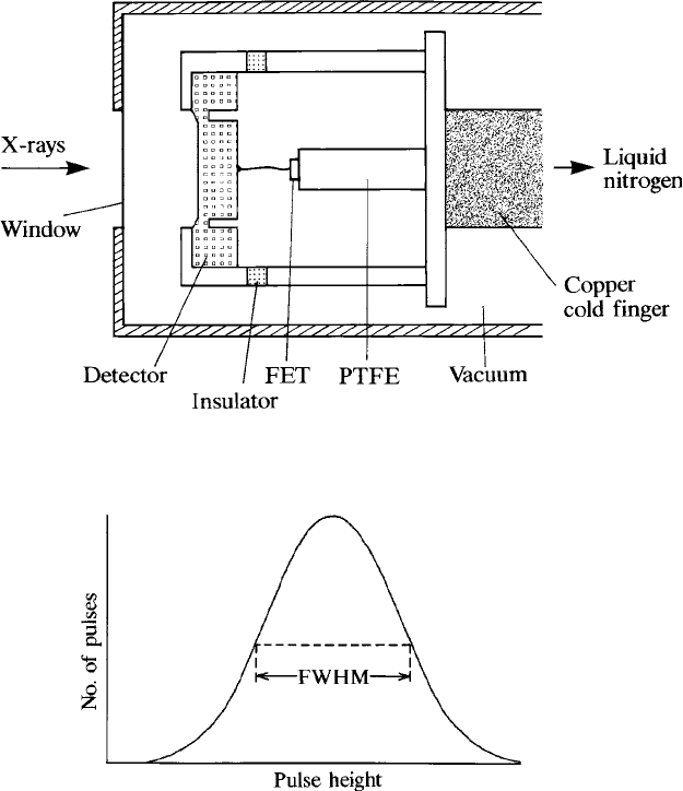
effect transistor (FET), which acts as a preamplifier. The detector and FET are
mounted on a copper rod, the other end of which is immersed in liquid
nitrogen, and the whole assembly is sealed inside an evacuated housing, or
‘cryostat’ (Fig. 5.1). (Mechanical refrigeration can be used instead to obviate
the need for liquid nitrogen.) The detector must be protected from damage
resulting from warming up while the bias voltage is on, by means of a tem-
perature sensor that switches off the voltage.
X-rays reach the detector via a ‘window’ capable of withstanding atmos-
pheric pressure, so the vacuum chamber of the instrument to which it is
attached can be vented to air safely. A typical beryllium window about 8 mm
thick absorbs X-rays of energy below about 1 keV, but low-energy X-rays can
be detected if a thin polymer window is used instead. To provide sufficient
strength to withstand atmospheric pressure, such windows are supported by a
grid. Thin-window detectors are sensitive to light, and in instruments with a
microscope the lamp must be switched off for X-ray spectrum acquisition.
An alternative type of detector is the ‘silicon drift detector’ (‘SDD’), which is
different in construction and does not involve Li compensation (‘drift’ in this
context refers to the motion of the charge carriers). The main advantages are the
ability to operate at count-rates above 10
5
counts s
1
, and the fact that moderate
thermo-electric cooling is sufficient, obviating the need for liquid nitrogen.
5.2 Energy-dispersive spectrometers 79

5.2.2 Energy resolution
The number of electron–hole pairs as calculated in the previous section is a mean
value. In reality the number is subject to statistical fluctuations, and X-rays of a
given energy E produce a spread of pulse heights in the form of a Gaussian
distribution (Fig. 5.2). The width is represented by the full width at half max-
imum (‘FWHM’), which can be expressed in energy units as E,givenby
E
2
¼ kE þ E
2
n
; (5:1)
Fig. 5.2. A Gaussian pulse-height distribution: its width is specified by the full
width at half maximum (FWHM).
Fig. 5.1. Mounting arrangements for a solid-state detector used for energy-
dispersive spectrometry.
80 X-ray spect rometers

where the value of the constant k is 2.53 for Si (1.93 for Ge). The first term is
determined by statistics and the second represents the effect of electronic noise in
the detector and preamplifier. The latter varies somewhat between different
detectors and also increases with increasing count-rate. The variation of E
with E takes the form shown in Fig. 5.3. Resolution is conventionally defined
as the FWHM peak width of the Mn Ka line (5.89 keV): the lowest practical
values (obtainable at low count-rates only) are about 128 eV for Si and 115 eV
for Ge.
5.2.3 Detection efficiency
The X-ray collection efficiency of an ED spectrometer is determined primarily
by the solid angle subtended by the detector, which is given by the area of the
detector divided by the square of its distance from the source. A large solid
angle is desirable for applications requiring a low beam current, in which case
the detector should be as close as possible to the specimen. However, some-
times it may be necessary to reduce the efficiency: for example when using WD
and ED spectrometers simultaneously, where the former requires a relatively
high beam current. To avoid overloading the detector, the sensitivity can be
reduced by moving the detector away from the specimen, when a means for
doing this is available, or by introducing an aperture in front of the detector.
The efficiency of detection of X-rays reaching the detector is close to 100%
over a wide energy range. Above about 20 keV the efficiency of a Si detector of
normal thickness falls because only partial absorption within the detector
200
150
100
50
0246810
∆E (eV)
E (keV)
Fig. 5.3. The energy resolution of an ED spectr ometer as a function of the
X-ray energy (E): E is the full width at half maximum of the peak (see Fig.
5.2); the shaded area represents the range of values obtained with Si(Li)
detectors.
5.2 Energy-dispersive spectrometers 81

occurs (Ge detectors are more efficient in this region owing to their greater
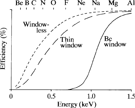
absorbing power). Where a beryllium window is fitted, efficiency falls below
about 2 keV, owing to absorption in the window, and is effectively zero below
about 0.7 keV, whereas useful efficiency is maintained even at low energies
when a thin window is fitted. Absorption also occurs in the gold contact layer
on the surface of the detector and in the ‘dead layer’ of silicon between the gold
layer and the active region. Figure 5.4 shows the detection efficiency as a
function of energy with various windows.
Sometimes absorption is increased by a film of vacuum-pump oil condensed
on the window (which is colder than its surroundings). The oil can be washed
off (following the manufacturer’s instructions) and condensation may be
avoided by installing a low-power heater to raise the temperature of the
detector housing slightly. This may also be used to remove ice formed from
water vapour diffusing through the window. The symptom of either of these
phenomena is reduced relative sensitivity to low-energy X-rays. The build-up
of ice or oil can be monitored by measuring the intensity ratio of suitably
chosen peaks (e.g. Cu La/Cu Ka) under standardised conditions.
5.2.4 Pulse processing and dead-time
The output pulses from the preamplifier are amplified to a size suitable for
pulse-height analysis. To minimise the effect of noise, the signal is averaged
over a time interval (typically a few tens of microseconds) defined by a
Fig. 5.4. ED detector efficiency in the low-energy region, with various
entrance windows (schematic only; the exact shape of the curve depends on
window thickness and composition).
82 X-ray spect rometers

parameter known as the ‘time constant’, or ‘process time’. While each pulse is
being processed the system is ‘dead’ – that is it does not respond to any further
pulses arriving from the detector. The time from the arrival of a pulse to the
moment when the system is ‘live’ again is the ‘system dead-time’ (t), which is
related to the time constant.
The dead-time o f an ED system is ‘exten dabl e’, m eani ng t hat, i f ano ther
pulse arrives during the processing of the preceding one, it extends the
dead pe riod by a further time t. F or such a system the input count-rate n
(the rate of ar rival of p ulses from the detec tor) a nd the o utput count-rate n
0
(the rate at which processed pulses ar e accumulated in the spectrum) are
related thus:
n
0
¼ n expðntÞ: (5:2)
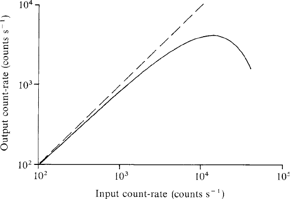
Equation (5.2) leads to the behaviour illustrated in Fig. 5.5: the output count-
rate increases with increasing input count-rate at first, reaches a maximum,
and then falls. At the maximum, n ¼ t
1
and n
0
¼ (et)
1
. A higher input rate is
pointless since the rate at which counts are accumulated in the spectrum then
falls below the maximum.
The ‘per cent dead-time’ represents the percentage of elapsed ‘real’ time for
which the system is dead, which is equal to 100[1 exp(nt)]. Normally a
warning is given if this exceeds 50%: this is partly because it is close to the
Fig. 5.5. Output versus input count-rate for a typical ED system: the
maximum output is governed by the extendable dead-time of the pulse-
processing electronics.
5.2 Energy-dispersive spectrometers 83

point of maximum ‘throughput’ (at 68% dead-time), but also because undesir-
able behaviour is liable to occur at higher count-rates.
The loss of effective counting time can be compensated automatically by
switching off the ‘clock’ controlling the counting time during the dead periods.
The spectrum is recorded for a live time selected by the user, the real time
which elapses during the recording period being extended to allow for the
dead-time. For example, with 30% dead-time a real time of 130 s is required in
order to accumulate a spectrum with a live time of 100 s.
The best energy resolution is obtained with a long time constant. As this is
reduced, E
n
in Eq. (5.1) increases and the resolution worsens, but pulses can
be recorded at a faster rate. The relationship between energy resolution and
maximum recording rate (as determined by the dead-time) for a typical system
is shown in Fig. 5.6. The energy resolution at a reasonably high count-rate is
more important for most purposes than the best possible resolution, which is
obtainable only at a low rate.
5.2.5 Spectrum display
The train of amplified pulses from the detector is converted into a spectrum by
means of a ‘multichannel pulse-height analyser’, which measures the height of
each incoming pulse and assigns it to a ‘channel’. The recorded spectrum takes
130
120
110
100
90
70
60
50
80
∆
E
n
(eV)
1
2
3
5
7
10
20
30
Max. throughput (10
3
counts s
–1
)
Fig. 5.6. The relationship between E
n
, the contribution of electronic noise
to the energy resol ution of the ED detector (see the text), and maximum
‘throughput’.
84 X-ray spect rometers
