Reed S.J.B. Electron microprobe analysis and scanning electron microscopy in geology
Подождите немного. Документ загружается.

the form of an array of numbers representing the contents of each channel and
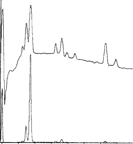
is displayed as a histogram (Fig. 5.7). Most X-ray lines of interest fall within
the range 0–10 keV, requiring 1000 channels of width 10 eV, or 2000 of width
5 eV. Correct energy calibration can be obtained by adjustment of zero and
gain controls so that known X-ray lines (a minimum of one at low energy and
one at high energy) appear at the correct points on the energy scale.
Various display facilities are provided: for example, the spectrum can be
expanded on either axis to facilitate inspection of particular features. As well
as the normal linear intensity scale, a logarithmic mode is an optional alter-
native: this has the advantage that large and small peaks are visible simultan-
eously. Markers showing the positions of elemental peaks can be inserted to
assist identification. A useful feature is the capability to display two spectra
simultaneously so that similarities and differences can be studied.
In addition to the spectrum itself, it is usual to have additional data dis-
played on the screen: typically these include current set-up information such as
electron volts per channel, energy range, type of display (logarithmic or linear),
intensity range, count-rate, preset live-time, elapsed time and per cent dead-
time, together with information such as date, time and spectrum label.
02468
Energy (keV)
Counts
Mg K
α
Al K
α
Si K
α
K K
α
Ca K
α
Ca K
β
Ti K
α
Fe K
α
Fe K
β
Fig. 5.7. The energy-dispersive X-ray spectrum of a silicate mineral, consisting
of a histogram of counts per channel, using logarithmic and linear scales (upper
and lower curves, respectively).
5.2 Energy-dispersive spectrometers 85
5.2.6 Artefacts in ED spectra
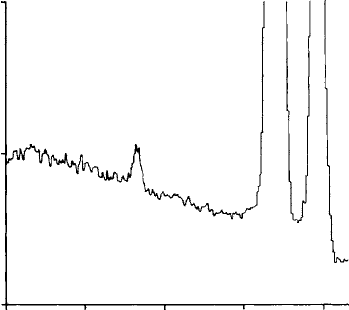
There are certain artefacts in ED spectra of which the user should beware. One
of these is the ‘escape peak’ – a small ‘satellite’ appearing 1.74 keV below its
‘parent’ peak in the case of a Si(Li) detector (Fig. 5.8), which is produced by the
following mechanism. After absorption of an X-ray photon in the detector, a
Si K photon may be emitted and, though usually this will be absorbed within
the detector, there is a finite probability that it may escape. If it does, the
output pulse height is reduced owing to the loss of the energy carried by the
Si K photon. The probability of escape depends on the energy of the incident
photon (which determines how deeply it penetrates the detector) but is gen-
erally less than 1%, so escape peaks are significant only when the parent peak
is large. With a Ge detector escape peaks are not usually observable.
Another related phenomenon occurring with S i(Li) d etectors is the
appearance of a spurious Si K peak in the spectrum, giving t he impression
ofthepresenceofasmallamountofSi(afractionof1%)eveninsamples
containing none. The size of the Si peak varies with the content of the
spectrum.
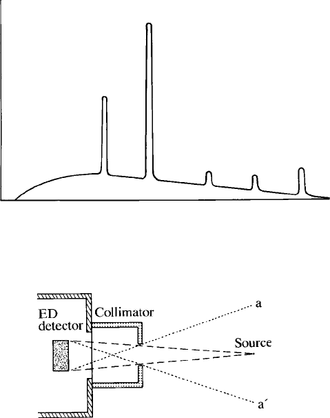
Small artefact peaks may also appear at energies corresponding to the sums
of the energies of major peaks in the spectrum (Fig. 5.9). These ‘sum peaks’ are
caused by pairs of pulses arriving so close together in time that the pulse
processor sees them as one. This effect is greatly reduced by electronic ‘pile-
up rejection’, but can never be eliminated totally. The probability of such
Cr peaks
K
βKα
Cr Kα
escape peak
Counts (10
3
)
2
1
0
23456
Energy (keV)
Fig. 5.8. The ‘escape peak’ in the ED spectrum of Cr, occurring 1.74 keV
below the main ‘parent’ peak (see the text for explanation).
86 X-ray spect rometers
coincidence between pulses is a function of the count-rate: sum peaks are
therefore significant only at high count-rates and, if troublesome, can be
reduced by decreasing the count-rate.
ED detectors have a wide angle of acceptance and do not discriminate between
X-rays produced at the point of impact of the electron beam and those generated
elsewhere by stray electrons. The spectrum may thus contain an unwanted
contribution from other regions of the specimen, the specimen holder, etc. This
can be minimised by means of a collimator in front of the detector, which restricts
the range of angles accepted (Fig. 5.10). However, it is impossible to achieve
perfect discrimination, and small spurious peaks may still occur.
Some backscattered electrons may have enough energy to penetrate the
detector window (especially if this is of the ultra-thin type) causing spurious
output pulses that increase the background level. This effect (which is greatest
with a high accelerating voltage) can be prevented by fitting an electron trap in
the form of a permanent magnet mounted in front of the detector.
A
B
A
+ A
A
+ B
B
+ B
Ener
gy
Number of counts
Fig. 5.9. ‘Sum peaks’ in the ED spectrum, occurring at energies equal to the
sums of the energies of the main peaks.
Fig. 5.10. A collimator attached to the front of an ED detector, limiting
acceptance to X-rays originating between a and a
0
.
5.2 Energy-dispersive spectrometers 87
5.3 Wavelength-dispersive spectrometers
Wavelength-dispersive (WD) spectrometers are distinguished from the energy-
dispersive (ED) type by the fact that the X-rays are ‘dispersed’ according to
their wavelength by means of Bragg reflection. WD spectrometers give high
spectral resolution but generally lower intensity for a given beam current than
the ED type: the two are thus complementary, as discussed in more detail in
Section 5.4.
5.3.1 Bragg reflection
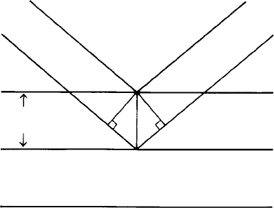
In certain directions waves scattered from successive layers of atoms in a
crystal are in phase and their intensity is enhanced. This is illustrated in
Fig. 5.11, where the difference in path length between rays ABC and A
0
B
0
C
0
is an integral multiple of the wavelength (l). This condition results in the
reflection of X-rays of a given wavelength by atomic layers with interplanar
spacing d at a certain angle of incidence and reflection known as the ‘Bragg
angle’ (). The relationship between these variables is given by Bragg’s law,
nl ¼ 2d sin ; (5:3)
which follows directly from the difference in path lengths between successive
planes. The integer n is the ‘order’ of reflection. The most intense reflections,
which are normally used in WD analysis, are those of the first order
(n ¼ 1). Higher orders add unwanted peaks to the spectrum, but their
intensity is relatively low and they can be suppressed by pulse-height analysis
(Section5.3.4).
C′
C
θ
θ
θθ
A
A
′
d
B
B
′
Atomic planes
Fig. 5.11. Bragg reflection: diffracted rays are in phase when distance A
0
B
0
C
0
differs from ABC by an integral number of wavelengths.
88 X-ray spect rometers

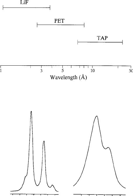
It follows from Eq. (5.3) that the wavelength range (for n ¼ 1) is limited for a
given value of 2d, and several crystals of different spacings are therefore
needed in order to cover the whole range of wavelengths of interest. The
crystals normally used are listed in Table 5.1 and their wavelength coverage
is shown in Fig. 5.12. Since the wavelength ranges overlap there is sometimes a
choice between two crystals for a given X-ray line (for example, the Ca Ka line
is reflected by LiF at 56.58 and by PET at 22.68). Where a given wavelength is
covered by two crystals, the one with the larger d and hence lower Bragg angle
gives higher intensity but poorer resolution (see Fig. 5.13).
Crystals with spacings larger than that of TAP are not available, but
artificial layered structures, or ‘pseudo-crystals’, enable the K lines of light
elements down to Be (atomic number 4) to be covered. In lead stearate, layers
of Pb atoms are separated by hydrocarbon chains, giving 2d ¼ 100 A
˚
, and
similar pseudo-crystals with larger spacings also exist. However, ‘multilayers’
produced by evaporating alternate layers of heavy and light elements (e.g. W
and Si) with contrasting X-ray scattering powers give considerably higher
intensities (but worse resolution) and have the additional advantage that
reflections of order higher than 2 are very weak, reducing the likelihood of
interference from lines of shorter wavelength. For light-element analysis it is
preferable to use multilayers optimised for different wavelengths by suitable
choice of elements and layer thicknesses (see Table 5.2).
Table 5.1. Crystals used in WD
spectrometers, with values of 2d
Crystal 2d (A
˚
)
LiF 4.026
PET 8.742
TAP 25.9
Table 5.2. Evaporated multilayers for long-wavelength
WD spectrometry
2d (A
˚
) Components K lines detected
60 W–Si F, O
100 Ni–C C, B
160 Mo–B
4
C Be, N
5.3 Wavelength-dispersive spectrometers 89
5.3.2 Focussing geometry
Bragg reflection typically occurs over a range of angles of less than 0.018 and,
with a point source of X-rays and a flat crystal, reflection of a particular
wavelength occurs over only a small part of the crystal. A larger reflecting
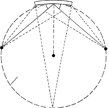
area can be obtained, however, if the crystal is curved. In the usual form of WD
spectrometer, source, crystal and detector are located on an imaginary
‘Rowland circle’ (Fig. 5.14). The atomic planes are curved to twice the radius
of this circle (r), which makes the Bragg angle the same at all points. Ideally the
surface of the crystal should lie on the Rowland circle, requiring the surface to
be ground to a radius r (‘Johansson geometry’). It is relatively easy to grind
Fig. 5.12. Wavelength coverages of crystals used in WD spectrometers.
Ce Lβ
1
Ce Lβ
1
Nd Lα
1
Nd Lα
1
0.580 0.592
sin
θ
(a)
sin
θ
(b)
0.267
0.272
Fig. 5.13. The effect of choice of crystal on resolution in a WD spectrum:
peaks are resolved with LiF (a) but not with PET (b).
90 X-ray spect rometers
LiF crystals, but PET and TAP are more difficult. ‘Johann geometry’, in which
the crystal is curved to the appropriate radius but not ground, is easier to
achieve but entails some sacrifice in resolution.
By placing a narrow slit in front of the counter, the peaks can be made
sharper (at the expense of reduced intensity). The enhanced resolution is
advantageous only in rare cases in which lines are so close that they are not
normally resolved. Otherwise it is better not to use a narrow slit (especially for
quantitative analysis), because it makes the peak intensity more sensitive to
error in the Bragg-angle setting.
Crystals of larger than normal dimensions are sometimes available as an
option, offering enhanced intensity, with some sacrifice in resolution. In many
applications resolution is not critical, but it affects the peak-to-background
ratio, which cancels out some of the benefit of the increased intensity, as far as
trace-element detection is concerned (Section 8.5).
Defocussing effects
The focussing geometry of a WD spectrometer is correct only with the X-ray
source in its normal position, that is, on the axis of the column and with the
surface of the specimen in the correct plane. Movement of the source in the z
direction causes a change in the Bragg angle (Fig. 5.15(a)), which has the effect
of shifting the position of the peak relative to the wavelength scale, which is
particularly important in quantitative analysis. In an electron microprobe the
Crystal
θθ
Source
Rowland
Circle
Detecto
r
r
Fig. 5.14. Rowland circle geometry: a constant Bragg angle is obtained when
the source, crystal and detector lie on the circumference of a circle.
5.3 Wavelength-dispersive spectrometers 91

specimen can be brought to the focus of the optical microscope by means of the
stage z movement, thereby avoiding the defocussing effect. In an SEM the
specimen plane is less well defined, the depth of focus in a scanning image
being much greater. A similar effect occurs if the beam is moved in a radial
direction relative to the Rowland circle (Fig. 5.15(b)). It is therefore desirable
to move the specimen rather than the beam when selecting different points for
analysis. Also, scanning images are liable to show intensity loss at the edges
(see Section 6.5).
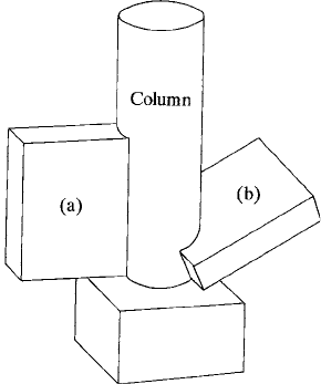
In Fig. 5.15 the spectrometer is assumed to be mounted vertically. The effect
of specimen-height variation is greatly reduced if the spectrometer is mounted
in an ‘inclined’ configuration (Fig. 5.16). This orientation is favoured when the
specimen height is poorly constrained, as in the case of SEMs. It is less usual in
electron microprobes, where it reduces the number of spectrometers that can
be fitted.
5.3.3 Design
In practical spectrometer designs the centre of the Rowland circle (Fig. 5.14)is
not in a fixed position. Instead the crystal moves along a linear track (aligned
with the X-ray source), the correct Bragg angle () being obtained by means of a
mechanical linkage, which also moves the detector along the appropriate path.
The source–crystal distance (x) is related to by the expression x ¼ 2r sin ,
where r is the radius of the Rowland circle. Since sin ¼ l/(2d) (for n ¼ 1), it
follows that x ¼ (r/d )l. The source–crystal distance is thus a linear function of
wavelength and the required wavelength is obtained by moving the crystal along
its track. The scale is calibrated by reference to a known X-ray line. WD
Electron
beam
Specimen
Crystal
(a) (b)
2r
sin
θ
θ
∆θ
∆θ
ψ
∆z
∆x
Fig. 5.15. Changes in Bragg angle () caused by displacement of the X-ray
source from its correct position: (a) in the z direction (height); and (b) in the x
direction (lateral shift).
92 X-ray spect rometers
spectrometers are usually provided with two or more crystals mounted on a
turret, in order to extend the wavelength range.
Normally WD spectrometers are evacuated to eliminate X-ray absorption
by air, which, though not serious for X-rays at the short-wavelength end of the
range of interest, is prohibitive at the long-wavelength end. High vacuum is not
essential, and in some instruments the spectrometers are pumped by a rotary
pump only, with the spectrometer separated from the column by a thin
window.
Electron microprobe instruments are commonly fitted with up to five ver-
tical WD spectrometers around the column. This has the advantage that
crystal changes can be avoided and time saved in multi-element analyses.
Usually SEMs can be fitted with only one spectrometer and therefore are
less efficient for WD analysis.
An important parameter is the ‘X-ray take-off angle’, which is defined as the
angle between the surface of the specimen and the X-ray path to the spectrometer.
If this is too low, the absorption of emerging X-rays is excessive (Section 7.7.2).
On the other hand, a high angle conflicts with other considerations, including the
desirability of a short final lens working distance. A reasonable compromise is a
value of about 408, which is typical for contemporary instruments (though note
that the effective take-off angle in SEMs is dependent on specimen tilt).
Alternatives to the conventional ‘fully focussing’ type of WD spectrometer
described above have been developed specifically for SEMs. In the ‘semi-focussing’
Fig. 5.16. Ori entation of WD spectrometers: (a) vertical – occupies less space
but is sensitive to specimen-height variations; and (b) inclined to the
horizontal – insensitive to specimen height, but occupies more space, so
fewer spectrometers can be fitted.
5.3 Wavelength-dispersive spectrometers 93
type, the source–crystal distance is fixed and interchangeable crystals are provided.
Each of these has a radius of curvature that gives satisfactory results over a limited
range of wavelengths. There is some cost saving compared with the fully focussing
type, at the expense of sacrificing full wavelength coverage.
In the ‘parallel-beam’ type of WD spectrometer, the X-rays are collimated
by means of focussing optics making use of the phenomenon of low-angle total
reflection. The resulting parallel beam is reflected by a flat crystal (or multi-
layer), giving higher intensities than in a conventional WD spectrometer for
long wavelengths (the efficiency of the focussing optics decreases with decreas-
ing wavelength). It is necessary for the specimen surface to lie in the correct
plane relative to the focussing optics: in an SEM lacking an optical micro-
scope, the appropriate location can be obtained by setting the spectrometer on
a known X-ray line and adjusting the specimen height for maximum intensity.
5.3.4 Proportional counters
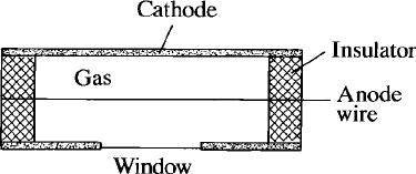
In a WD spectrometer X-rays are detected with a ‘proportional counter’
consisting of a gas-filled tube with a coaxial wire held at a positive potential
between 1 and 2 kV (Fig. 5.17). Ionisation of the gas atoms by X-rays generates
free electrons and positive ions, which are attracted respectively to the anode
wire and to the body of the counter (acting as cathode). The accelerated
electrons cause further ionisation, creating an ‘avalanche’, which results in a
pulse of electrical charge appearing on the anode. The size of the pulse is
dependent on the initial number of ions produced by the X-ray photon and,
since this number is proportional to the energy of the absorbed photon, the
pulse height is proportional to this energy. Electron multiplication in the
counter gas is strongly dependent on the anode voltage: as this is varied the
absolute pulse heights for all energies therefore change, while the relative
heights retain a constant relationship.
Fig. 5.17. A proportional counter as used in WD spectrometers: an X-ray
photon entering the window causes ionisation of the gas; the electric field
around the anode wire causes multiplication of ions and electrons, giving an
amplified output pulse proportional in height to the X-ray energy.
94 X-ray spect rometers
