Qin Y. Micromanufacturing Engineering and Technology
Подождите немного. Документ загружается.

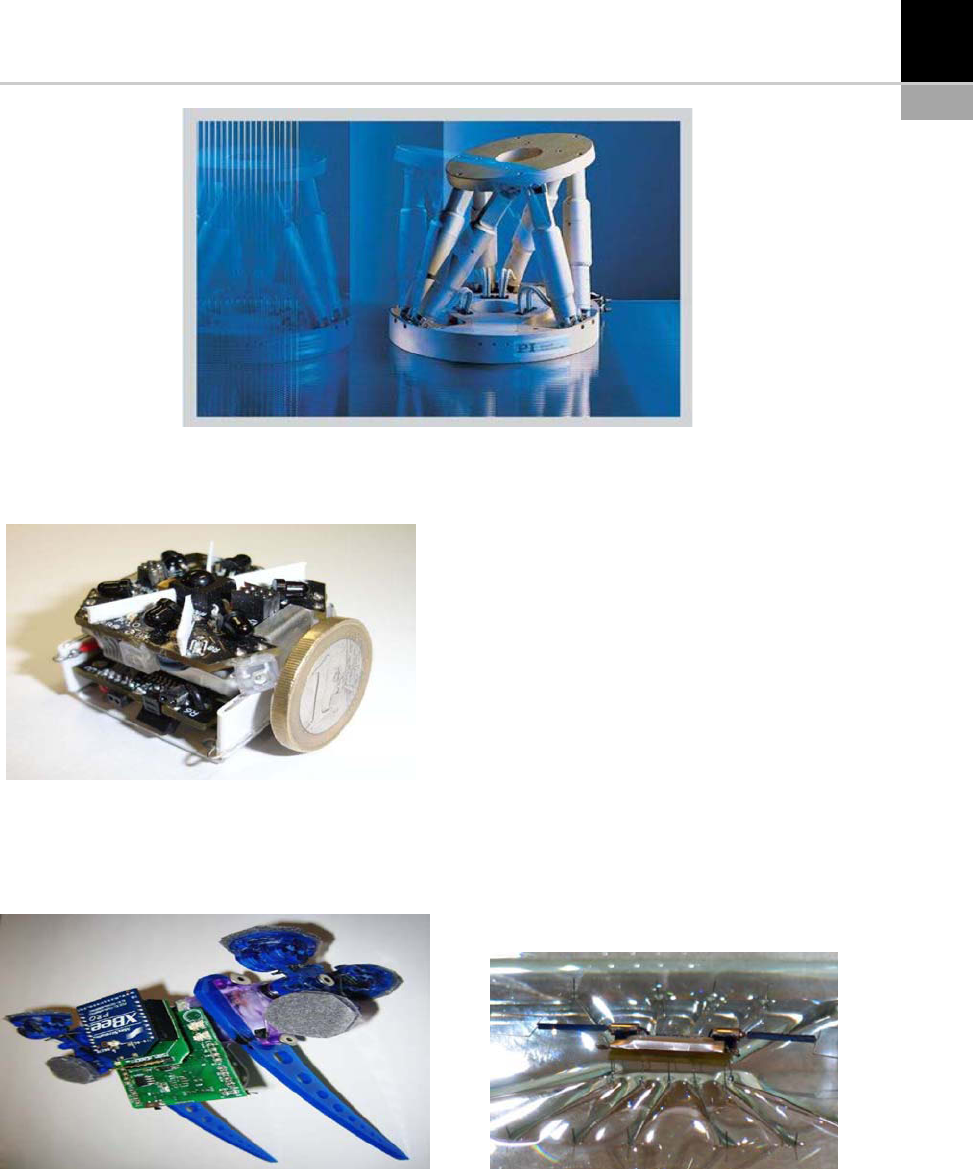
Using two actuated legs with rotary motion and
two passive revolute joints at each foot, this robot
can climb and steer in any orientation. The main
applications for this robo t are inspection and sur-
veillance, and space missions.
Strider Micro-robot [20]
This micro-robot (also developed at Nano-
Robotics Laboratory, Carnegie Mellon Univer-
sity) was inspired by water-strider insects
(Fig. 19-10 ). The robot ( as with the insects) uses
surface-tension force to balance its weight on
water by using hydrophobic Teflon-coated wire
legs. The maximum forward speed has been
measured to be 3 cm/s and its rotational speed
is 0.5 rads/s.
FIGURE 19-7 The Hexapod M-850.
(Source: courtesy of Physik Instrumente (PI) GmbH & Co. KG.)
FIGURE 19-8 The Jasmine III Robot.
(Source: courtesy of Open-source micro-robotics project.)
FIGURE 19-9 The Waalbot Robot.
(Source: courtesy of Metin Sitti, Nano-Robotics Laboratory,
Carnegie Mellon University.)
FIGURE 19-10 The Strider Micro-robot.
(Source: courtesy of Metin Sitti, Nano-Robotics Laboratory,
Carnegie Mellon University.)
CHAPTER 19 Robotics in Micro-Manufacturing and Mi cro-Robotics 321
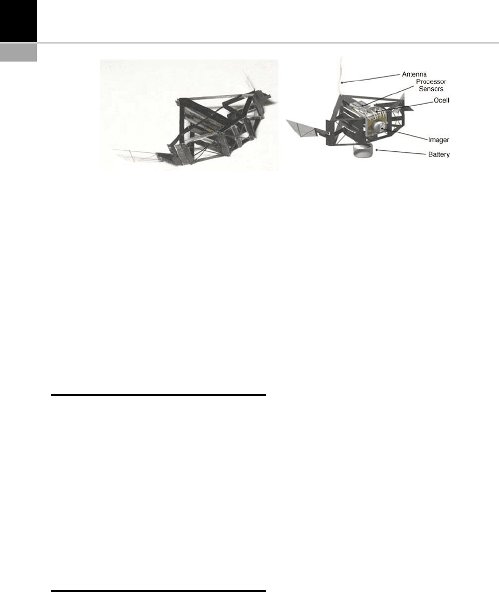
MFI Project
Closely related to the strider micro-robot
(inspired by insects) and the micro-air-vehicle
(autonomous flying micro-robot) is the Micro-
mechanical Flying Object (MFI) developed at
the Biomimetic Millisystem Lab. at UC Berkeley
(Fig. 19-11).
The goal of the project is to de velop a very
small device capable of sustained autonomous
flight, based on the flight performance of flies
and by means of piezoelectric actuators and flex-
ible thorax struc tures.
CONCLUSIONS
As has been seen throughout this chapter, micro-
robotics is a relatively new field with a large
amount of research still to be done. Numerous
laboratories and investigation centers are concen-
trating their efforts towards obtaining increas-
ingly more sophisticated and smaller robotic
devices.
Future applications of micro-robots are numer-
ous and extend from searc hing for survivors dur-
ing rescue missions to extremely precise surgical
operations (even autonomously by a micro-robot
inside the patient’s body) all the way up to top-
secret espionage missions.
REFERENCES
[1] M.B. Cohn, K.F. B
€
ohringer, J.M. Novorolski, A.
Singh, C.G. Keller, K. Goldberg, R.T. Howe, Micro-
assembly technologies for MEMS, Proc. SPIE Micro-
machining and Microfabrication, Conference on
Micromachining and Microfabrication Process Tech-
nology IV, Santa Clara, CA (Sept. 21–22, 1998)
2–16.
[2] G. Reinhart, O. Anton, M. Ehrenstrasser, C. Patron,
B. Petzold, Framework for teleoperated microassem-
bly systems, Proc. SPIE 4570, pp. 86-96, Telemani-
pulator and Telepresence Technologies VIII,
Matthew R. Stein (ed.) (2002).
[3] K. Kaneko, H. Tokashiki, K. Tanie, K. Komoriya,
Impedance shaping based on force feedback bilateral
control in macro-microteleoperation system, Proc.
ICRA (1997) 710–717.
[4] K. Shimamura, Y. Sasaki, M. Fujii, Low cost micro-
assembly machine, FUJITSU Sci. Tech. J 43 (1) (Jan.
2007) 59–66.
[5] G. Danuser, I. Pappas, B. V
€
ogeli, W. Zesch, J. Dual,
Manipulation of microscopic objects with nanometer
precision: potentials and limitations in nano-robot
design, Int. J. of Robotics Research (1997) 1–47.
[6] M.B. Cohn, Y.C. Liang, R.T. Howe, A.P. Pisano,
Wafer-to-wafer transfer of microstructures for vac-
uum packaging, 1996 Solid-State Sensor and Actua-
tor Workshop, Hilton Head Island, SC, USA (June
26, 1996).
[7] Q. Zhou, A. Aurelian, B. Chang, C. del Corral, H.N.
Koivo, Microassembly system with controlled envi-
ronment, J. of Micromechatronics 2 (3) (2004)
227–248.
[8] B. Kim, H. Kang, D.H. Kim, J.O. Park, A
flexible microassembly system based on hybrid manip-
ulation scheme for manufacturing photonics compo-
nents, The Int. J. of Advanced Manufacturing Tech-
nology (Springer London) 28 (3–4) (2005) 379–386.
[9] T. Zhu, D. Tan, Research on microrobot-based
microassembly system, Intelligent Control and Auto-
mation 2 (2002) 1236–1240.
[10] S. Fatikow, J. Seyfried, S. Fahlbusch, A. Buerkle,
F. Schmoeckel, A flexible microrobot based micro-
assembly station, J. of Intelligent and Robotic Sys-
tems 27 (1–2) (Jan. 2000) 135–169.
FIGURE 19-11 The MFI Project at UC Berkeley.
322 CHAPTER 19 Robotics in Micro-Manufacturing and Micro-Robotics

[11] N. Dechev, W.L. Cleghorn, J.K. Mills, Construction
of 3D MEMS microstructures using robotic micro-
assembly, International Conference on Robots and
Intelligent Systems (IEEE/RSJ IROS 2003), Las
Vegas, Nevada, USA (Oct. 27–31, 2003).
[12] I. Karjalainen, T. Sandelin, J. Uusitalo, R. Tuokko,
Robotic assembly and joining of miniature and
MEMS components using adhesive films – test envir-
onments and experiences, J. Assembly Automation
24 (1) (2004) 58–62.
[13] S. Henein, M. Thurner, A. Steinecker, Flexible micro-
gripper for micro-factory robots, Created by SHe/
Rev (July 2003).
[14] R. Keoschkerjan, H. Wurmus, A novel microgripper
with parallel movement of gripping arm, Proceedings
of the Eighth International Conference on New Actua-
tors, Bremen, Germany (June 10–12, 2002) 321–324.
[15] M. Goldfarb, A. Strauss, E.J. Barth, Overview of
mechatronics. Chapter 5: An introduction to micro
and nanotechnology, CRC Press LLC (2002).
[16] C.B. Sippola, C.H. Ahn, A thick film screen-printed
ceramic capacitive pressure microsensor for high
temperature applications, J. of Micromechanics and
Microengineering 16 (5) (2006) 1086–1091.
[17] E. Dereine, B. Dehez, D. Grenier, B. Raucent, A sur-
vey of electromagnetic micromotors, Proceedings of
the 1st International Precision Assembly Meeting
(IPAS 2003), Bad Hofgastein, Austria (March 17–
19, 2003) 85–94.
[18] R.J. Wood, S. Avadhanula, E. Steltz, M. Seeman, J.
Entwistle, A. Bachrach, G. Barrows, S. Sanders, R.S.
Fearing, An autonomous palm-sized gliding micro air
vehicle, IEEE Robotics & Automation Magazine
(2007) 82–91.
[19] M.P. Murphy, Waalbot: an agile small-scale wall-
climbing robot utilizing dry elastomer adhesives,
IEEE/ASME Transaction on Mechatronics 12 (3)
(2007) 330–338.
[20] Y.S. Song, M. Sitti, Surface tension driven biologi-
cally inspired water strider robots: theory and experi-
ments, IEEE Trans. on Robotics 23 (3) (June 2007)
578–589.
FURTHER READINGS
[1] P.J. McKerrow, Introduction to Robotics, Ed. Addi-
son Wesley (1990).
[2] A.J. Sanchez, R. Lopez, R. Guzman, C. Ricolfe, Recent
development in micro-handling systems for micro-
manufacturing, J. of Materials Processing Technology
167 (22) (2005) 499–507.
[3] G. Lin, R.A. Lawton, 3D MEMS in standard pro-
cesses: fabrication, quality assurance and novel mea-
surement microstructures, NASA Technical Report
(2000).
[4] J. Bell, Automated packaging of MEMS devices, J. of
SMT 16 (2) (2003) 22–26.
CHAPTER 19 Robotics in Micro-Manufacturing and Mi cro-Robotics 323

20
Optical Coherence
Tomography for the
Characterization of
Micro-Parts and -Structures
David Stifter
INTRODUCTION
Optical coherence tomography (OCT) was pre-
sented in 1991 for the first time as a powerful
technique for applications in the field of medical
diagnostics [1]. It was demonstrated that with
OCT high resolution cross-sectional images of bio-
logical tissue can be obtained in a contactless and
non-invasive way. The investigation of retinal dis-
eases, such as glaucoma, belonged to the first appli-
cations, with a main consequence being that com-
mercial retinal OCT scanners are already available
andusedineyeclinicsandhospitals.TheOCT
method has been further refined in the meantime
and a multitude of new developments and exten-
sions for this technique have been introduced, as
also summarized in several books and reviews (e.g.
[2–3]). The main applications and driving forces
for these developments in the field of OCT research
can still be found in the area of biomedical diag-
nostics, ranging from the investigation of the eye
(e.g. also cornea), skin (e.g. melanoma), teeth (car-
ies) or of interior organs and vessels (e.g. by OCT
endoscopy) to life science applications providing
image details down to the sub-cellular level.
Although the main route of OCT develop-
ments is for biomedical purposes, the potential
of OCT for contactless and non-destructive eval-
uation of non-biological materials and compo-
nents has been recognized due to the fact that
depth-resolved structural information can be
obtained with high accuracy in a fast and easy
way from the interior of materials, even for those
of a highly scattering na ture. Consequently, a
variety of technical applications for OCT have
now emerged, as compiled in a recent compr ehen-
sive review [4], anticipating a future increase in
their number with respect to their biomedical
counterparts.
In this chapter, initially, an introduction is
provided to the underlying measurement princi-
pleofOCTandashortoverviewofdifferent
alternative and advanced OCT measurement
concepts. A selection of proven applications of
classical and advanced OCT techniques for the
evaluation of micro-structures is then presented.
It is worth noting that due to the novelty of the
herein-introduced OCT techniques and results,
routine testing and evaluation of micro-struc-
tures by OCT is not yet standard but it is
expected to fully emerge in the next few year s,
as OCT measurement technology further devel-
ops and matures.
CHAPTER
324
MEASUREMENT PRINCIPLES OF
STANDARD AND SELECTED
ADVANCED OCT TECHNIQUES
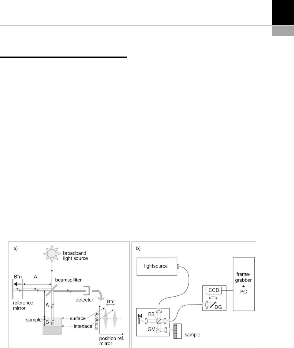
The basic physical principle of OCT is low coher-
ence interferometry (LCI): an interferom eter,
mostly built in Michelson geometry, is illumi-
nated with spectrally broad light, as depicted in
Figure 20-1(a). The sample is placed in one arm of
the interferometer, the reference arm itself is
equipped with a movable mirror. Since low coher-
ence light is used, interference is only observed if
the length of the optical path between the beams-
plitter and a backscattering feature within the
sample, such as a buried interface, equals the opti-
cal path length of the reference arm. In this way,
the absolute positions of back-scattering and
-reflecting features can be determined. In the
example of Figure 20-1(a), two interference peaks
are observed when moving the reference mirror
(from surface and interface). Usually, the enve-
lope of the interference signal is taken and it repre-
sents a reflectivity depth profile of the sample at a
fixed lateral position (so-called A-scan). By mea-
suring several depth profi les at adjacent positions,
e.g. by scanning the focused light beam over the
sample, cross-sectional images (B-scans) and 3D
volume data can be acquired.
The width of the peaks in a depth profile, deter-
mining the axial resolution of the system, is given
by the coherence length of the used light source:
the broader the spectrum, the higher the axial
resolution [5]. Standard OCT systems exhibit
axial resolutions in the 5–15 mm range, ultra-high
resolution OCTs (UHR-OCT [6]) have been
reported showing even sub-micron resolutio n
with very broadband light sources, like thermal
or super continuum sources. The light sources are
spectrally situated mostly in the near-infrared
region (800 nm–1500 nm) with an average power
in the mW range, ensuring damage-free (non-
invasive) inves tigation of living tissue. Conse-
quently, no special safety precautions have to be
taken when handling OCT apparatus, as is man-
datory for, e.g., X-ray computed tomography.
For OCT the axial resolution is decoupled
from the lateral one, which is determined by the
spot-size of the focused light beam on the sample.
Due to the decoupling, a high axial resolution can
be maintained even for long working distances, in
contrast to confocal microscopy where high
numerical aperture (NA) optic s have to be used
for high depth discrimination [7]. The axial reso-
lution in OCT should not be mi staken for the
accuracy with which the position of the envelope
peaks can be determined: on smooth surfaces
FIGURE 20-1 (a) Schematic sketch of a standard (time-domain) OCT set-up with a broadband light source illuminating
a Michelson interferometer. A, B: path lengths, n: refractive index of sample layer; (b) layout of a spectral-domain OCT
set-up. Abbreviations: beamsplitter (BS), reference mirror (M), galvano-scanner mirror for lateral scanning (GM), diffraction
grating (DG), line camera (CCD).
CHAPTER 20 Optical Coherence Tomography for the Characterization of Micro-Parts and -Structu res 325

and interfaces, precision in the nanometer range is
feasible. In this context, scannin g white-light
interferometry (SWLI) and coherence probe
microscopy (CPM) shall be mentioned, since these
methods share the same principle (LCI) and a
similar set-up as OCT [8]: in contra st to conven-
tional OCT, mostly whole surface areas are illu-
minated at once and area cameras are used to
register the interference signal when moving a
reference mirror. Such systems were optimized
for the measurement of surface topographies with
sub-nanometer accuracy and are now widely
employed in the semiconductor industry for the
inspection of integrated circuits [9] or for the
characterization of micro-electromechanical
structures (MEMS) [10] . Although transparent
layers can also be meas ured with these systems
(e.g. thickness), only OCT is also capable of deter-
mining the internal structure of scattering and
turbid media with reasonable penetration depth
(in the millimeter range). However, the border
between the different techniques start to blur, as
a combination of the CPM technique and OCT
leads, e.g. to full-field OCT using high NA optics,
area illumination and area cameras [11].
The concept of obtaining depth information by
moving the reference mirror (Figure 20-1(a))is
referred to as time-domain (TD) OCT configura-
tion. A new trend is to build OCT devices prefer-
ably in the Fourier-domain (FD) configuration. In
FD-OCT the reference mirror is fixed and the
wavelength of a narrow-band light source is rap-
idly tuned (swept-source OCT) or the spectrum of
a broadband light source is acquired by a spec-
trum analyzer as depicted in Figure 20-1(b) (spec-
tral-domain OCT (SD-OCT)). The obtained spec-
tral data is Fourier transformed to obtain at once
the desired depth information in the form of a
whole A-scan. The main advantages of FD-OCT
over TD-OCT can be found in the fact that no
movable parts are needed for depth scanning, in
the increased system sensitivity and in the high
measurement speed with A-scan rates of more
than 100 kHz (e.g. [12,13]).
Besides measuring the intensity of the back
reflected light to gain structural information,
further sample properties and enhanced con-
trast can be obtained by taking additional phys-
ical phenomena into account with advanced
OCT extensions: a determination of the fre-
quency shift of the reflected light from moving
particles leads to optical Doppler tomography
(ODT) for the depth-resolved measurement of
flow veloci ties [14]. Finally, polarization-
sensitive OCT (PS-OCT) shall be mentioned:
the evaluation of the polarization state of the
light gives insight into the birefringence prop-
erties of a material [15].
APPLICATIONS OF OCT FOR THE
EVALUATION OF STRUCTURES ON
THE MICRON SCALE
In the following, a short selection of instructive
examples for OCT recently applied by the author
to measurement tasks in micro- and miniature
manufacturing are given, especially with the
intention to familiarize the reader with the poten-
tial of the novel OCT techniques for these kinds of
applications and to promote OCT for future rou-
tine tasks in the characterization and testing of
micro- and miniature structures.
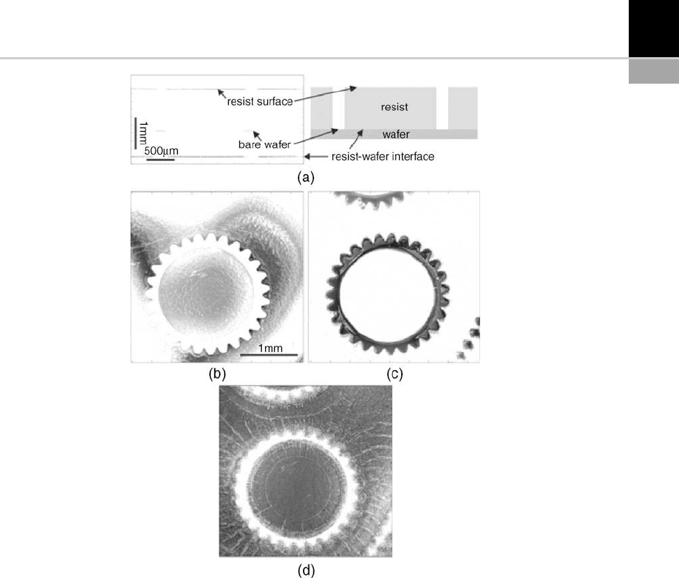
At first, an OCT study on photoresist molds for
the production of miniature gear wheels with the
LIGA molding technology (LIGA, German acro-
nym for lithography-electroplating-molding) is
presented, with the results depicted in Fig. 20-2.
High aspect ratio trenches exhibiting widths
down to 30 mm were etched in thick photoresist
layers deposited on gold-coated silicon wafers.
Residual parti cles in the trenches as well as the
defects of the resist/wafer interface are of primary
concern since they affect the quality of the molds,
crucial for the subsequent electroplating step. As
can be seen from the OCT cross-sectional image in
Fig. 20-2(a), the thickness of the photoresist layer
can easily be determined. However, since the opti-
cal path length within the material is different to
the path length in the (air-filled) trenches, a virtual
step is observed at the wafer surface. From the
height of this virtual step the refractive index of
the material can be determined and therefore
also the geome trical (and not only the optical)
thickness of the resist layer can be obtained.
326 CHAPTER 20 Optical Coherence Tomography for the Characterization of Micro-Parts and -Structures
Furthermore, it should be noted that the rather
low NA optics of OCT is of significant advantage
in order to access the bottom of the high aspect
ratio trenches.
By using so-called en-face scann ing OCT [16],
single planes parallel to the sample surface can
be at once imaged at defined depth locations,
as shown in Fig. 20-2(b)–(d). In addition to
the geometrical form of the whee l, surface corru-
gations with a maximum height of less than
100 nm (Fig. 20.2(b)), residual particles in the
trenches (white spots in the black wheel structure,
Fig. 20-2(c)) and a network of ridges (most prob-
ably caused by undercutting, Fig. 20-2(d)), could
be observed and evaluated.
A second example is presented in Fig. 20-3,
where a drug-elut ing medical stent has been inves-
tigated. The stent structure is essentially a thin
metallic network in the shape of a tube (see also
the schematic sketch in the inset of Fig. 20-3(a))
FIGURE 20-2 (a) Cross-sectional scan and schematic drawing of a mold for a miniature wheel in a 1.3 mm thick photoresist
layer on a gold coated wafer; (b)–(d) 3 3mm
2
en-face scans of the structure. In (a), only the surfaces of the bare resist and
the wafer, and the resist/wafer interface can be distinguished. In (b)–(d), the full geometric information of the structure at
these levels is obtained. In (b) the resist surface is imaged, (c) and (d) were recorded at depth positions of the optical path
length corresponding to the bare wafer surface and the resist/wafer interface (as shown in (a)), respectively.
From [16], Optical Society of America 2005.
CHAPTER 20 Optical Coherence Tomography for the Characterization of Micro-Parts and -Structu res 327
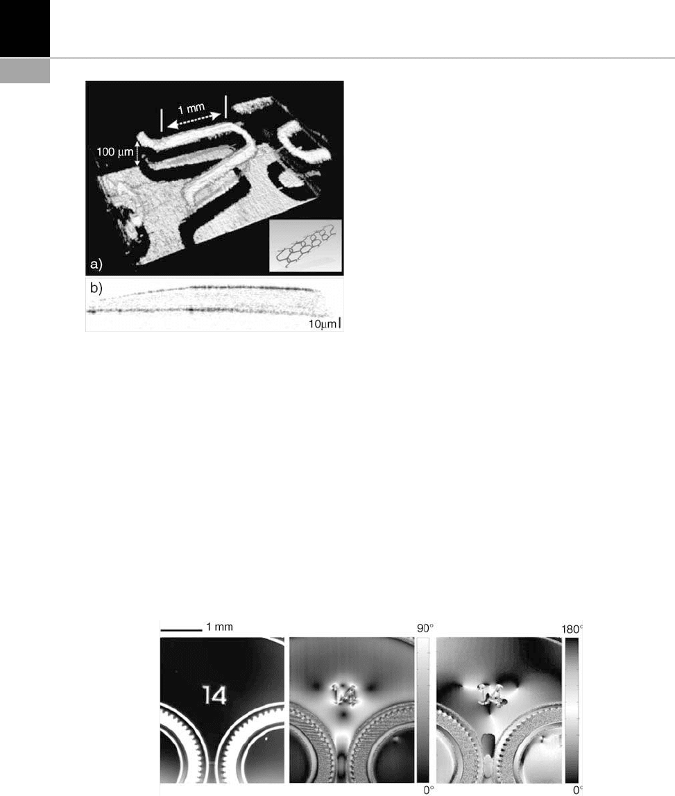
which is inserted, e.g. in blood vessels, and dilated
to counteract a disease-induced decrease of vessel
and duct diameter and helps in this way to main-
tain the flow. Of interest is the topography and
roughness of the individual metallic meanders
(Fig. 20-3(a)). The stent structure is also coated
with a thin (5–20 mm) drug-containing polymer
layer. Its homogeneity and thickness are crucial
for a determined delivery of a drug – enclosed in
the polymer layer – to the surrounding tissue in
order to hinder the formation of thick tissue at the
interior of the dilated vessel, which could again
obstruct the blood flow. As can be seen in the
cross-section of Fig. 20-3(b), taken along one of
the linear segmen ts of the meander structure, the
polymer layer can easily be resolved with OCT on
the rough metal surface and exhibits inhomoge-
neous regions with thickness variations of more
than 100%.
The extension of OCT towards PS-OCT leads
to additional contrast and information, exempli-
fied in Fig. 20-2. En-face PS-OCT has been per-
formed on resist molds, similar to those depicted
in Fig. 20-4: PS-OCT is capable of detecting the
birefringence caused by residual stress in the pho-
toresist layer allowing in this way high resolution
strain/stress mapping within the wheel mold
structures. In addition to the standard intensity
image, a retardation image (middle image in Fig.
20-4) is obtained. This image is grayscale coded
and gives the phase lag of the reflected light of one
polarization direction with respect to the other,
orthogonal one. Highly strained areas show
strong birefringence leading to higher optical re-
tardation and are depicted in light gray and white
in the image. By calibrat ion pro cedures, i.e.
determining the stressoptical coefficient of the
material under investigation, even a quantitative
evaluation of the strain distribution is possible
FIGURE 20-3 OCT inspection of a coated (drug-eluting)
medical stent: (a) topography of one stent segment with
schematic structure of the whole stent in the inset; (b) cross-
section of the polymer coating on the metal surface of the
stent (scan taken along the straight part of the segment as
indicated in (a)); measurements performed in cooperation
with K. Wiesauer, UAR GmbH.
FIGURE 20-4 En-face images of photoresist molds for miniature gearwheels. Left: OCT intensity image; middle: optical
retardation image (phase lag grayscale coded from 0
to 90
); right: orientation of optical axis indicating the direction of the
internal stress (orientation grayscale coded from 0
to 180
).
From [18], Oldenbourg Verlag 2007.
328 CHAPTER 20 Optical Coherence Tomography for the Characterization of Micro-Parts and -Structures

[17]. Furthermor e, the orientation of the optical
axis, which is related to the orientation of the
internal stress within the sample, can also be mea-
sured, as shown in the right image of Fig. 20-4 .
With the help of PS-OCT valuable insight for the
optimization of the production process with its
individual photolithographic steps can conse-
quently be gaine d, especially focusing on the min-
imization of internal stress, which can cause dis-
tortions of the wheel geometries and may lead to
detrimental cracks at the wafer/resist interface
and between trenches.
This section is conclud ed on selected measure-
ment applications for micro-structures by recal-
ling the evaluation of micro-fluidic devices with
OCT: the knowledge of the flow characteristics in
micro-fluidic networks and devices, such as
micro-mixers and lab-on-a-chip systems, helps
to assess the predicted performance and verify
the functionality of novel fluidic chip designs. In
this view, the flow behavior of micro-mixers was
studied by conventional OCT imaging: two
liquids with a different concentration of scatterers
were used to visualize flow patterns in the OCT
images, giving in this way information on the
status of intermixing and the spatial distribution
of vortices in the fluidic structure [19]. Besides the
retrieval of geometrical information of the flow
structure, ODT can now be applied to measure
even the flow velocity profiles in the micro-chan-
nels in a spatially resolved way, as recently
reported in, e.g. ref. [20].
CONCLUSIONS AND OUTLOOK
Classical OCT and advanced OCT techniques,
providing high resolution and taking advantage
of different contrast mechanisms such as birefrin-
gence or flow velocity, have been introduced for
the evaluation of micro- structures and -parts. The
fact that depth resolved information can be
obtained – even from the interior of scattering
materials – in a contactless way by OCT using
harmless infrared light renders this method espe-
cially promising for future routine applications in
the metrology of micro-structures. From the tech-
nological point of view the advantage of FD-OCT
with respect to robustness, speed and achievable
sensitivity will promote its breakthrough. Cur-
rently, the potential is recognized by companies
which offered up to now only commercial bio-
medical OCT systems but are preparing their pro-
ducts for the industrial metrology market, such as,
e.g. automated inspection of thin multilayer sys-
tems. Routine applications for advanced OCT
techniques such as PS-OCT or for phase-resolved
OCT microscopy providing sub-nanometer accu-
racy [21] are yet to come. Finally, it shall be con-
sidered that hybrid techniques based on SWLI,
CPM and OCT will allow tailoring the char-
acteristics of measurement systems to the exact
requirements imposed by the specific micro-
structure evaluation tasks.
ACKNOWLEDGMENTS
Financial support by the Austrian Science Fund
FWF (projects: L126-N08 and P19751-N20) is
acknowledged.
REFERENCES
[1] D. Hu ang, E.A. Swan son, C.P. Lin, J.S. Sc human,
W.G. Stinson, W. Chang, M.R. Hee, T. Flotte,
K. Gregory, C.A. Puliafito, J.G. Fujimoto, Optical
coherence tomography, Science 254 (1991)
1178–1181.
[2] B.E. Bouma, G.J. Tearney, (eds), Handbook of
Optical Coherence Tomography, Marcel Dekker
Inc, New York (2002).
[3] A.F. Fercher, C.K. Hitzenberger, Optical coherence
tomography, Progr. Opt 44 (2002) 215–301.
[4] D. Stifter, Beyond biomedicine: a review of alterna-
tive applications and developments for optical coher-
ence tomography, Appl. Phys. B 88 (2007) 337–357.
[5] E.A. Swanson, D. Huang, M.R. Hee, J.G. Fujimoto,
C.P. Lin, C.A. Puliafito, High-speed optical coher-
ence domain reflectometry, Opt. Lett 17 (1992)
151–153.
[6] W. Drexler, U. Morgner, F.X. Kartner, C. Pitris, S.A.
Boppart, X.D. Li, E.P. Ippen, J.G. Fujimoto, In vivo
ultrahigh-resolution optical coherence tomography,
Opt. Lett 24 (1999) 1221–1223.
[7] C.J.R. Sheppard, Confocal Laser Scanning Micros-
copy (1997) Springer, New York.
[8] C.J.R. Sheppard, M. Roy, M.D. Sharma, Image for-
mation in low-coherence and confocal interference
microscopes, Appl. Opt 43 (2004) 1493–1502.
CHAPTER 20 Optical Coherence Tomography for the Characterization of Micro-Parts and -Structu res 329

[9] M. Davidson, K. Kaufman, I. Mazor, F. Cohen, An
application of interference microscopy to integrated
circuit inspection and metrology, Proc. SPIE 775
(1987) 233–247.
[10] C. O’Mahoni, M. Hill, M. Brunet, R. Duane, A.
Mathewson, Characterization of micromechanical
structures using white-light interferometry, Meas.
Sci. Technol 14 (2003) 1807–1814.
[11] A. Dubois, A.C. Boccara, M. Lebec, Real-time reflec-
tivity and topography of depth-resolved microscopic
surfaces, Opt. Lett 24 (1999) 309–311.
[12] M.W. Jenkins, D.C. Adler, M. Gargesha, R. Huber,
F. Rothenberg, J. Belding, M. Watanabe, D.L.
Wilson, J.G. Fujimoto, A.M. Rollins, Ultrahigh-
speed optical coherence tomography imaging and
visualization of the embryonic avian heart using a
buffered Fourier Domain Mode Locked laser, Opt.
Express 15 (2007) 6251–6267.
[13] B. Potsaid, I. Gorczynska, V.J. Srinivasan, Y. Chen,
J. Jiang, A. Cable, J.G. Fujimoto, Ultrahigh speed
spectral/Fourier domain OCT ophthalmic imaging
at 70,000 to 312,500 axial scans per second, Opt.
Express 16 (2008) 15149–15169.
[14] Z. Chen, T.E. Miller, S. Srinivas, X.J. Wang, A. Mal-
ekafzali, M.J.C. van Gemert, J.S. Nelson, Noninva-
sive imaging of in vivo blood flow velocity using
optical Doppler tomography, Opt. Lett 22 (1997)
1119–1121.
[15] C.K. Hitzenberger, E. G
€
otzinger, M. Sticker, M.
Pircher, A.F. Fercher, Measurement and imaging of
birefringence and optic axis orientation by phase
resolved polarization sensitive optical coherence
tomography, Opt. Express 9 (2001) 780–790.
[16] K. Wiesauer, M. Pircher, E. G
€
otzinger, S. Bauer, R.
Engelke, G. Ahrens, G. Gr
€
utzner, C.K. Hitzenberger,
D. Stifter, En-face scanning optical coherence
tomography with ultra-high resolution for mate-
rial investigation, Opt. Exp ress 13 (2005)
1015–1024.
[17] K. Wiesauer, A.D. Sanchis Dufau, E. G
€
otzinger,
M. Pircher, C.K. Hitzenberger,D.Stifter,Non-
destructive quantification of internal stress in poly-
mer materials by polarisation sensitive optical
coherence tomogra phy, A cta M ater ialia 53 (2005)
2785–2791.
[18] D. Stifter, K. Wiesauer, M. Pircher, E. G
€
otzinger, R.
Engelke, G. Ahrens, G. Gr
€
utzner, C.K. Hitzenberger,
Optische Koh
€
arenztomografie als neues Werkzeug
f
€
ur die zerst
€
orungsfreie Werkstoffpr
€
ufung, TM 74
(2007) 51–56.
[19] C. Xi, D.L. Marks, D.S. Parikh, L. Raskin, S.A. Bop-
part, Structural and functional imaging of 3D micro-
fluidic mixers using optical coherence tomography,
Proc. Nat. Acad. Sci 101 (2004) 7516–7521.
[20] Y.C. Ahn, W. Jung, J. Zhan, Z. Chen, Investiga-
tion of laminar dispersion with optical coherence
tomography and optical Doppler tomography, Opt.
Express 13 (2005) 8164–8171.
[21] C. Joo, T. Akkin, B. Cense, B.H. Park, J.F. de Boer,
Spectral-domain optical coherence phase microscopy
for quantitative phase-contrast imaging, Opt. Lett 30
(2005) 2131–2133.
330 CHAPTER 20 Optical Coherence Tomography for the Characterization of Micro-Parts and -Structures
