Murray J. Clifford. Angiogenesis Protocols - Methods in Molecular Medicine, Vol. 46
Подождите немного. Документ загружается.

Disc Angiogenesis 75
15. Sandison, J. C. (1924) A new method for the microscopic study of living growing
tissues by the introduction of a transparent chamber in the rabbit’s ear. Anat. Rec.
28, 281–287.
16. Reinhold, H. S., Blachiewicz, B., and Berg-Blok, A. (1979) Reoxygenation of
tumors in “sandwich” chambers. Eur. J. Cancer 15, 481–489.
17. Warren, B. A. and Shubik, P. (1966) The growth of the blood supply to melanoma
transplants in the hamster cheek pouch. Lab. Invest. 15, 464–478.
18. Proia, A. D., Chandler, D. B., Haynes, W. L., Smith, C. F., Suvarnamani, C.,
Erkel, F. H., and Klintworth, G. K. (1988) Quantitation of corneal
neovascularization using computerized image analysis. Lab. Invest. 58, 473–479.
19. Young, C. L., Adamson, T. C., III, Vaughan, J. H., and Fox, I. R. (1986) Immuno-
histologic characterization of synovial membrane lymphocytes in rheumatoid
arthritis. Arthritis Rheum. 27, 32–39.
20. Hirata, S., Matsuhara, T., Saura, R., Tateishi, H., and Hirohata, K. (1989) Inhibi-
tion of in-vitro vascular endothelial cells proliferation and in-vivo vascularization
by low-dose methotrexate. Arthritis Rheum. 32, 1065–1069.
21. Allison, A. C. and Kowalski, W. J. (1989) Prostaglandins as transducers of prolif-
eration signals in microvascular endothelial cells and the pharmacological control
of angiogenesis. In Vascular Endothelium-Receptors and Transduction Mecha-
nisms (Catravas, J. D., Gillis, C. N., and Ryan, U. S., eds.), Plenum Press, New
York, pp. 99–110.
22. Page, R. C. (1986) Gingivitis. J. Clin. Periodontol. 13, 345–353.

Sponge Model 77
77
From:
Methods in Molecular Medicine, Vol. 46: Angiogenesis Protocols
Edited by: J. C. Murray © Humana Press Inc., Totowa, NJ
6
Sponge Implant Model of Angiogenesis
Silvia P. Andrade
1. Introduction
The process of capillary growth, angiogenesis, is an integral part of wound
healing and repair mechanisms. When it occurs during these conditions, it is
tightly controlled and strictly delimited. However, in tumor growth and in a
variety of vascular diseases, unrestrained angiogenesis can contribute signifi-
cantly to the pathology and persistence of these manifestations (1). Research
on angiogenesis was initiated with the development of several bioassays that
have permitted direct observations of the microvasculature in the living ani-
mal. The bioassays have been used for a variety of purposes; for example, to
detect angiogenesis activity in malignant and normal cells and tissue, to screen
purified test substances for angiogenic activity, and to elucidate the cellular
events that accompany vessel growth. The response observed after the introduction
of an appropriate stimulus such as mechanical injury or injection of neoplastic
tissue implants has allowed the cataloging of the main events of the angiogenic
cascade as well as the characterization of pro- and antiangiogenic factors.
The in vivo assays now available to study blood vessel formation include
implantation of a variety of synthetic matrices in which determination of sev-
eral quantitative and qualitative components of the fibrovascular tissue that
infiltrate the sponge compartment can be assessed. The common principle
underlying this technique is that injury caused by introduction of the device
elicits within the area circumscribed by the implant a response that mimics the
stages of wound repair; therefore, the angiogenic response in inflammatory
tissue can be evaluated (2,3). Additionally, spongy implants have also been
used as a framework to host different tumor cell lines in rodents (4–6) for study-
ing tumor angiogenesis. The advantage of implantation technique for the pur-
78 Andrade
pose of investigating tumor-induced angiogenesis is that assessment of the rela-
tive contributions of the tumor cells to early changes in the implant blood-flow
can be detected even before visible growth of the tumor mass is evident.
The development of vasoactive regulatory systems and pharmacological
reactivity of the neovasculature have also been investigated by means of sponge
implantation technique (6–9). Yet, the cannulated sponge implant model has
emerged as an alternative biological route for site-specific and systemic drug
delivery in cases where repeated injections at the conventional sites are not
feasible. For example, the tails and skin of experimental animals are too fragile
to stand daily injections. Several parameters derived from physiological, bio-
chemical, and morphological approaches have been used for defining the com-
ponents of the repair processes as well as for quantitating the implant
fibrovascular infiltration of the host tissue. By employing radioactive isotopes
and fluorogenic-dyes washout techniques, measurement of blood-flow in the
wound compartment can be performed, which in turn indicates the functional
state of the neovasculature and the interaction between the angiogenic site and
the systemic circulation (3,8). Biochemical determination of several compo-
nents of the fibrovascular tissue, such as wet and dry weight, DNA, protein,
extracellular matrix components, hemoglobin, enzyme activity and others, can
provide assessment of cellular proliferation kinetics and extracellular matrix
components involved in the process (7,10–14). Utilizing morphological or
morphometric approaches, the sequence of histological changes and vascular
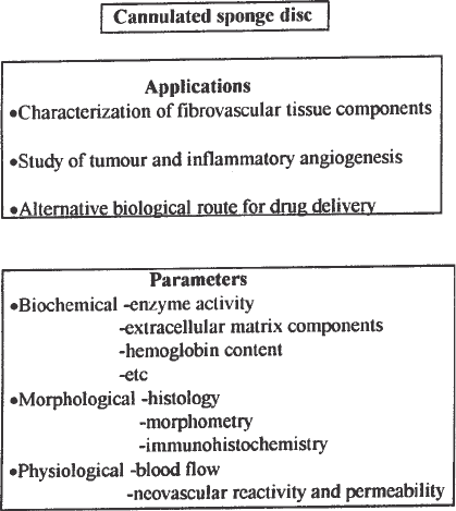
density can be determined (3,8,10,11). Figure 1 shows a schematic representa-
tion of several approaches and parameters that might be analyzed in the cannu-
lated sponge model of angiogenesis. Analysis and modulation of various
aspects of the inflammatory response that accompanies implantation are facili-
tated by the readily accessible location of the device and have contributed to
new insights and strategies towards better understanding the angiogenic pro-
cess. This chapter is an account of the usefulness of the sponge implant model
of angiogenesis and a detailed description of the methodology.
2. Materials
1. Sponge matrix: A number of different sponge matrices have been used for induc-
ing fibrovascular growth and as host to implanted tumor cells. The synthetic
materials are mainly polyvinyl alcohol, cellulose acetate, polyester, polyether,
and polyurethane alone or in combination. In our laboratory we use sponge discs
made of polyether polyurethane. This type of material possesses the following
characteristics: uniform pore size and intercommunicating pore structure, ability
to resist chemical treatment, and biocompatibility.
2. Polythene tubing for cannula, 1.4 mm internal diameter, 1.2 cm long.
Sponge Model 79
3. Polythene tubing for plug, 1.2 mm internal diameter, 0.6 cm long.
4. 5-0 silk sutures for attachment of the cannula to the center of the sponge discs,
for holding the sponge disc in place following implantation, and for closing the
surgical incision.
5. Clippers.
6. Ethanol solution (70% in distilled water).
7. Sterilized surgical gauze.
8. Blades.
9. Curved scissors.
10. Forceps.
3. Methods
3.1. Preparation of Cannulated Sponge Implants
1. Circular sponge discs are cut from a sheet of sponge using a cork-borer. Usually
the diameter and thickness of the disc depend on the animal used. For mice and
rats, the recommended minimum dimensions are 8 mm × 4 mm and 12 mm × 6 mm,
respectively.
Fig. 1. Applications and parameters that may be analyzed using the cannulated
sponge implant model of angiogenesis.
80 Andrade
2. A segment of polythene tubing (1.4 mm internal diameter) is secured to the inte-
rior of each sponge disc (i.e., midway through its thickness) by three 5-0 silk
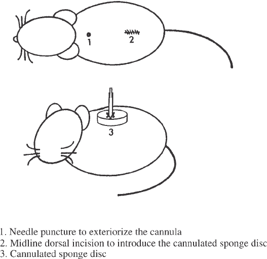
sutures, in such a way that the tube is perpendicular to the disc face (see Fig. 2).
3. The open end of the tube is sealed with a removable plastic plug made of a smaller
polythene tubing (1.2 mm internal diameter). This cannula allows accurate injec-
tion of tracers, tests substances, and withdrawal of fluid into and from the interior
of the implant.
4. The cannulated sponge discs are sterilized by boiling in distilled water for 10 min,
then placed in sterile glass petri dishes, and irradiated overnight under ultraviolet
light in a laminar flow hood.
3.2. Anesthesia
1. Rats are anesthetized with intramuscular injection of 0.5 mL/kg of 0.315 mg/kg
fentanyl citrate and 10 mg/mL fluanisone.
2. Mice are anesthetized using the combination of fentanyl citrate and fluanisone
acetate plus 5 mg/mL of midazolam hydrochloride each at a dose of 0.5 mL/kg.
Ether inhalation can also be used.
Fig. 2. Schematic representation of the cannulated sponge disc and its arrangement
following implantation.
Sponge Model 81
3.3. Surgical Procedure for Sponge Disc Implantation
Implantation of sponge discs is performed with aseptic techniques follow-
ing induction of anesthesia:
1. The hair on the dorsal side is shaved and the skin wiped with 70% (v/v) ethanol in
distilled water.
2. A 1 cm midline incision is made, and through it a subcutaneous pocket is pre-
pared by blunt dissection using curved scissors.
3. The sterilized sponge implant is inserted into the pocket, its cannula pushed
through a small incision previously made on the cervical side of the pocket.
4. The base of the cannula is sutured to the animal skin to immobilize the sponge
implant.
5. The cannula is plugged with a smaller piece of sealed polythene tubing to prevent
infection.
6. The midline incision is closed by three interrupted 5-0 silk stitches.
7. The animals are kept singly with free access to food and water after recovery
from anesthesia.
The cannulated sponge discs can be left in situ for periods ranging from days
to weeks. A schematic representation of the cannulated sponge disc and its
arrangement after subcutaneous implantation is shown in Fig. 2. (See Notes 1–5.)
3.4. Estimation of Blood Flow in the Sponge Implant
by the
133
Xe Washout Technique
The sequential development of blood-flow in the implanted sponges, origi-
nally acellular and avascular, can be determined by measuring the washout rate
of
133
Xe injected into the implants, a technique developed to measure blood
flow and thus monitor the vascular changes indirectly. This is based on the
principle that the amount of a locally-deposited radioactive tracer decreases at
a rate proportional to the blood flow at the site of the injection. The decrease in
radioactive counts should be exponential, and t
1/2
(time taken for the radioac-
tivity to fall to 50% of its original value) for the washout inversely related to
the local blood flow (15):
1. Anesthetize the animals as in Subheading 3.2..
2. Inject
133
Xe (10 µL containing approx 1 × 10
6
cycles per second) into the implant
via the cannula. Immediately after injection, plug the cannula to prevent evapora-
tion of the tracer.
3. Monitor the washout of radioactivity from the implant using a collimated gamma-
scintillation detector for 6 min. The detector, a sodium iodide thallium activated
crystal (1 in × 1 in) should be positioned directly above the site of injection.
4. Record radioactivity for 40-sec periods and print on a scalar ratemeter.
82 Andrade
5. The radioactivity-vs-time data should be fitted to an exponential decline curve to
derive t
1/2
(half-time in minutes), after deduction of background radioactivity.
A particular advantage of the
133
Xe-t
1/2
assay is that it allows nondestruc-
tive, and thus repeatable, measurements of blood flow in the same animal over
the period of neovascularization of the sponge. This combination of techniques
requires fewer animals and also allows an estimate of variability in individual
animals.
3.5. Estimation of Blood Flow by Efflux of Sodium Fluorescein
from an Implanted Sponge
Low-molecular fluorochrome-complexed tracers or fluorogenic dyes pro-
vide additional methods for detecting new blood vessels. Compared with
radioactive isotope compounds, the advantages of fluorescent dyes are obvi-
ous; fluorescence is relatively nontoxic, nonradioactive and inexpensive (16).
The measurement of fluorochrome-generated emission in the bloodstream fol-
lowing its application to the sponge implant compartment at various intervals
postimplantation reflects the degree of local blood-flow development and the
interaction of the angiogenic site with the systemic circulation (8). This
approach can be used to study sponge-induced angiogenesis quantitatively and
to investigate the pharmacological reactivity of the neovasculature. Measure-
ments of the extent of vascularization of sponge implants can be made by esti-
mating t
1/2
(min) of the fluorescence peak in the systemic circulation following
intraimplant injection of sodium fluorescein at fixed time intervals (for
example; d 1, 4, 7, 10 and 14) postimplantation.
1. Anesthetize the animal.
2. Determine blood background fluorescence by piercing the extremity of the tail
and collecting 5 µL of blood with a heparinized yellow tip. Transfer the blood
sample to a centrifuge tube contained 1 mL of isotonic saline (0.9%).
3. At time 0, administer sodium fluorescein (50 µL of a sterile solution of 10%
sodium fluorescein per kg weight) to anesthetized animals.
4. After 1 min collect the first blood sample following dye injection as in step 2.
5. At 3 min collect a second blood sample. Repeat this procedure every 2–3 min for
25–30 min.
6. Centrifuge blood samples for 10 min at 1400g (2000 rpm). Keep the supernatant
for fluorescence determination (excitation 485nm/emission 519nm).
7. From the fluorescence values estimate the time taken for the fluorescence to peak
in the bloodstream (absorption) and the time required for the elimination of the
dye from the systemic circulation (elimination). These parameters are expressed
in terms of half-time (t
1/2
; time taken for the fluorescence to reach, or to decay to,
50% of the peak value in the systemic circulation).
Sponge Model 83
3.6. Biochemical Analysis of Implanted Sponges
Quantitation of various biochemical parameters further supports the func-
tional characterization of the fibrovascular tissue that infiltrates the implants
and has been used to corroborate assessment of angiogenesis (7,10–14):
1. Remove the implants at any time postimplantation as required.
2. Immediately upon removal, weigh, homogenize, and centrifuge the tissue in iso-
tonic physiological solution (saline 0.9% or phosphate-buffered saline).
3. Store the supernatant of the homogenate at –20°C for later analysis.
3.7. Histological Analysis of Implanted Sponges
To further establish the sequential development of granulation tissue and
blood vessels in the implants, several histologic techniques have been employed:
1. Kill the animals bearing implants and dissect the implants free of adherent tissue.
2. Fix implants in formalin (10% w/v in isotonic saline) and embed in paraffin.
3. Cut sections (5–8 µm) from halfway through the sponge thickness.
4. Stain and process for light microscopy studies.
Figure 3 shows examples of sponge implants at 7 and 14 d postimplantation.
The implants can be photographed by transillumination using an inverted
microscope, which allows qualitative and morphometric analysis of the vascu-
lar pattern within the sponge matrix (Fig. 4).
4. Notes
1. The attachment of the cannula to the center of the sponge disc is facilitated by
making in one end of the polythene tubing ‘tooth-like’ structures (usually four),
in such a way that the thread is secured in one of them and then sutured to the
sponge.
2. To avoid leakage following injection of test substances via the cannula, it is
important to hold the base of the cannula with a forceps. Sometimes it is neces-
sary to force in the injected substances by applying pressure to the cannula.
3. To avoid infection, plugs should be changed every time they are removed from
the cannula.
4. Administration of drugs should be performed 2–3 d postimplantation to allow
time for encapsulation of the sponges.
5. To eliminate acute effects, vasoactive substances (vasodilators or vasoconstric-
tors) should be given 6–8 h prior to blood-flow measurement.
Acknowledgements
This work was supported by grants from CNPq, FAPEMIG and PRONEX-
Brazil.

84 Andrade
Fig. 3. Sequence of histological changes during angiogenesis in the rat sponge
granuloma. Sections of implants removed on d 7 or 14 postimplantation, fixed in for-
malin, embedded in paraffin and 5 µm sections stained (H&E ×500). The sponge
matrix is seen as triangular shapes. (A) At d 7, the pores of the implants are filled with
a fibrous network, numerous polymorphonuclear leukocytes and spindle-shaped cells
(fibroblasts). (B) By d 14 postimplantation, a uniformly dense, more highly organized
fibrous matrix, with numerous spindle-shaped cells and capillaries interspersed within
the sponge, can be seen.
Sponge Model 85
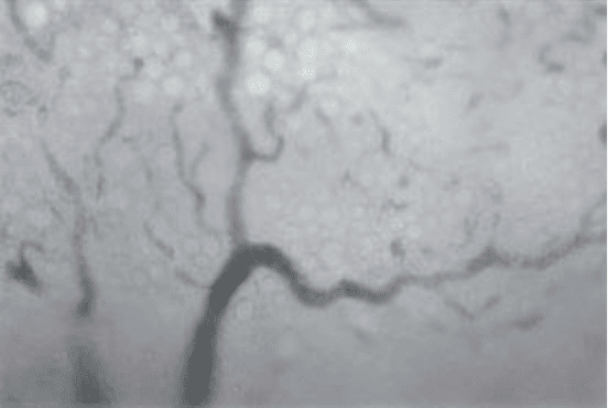
Fig. 4. Photomicrograph of the vascular pattern of a trans-illuminated sponge
implant at d 7 postimplantation. Tortuous, dilated, and saccular capillaries are seen
infiltrating the polyether-polyurethane sponge matrix, which appears as a beehive-like
structure (magnification ×7). The implant was photographed with an Olympus-IM
inverted microscope equipped with a 35 mm camera.
References
1. Folkman, J. and Shing, Y. (1992) Angiogenesis. J. Biol. Chem. 267, 10,931–10,934.
2. Edwards, R. H., Sarmenta, S. S., and Hass, G. M. (1960) Stimulation of granula-
tion tissue growth by tissue extracts; study by intramuscular wounds in rabbits.
Arch. Path. 69, 286–302.
3. Andrade, S. P., Fan, T. P. D., and Lewis, G. P. (1987) Quantitative in vivo studies
on angiogenesis in a rat sponge model. Br. J. Exp. Path. 68, 755–766.
4. Thiede, K., Momburg, F., Zangemeister, U., Schlag, P., and Schirrmacher, V.
(1988) Growth and metastasis of human tumors in nude mice following tumor-
cell inoculation into a vascularized polyurethane sponge matrix. Int. J. Cancer 42,
939–945.
5. Mahadevan, V., Malik, S. T. A., Meager, A., Fiers, W., Lewis, G. P., Hart, I. R.
(1990) Role of tumor necrosis factor in flavone acetic acid-induced tumor vascu-
lature shutdown. Cancer Res. 50, 5537–5542.
6. Andrade, S. P., Bakhle, Y. S., Hart, I., and Piper, P. J. (1992) Effects of tumor
cells and vasoconstrictor responses in sponge implants in mice. Br. J. Cancer 66,
821–826.
7. Andrade, S. P., Vieira, L. B. G. B., Bakhle, Y., Piper, P. J. (1992) Effects of
platelet activating factor (PAF) and other vasoconstrictors on a model of angio-
genesis in the mouse. Int. J. Exp. Path. 73, 503–513.