Murray J. Clifford. Angiogenesis Protocols - Methods in Molecular Medicine, Vol. 46
Подождите немного. Документ загружается.

54 Ko et al.
outcome may have resulted only from the tissue manipulation, wound healing,
or ischemia.
This chapter describes a technique using a slow-release system for angio-
genic growth factor, one that permits induction of angiogenesis virtually any-
where the system is applied. We utilize a microbead alginate delivery system
(1–3). The advantage of this system is that it can be constructed with any type
of growth factor [we have used it jointly with a nerve growth factor, brain-
derived neurotrophic factor [BDNF], epidermal growth factor, and others], and
the system is quite simple to apply in vivo. We have also found that subsequent
molecular, histological, and/or immunological investigation of the tissue can
be performed quite easily. This system, which does utilize tissue manipulation
during its application, has been demonstrated to induce angiogenesis beyond
that which would occur solely by tissue manipulation or ischemia, etc. More-
over, this system produces consistent results for a given tissue.
2. Materials
1. Calcium alginate: Keltone TM LV (Kelco, San Diego, CA). The alginate,
derived primarily from Macrocystis pyrifera, contains approx 61% mannuronic
acid and 39% guluronic acid.
2. NaCl: 0.9% solution.
3. CaCl
2
: 1.5% solution.
4. Ultraviolet (UV) light for sterilization.
5. Compressed air supply.
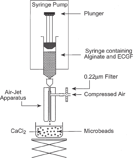
6. Droplet-generating apparatus: A schematic representation of the droplet-generating
apparatus used for this application is shown in Fig. 1. It consists of a syringe
attached to an air-jet nozzle device. The nozzle consists of a solid brass cylinder
that has been drilled along its axis to allow the placement of a 22- or 23-gauge
needle through it. The hole is tapered from the top to the bottom in such a way
that at the bottom of the nozzle there is a 1-mm circular clearance around the
needle once it is in place. The top of the needle should be snug, so as to prevent
movement of the needle once it is in place. The needle is not beveled but cut at
right angles, with care to ensure that the cut edges are smooth. Placement screws
are located near the middle of the nozzle to hold the needle in place. Also, these
screws can be used to adjust the placement of the needle so that it is exactly in the
center of the hole. The nozzle also consists of a second lateral hole that is drilled
at a right angle, near the top part of the nozzle, that connects with the longer
tapered hole that runs along the axis of the nozzle. The lateral hole is connected
to a compressed air supply such that a constant stream through the air-jet nozzle
flows coaxially to the needle. After the syringe is attached to the air jet it is
placed in a syringe pump as shown (Fig. 1). The material to be encapsulated is
pushed through the needle and as soon as it emerges it is plucked by the coaxial
air-flow and is collected in the CaCl
2
solution. The mixture extruded through the
droplet-generating apparatus forms microbeads of approx 300–700 µm diameter.
Alginate Release 55
Larger and smaller diameter microbeads may also be prepared by using a smaller
gauge needle and by adjusting air-flow rate through the nozzle. The optimal air-
flow rate with our device is approximately 65–70 mL/sec. (See Note 1).
3. Methods
3.1. Alginate Preparation
1. Prepare a 3% solution of sodium alginate in sterile 0.9% NaCl.
2. Centrifuge the solution at 20,000g for 3 h to remove all particulate debris.
3. Aliquot the sodium alginate solution into sterile tubes and leave overnight under
UV light to insure complete sterilization.
3.2. Calcium Alginate-Angiogenic Growth Factor Release System
The amount of growth factor used will depend on the activity of the prepara-
tion. The total amount of alginate used will be in a volume of 4 mL. This
amount should be kept in mind when calculating the final concentration of
growth factor (e.g., if the final concentration requires 1 mg/mL, then 4 mg is
needed). Once this is calculated, the required amount of growth factor is dis-
Fig. 1. Schematic demonstrating the microencapsulation apparatus used to create
microbeads for the alginate microbead release system.
56 Ko et al.
solved in 0.25 mL of 0.9% NaCl solution. The freshly dissolved angiogenic
growth factor is then added to 3.75 mL sodium alginate.
To construct microbeads, the mixture is extruded through the droplet-generating
apparatus to form microdroplets of approximately 300–700 µm diameter. In
this reaction, the free-flowing aqueous sodium alginate microdroplets
immediately form solid calcium alginate hydrogel microbeads upon exposure
to 1.5% calcium chloride solution. The microbeads are quickly rinsed in a mini-
mal amount of calcium chloride solution.
3.3. In Vivo Placement of Alginate Release System
Our in vivo work thus far has been in the male Wistar rat weighing approx
300 g. We have placed the alginate release system in a variety of sites; how-
ever, probably the easiest and most reliable site is the fasciovascular groin flap
(4–5). The fasciovascular tissue is a translucent vascular film that adheres to
the vessels and surrounding groin connective and adipose tissues. The blood
supply to this tissue is supplied by the superficial inferior epigastric vessels,
which branch directly from the femoral vessels.
1. Make a transverse incision in the groin crease.
2. Carefully dissect down to the femoral vessels. Superior and slightly superficial to
these vessels is the groin fasciovascular tissue. Isolation of this fascial and fat
tissue with the superficial epigastric vessels is possible with meticulous dissec-
tion. Take care not to transect the epigastric vessels.
3. The pedicled flap produced is isolated to an approximate size of 3 by 4 cm.
4. Alginate microbeads containing growth factor are placed within the flap, either
by embedding them into the flap or by wrapping the flap around the beads and
suturing it closed like a sack. Other shapes may be easier to use in certain sites
(see Note 2).
4. Notes
1. When using the air jets, it is critical that the needle is placed at the center of the
nozzle opening; failing to do so will result in irregularly shaped microbeads. In
addition, alginate can sometimes adhere to the sides of the nozzle, thus clogging
it. The air-flow rate should also be monitored carefully. A slow rate of flow may
form a large droplet that can sometimes clog tip of the nozzle. Also, if the air-
flow rate is too high it may cause the CaCl
2
to splash up to the nozzle tip, thus
clogging it. The viscosity of the alginate is also a concern sometimes. A lower
viscosity alginate will flow faster through the nozzle and the air-flow rate should
be regulated to control microbead shape and size. We have found a 3% alginate
solution to be optimal for our purpose. Finally, the tip of the needle should opti-
mally be placed approx 1 cm from the surface of the CaCl
2
. Placing it higher (i.e.,
further from the CaCl
2
solution) will result in irregularly shaped microbeads.
Placing it lower risks splashing the CaCl
2
on to the nozzle tip, causing it to clog.
Alginate Release 57
It should be noted that this is a very convenient procedure for immobilizing
growth factors. The procedure is carried out at room temperature using gentle,
buffered solutions. Thus care should also be taken to see that the CaCl
2
is not ice cold.
2. Variations in microbead shape: In our initial experience with the alginate release
system, we utilized the microbead shape. However, when we started using the
system in vivo, we found that the beads were sometimes a bit too cumbersome to
use in certain situations, as they often would migrate with gravity or easily dis-
lodge with even the slightest tissue manipulation. Therefore, we started construct-
ing other shapes out of the identical alginate material. We have made more
practical delivery-system shapes, including a filamentous (string-shaped) and a
flat-sheet shaped design. We have found that both shapes can be more functional
in vivo in some anatomic regions. The filamentous configuration is advantageous
because it can be wrapped around certain structures, while the flat-sheet system
can be laid securely adjacent to appropriate tissues to target release.
To make the alginate filament system, the mixture of 3% alginate and growth
factor is extruded through a 25-gauge needle into the 1.5% calcium chloride
solution. To ensure uniform diameter of the string, a syringe pump apparatus is
utilized to apply uniform pressure to the syringe. To make the flat sheet alginate
system, our engineering department constructed a special device made of Lucite
that fits securely onto a syringe and extrudes a uniformly flat alginate sheet
through a slit at its endpoint. The slide dimensions are 25 mm × 0.25 mm. Again,
a mixture of 3% alginate and growth factor is extruded through this device into
the 1.5% CaCl
2
solution. To facilitate string and sheet formation, we found it
advantageous to submerge the device tip in CaCl
2
solution during construction.
References
1. Ko, C. Y., Dixit, V., Shaw, W., and Gitnick, G. (1995) In vitro slow release pro-
file of endothelial cell growth factor immobilised within calcium alginate
microbeads. Art Cells, Blood Subs. and Immob. Biotech. 23, 143–151.
2. Dixit, V., Darvasi, R., Arthur, M., Brezina, M., Lewin, K., and Gitnick, G. (1990)
Restoration of liver function in Gunn rats without immunosuppression using trans-
planted micro-encapsulated hepatocytes. Hepatology 12, 1342–1349.
3. Ko, C. Y., Dixit, V., Shaw, W., and Gitnick, G. (1997) Extensive in vivo angio-
genesis from the controlled release of endothelial cell growth factor: implications
for cell transplantation and wound healing. J. Controlled Rel. 44, 209–214.
4. Ko, C. Y., Dixit, V., Shaw, W., and Gitnick, G. (1995) Succesful xenotrans-
plantation of microencapsulated hepatocytes in the rat fasciovascular groin flap.
Presented at the Tenth World Congress of the International Society for Artificial
Organs, Taipei, Taiwan. Abstract book: 42.
5. Borud, L. J., Shaw, W., Passaro, Jr. E., Brunicardi, F. C., and Mullen, Y. (1994)
The fasciovascular flap: a new vehicle for islet transplantation. Cell Transplant.
3, 509–514.

Disc Angiogenesis 59
59
From:
Methods in Molecular Medicine, Vol. 46: Angiogenesis Protocols
Edited by: J. C. Murray © Humana Press Inc., Totowa, NJ
5
Disc Angiogenesis Assay
Anthony C. Allison and Luis-F. Fajardo
1. Introduction
The aim of our research was to develop a quantitative assay for angiogen-
esis in mammals, especially the mouse. This is a convenient experimental
animal because of its small size, which allows compact housing and experi-
mentation with angiogenic factors or inhibitors in limited supply. Mouse
genetics is an advanced discipline, resulting in the availability of many inbred
strains and histocompatible tumors. Recombinant growth factors and other
proteins are usually of human or mouse origin, and the desirability of using
proteins of the experimental animal under study has been demonstrated. Mice
genetically engineered to overproduce or not produce particular growth factors
or receptors are valuable experimental tools.
Growth factors can accelerate the proliferation and/or migration of endothe-
lial cells and surrounding connective tissue cells, and it is desirable to quantify
these processes independently. The disc angiogenesis assay arose from our
observations of the growth of blood vessels into a polyvinyl alcohol sponge
implanted sub-cutaneously in the mouse (1). The design was improved to allow
quantification of vascular in-growth from the circumference of a disc, as well
as incorporation of labeled thymidine into endothelial and other connective
tissue cells (2). In this way effects of angiogenic factors or inhibitors on the
proliferation and migration of endothelial cells and surrounding connective
tissue cells could be monitored separately.
2. Materials
1. Polyvinyl alcohol sponge (Kanebo, PVA, Rippey Co., Santa Clara, CA); 2 mm
thickness.
2. Cell-impermeable filters (0.45 micron, HAWP 013, Millipore).
60 Allison and Fajardo
3. #1 Millipore glue (XX70 000 00, Millipore).
4. Acetate copolymer (Elvax, Dupont).
5. [Methyl-
3
H]-Thymidine (6.7 Ci/mmol, Dupont).
6. Photographic emulsion (NTB-2, Eastman Kodak Corp).
7. Leuconyl blue (BASF; distributed by Wyandotte Co., Hotland, MI).
3. Methods
3.1. Preparation of Sponge Disc
1. A disc of 13 mm diameter and 2 mm thickness is prepared from sterile materials,
under sterile conditions within a laminar-flow hood. Alternatively, nonsterile
discs can be sterilized by a variety of methods.
2. The flat circular sides of the disc are covered with the cell-impermeable filters
and sealed with glue (Fig. 1). This leaves only an external 2 mm rim for penetra-
tion or exit of cells.
3. When indicated, and prior to placement of the filters, a hole 3 mm in diameter is
bored in the center of the disc.
4. A pellet of sponge material 2 mm in diameter, containing an angiogenic agonist
or antagonist to be tested, is coated with acetate copolymer and placed in the
central hole. Each pellet of this size can hold up to 20 µL of the material to be
tested. Test substances we have used, providing a strong angiogenic stimulus,
include recombinant human epidermal growth factor (EGF; usually 20 µg per
disc) and recombinant human basic fibroblast growth factor (bFGF; usually 20 µg
per disc).
5. Care should be exercised to avoid physical or chemical damage to the sponges, or
to the angiogenic or inhibitory agents once loaded into the disc. Sponges tolerate
dry heat up to 120°C, but can be damaged by boiling water.
3.2. Implantation of Sponge Disc
1. The mouse is anesthetized by an intraperitoneal injection of ketamine hydrochlo-
ride (50 mg/kg), xylazine (5 mg/kg), and acepromazine maleate (1 mg/kg).
2. The shaved skin surface is sterilized with 70% ethanol, and a 1.5 cm long inci-
sion into the subcutis is made at least 1 cm away from the desired location of the
disc. Blunt dissection is used to produce a tunnel toward the site of implantation.
3. Phosphate-buffered saline (PBS) is dripped into the area with a Pasteur pipette.
Holding it gently with forceps, the PBS-moistened disc is then inserted through
the wound and into the tunnel up to the desired site.
4. The skin wound is closed with 3-4 metal clips.
5. The animals are housed as usual, with standard chow and water ad libitum, dur-
ing the period of the experiment, 7–30 d, most often 14 d (see Notes 1–4).
3.3. Assay for Cellular Proliferation in Sponge Disc
Uptake of tritiated thymidine (
3
HTdR) is used to evaluate DNA synthesis,
and therefore proliferation of endothelial and other cells (3). Total incorpora-
Disc Angiogenesis 61
tion of label is measured by scintillation counting while autoradiography iden-
tifies the different cell types synthesizing DNA.
1. Mice are injected intraperitoneally with [methyl-
3
H]-thymidine (66 µCi/25 g
body weight) at 24, 18, and 12 h before sacrifice (total = 200 µCi). Alternatively
radiolabel can be delivered continuously by osmotic minipumps (Alza Co., Palo
Alto, CA) implanted subcutaneously, for 7–14 d.
2. After sacrifice, tissues are removed, fixed, and paraffin-embedded.
3. Serial 6 µm-thick sections are cut.
4. For determination of total radioactivity, sections are subjected to alkali solubili-
zation and beta radiation activity determined in a scintillation counter. These
values are representative of DNA synthesis of all cells in the sample.
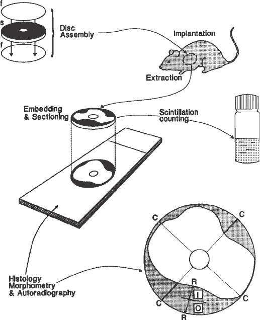
Fig. 1. Disc assembly, implantation, removal, embedding, sectioning, and analysis
(quantitative and qualitative). See Subheading 3.
62 Allison and Fajardo
5. For autoradiography, sections are prepared by dipping the slide-mounted sec-
tions in photographic emulsion. These slides are held at 4°C for 35 days and
developed. The developed slides are counterstained with hematoxylin and eosin.
In these autoradiographic preparations, we perform differential counting of
labeled vs. unlabeled cells using standard-size microscopic fields, two in the outer
and two in the inner zone of each disc. Cells containing 5 or more grains are
considered labeled. (In our experience, after 5 wk exposure the great majority of
labeled cells contain more than 10 grains per cell, while background is less than
1 grain per 100 µm
2
; Fig. 2.) Raw data were analyzed using RSI statistical analy-
sis software. Having obtained means, standard errors, and standard deviations for
each group of discs, comparisons of experimental and control groups are carried
out using Student’s t test. Other methods for identifying cells that have prolifer-
ated are available.
3.4. Identification of Blood Vessels in Sponge Discs
by Light Microscopy
1. An injection of Leuconyl blue (0.4 mL of a 40% solution in PBS) is given intra-
cardially to the mouse 15 min prior to euthanasia (4).
2. Mice are killed with a large intra-peritoneal dose of anesthetic followed by quick
cervical dislocation, or by placing the animal(s) in a concentrated CO
2
chamber.
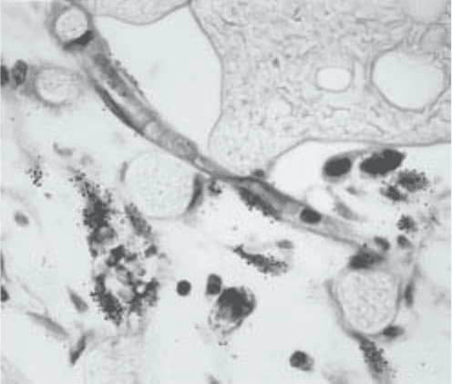
Fig. 2. Autoradiograph of paraffin section from disc stimulated with 20 mg of bFGF.
The diagonally-oriented capillary in the center has at least two endothelial cells labeled
by
3
HTdR. The labeled cells outside of the capillary are fibroblasts. Inner zone of
growth. Counter-stained with H&E ×460.
Disc Angiogenesis 63
3. A careful incision is made in the skin overlying the implanted disc, which is then
dissected gently from the surrounding tissues, and removed.
4. The filter is carefully dissected away from the disc with a sharp blade to permit
effective fixative penetration and dehydration.
5. The dissected disc is placed in 10% neutral formalin for 48–72 h. The disc is the
embedded flat in paraffin, in such an orientation that initial sections will be taken
from the uncovered side of the disc.
6. Multiple, 6-µm thick planar sections are cut (Fig. 1). For light microscopy and
measurement of radial growth, sections are stained with hematoxylin and eosin;
for measurement of total area growth, with toluidine blue. Sections are also cut
for autoradiography and scintillation counting.
3.4. Quantification of Angiogenesis
3.4.1. Centripetal Vessel Growth
Direct measurement of blood vessels can be performed by various methods,
including point counting on histologic sections (5) and determination of intra-
vascular volume, e.g., with radioactive isotopes (6). Such methods are tedious
and impractical when examining a large number of discs. We have devised a
simpler, indirect procedure based upon the centripetal growth of new vessels:
1. Project an image of the section at 100× magnification onto a horizontal surface
(we use a Bausch and Lomb microprojector).
2. Identify the innermost blood vessel.
3. Measure in centimeters the radial distance between the leading tip of the inner-
most vessel and the rim of the disc (Fig. 1).
We have named this end-point “centripetal vessel growth” (CVG). The CVG
is directly proportional to the total vessel growth and is therefore a reliable,
indirect measure of such growth (1,2).
3.4.2. Total Growth Area
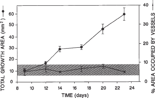
We have shown in Subheading 3.4.1. that the total growth area within the
disc is consistently proportional to the area occupied by blood vessels (Fig. 3).
Therefore, any measurement of the total growth area is an accurate, indirect
measurement of the area occupied by blood vessels. A method of determining
the total growth area is to measure the radial fibrovascular growth along two
perpendicular diameters (four radii) using the projection apparatus (Subhead-
ing 3.4.1., step 1; Fig. 1). The average value of the four measurements is then
obtained for each disc.
A precise system of measuring total growth area uses 6-µm paraffin sec-
tions stained with toluidine blue to obtain high contrast (Fig. 1). Toluidine blue
uniformly stains the area of growth and not the sponge trabeculae. The sections
64 Allison and Fajardo
(mounted on slides without cover slips) are placed on the stage of a microscope
fitted with a computer-assisted digital image analysis system using the NIH
Image program, which measures the entire area of growth automatically, in
pixels. Threshold values are standardized for a particular experiment, and pixel
values are converted to mm
2
.
3.4.3. Differential Cell Counting
Differential counting of the various cells that comprise the fibrovascular
growth is performed with microscope reticules. The relative proportions of
various cell types, as well as the absolute numbers of the cells per given area
are established by differential counting within standard-size areas of the vari-
ous zones of growth in the disc assay (Fig. 1). The space occupied by vessels
and other elements is determined using the intersection points of microscope
grids. Such procedures, which are time consuming, were used initially to ana-
lyze the biology of fibrovascular growth and to validate the more often used
quantitative methods described in the previous paragraphs.
3.6. Characteristics of Vascular Growth in the Disc Assay
Centripetal growth into the disc is composed of blood vessels and surround-
ing stroma (Figs. 1, 4) and is usually asymmetric. This asymmetry may be
related to the density of mother vessels (venules) in the surrounding host tis-
Fig. 3. Vessel growth is directly proportional to total fibrovascular growth (see
Subheading 3.4.). As the latter increases continuously (upper line) during the period
of observation, the proportion of the growth area occupied by blood vessels (lower
line) remains constant. Total growth area was measured by computer-assisted digital
image analysis, and vascular area by point counting (see text). Growth stimulated by
20 mg of EGF. Bars, standard error of mean.