Murray J. Clifford. Angiogenesis Protocols - Methods in Molecular Medicine, Vol. 46
Подождите немного. Документ загружается.


Pericyte Isolation and Culture 247
247
From:
Methods in Molecular Medicine, Vol. 46: Angiogenesis Protocols
Edited by: J. C. Murray © Humana Press Inc., Totowa, NJ
18
Bovine Retinal Microvascular Pericytes
Isolation, Propagation, and Identification
Ramesh C. Nayak and Ira M. Herman
1. Introduction
The growth of new capillaries from existing vessels (angiogenesis) is of
fundamental importance in wound healing and in pathological situations such
as proliferative diabetic retinopathy (1), rheumatoid arthritis (2), and tumor
growth. Consequently, considerable interest in vascular cell biology has arisen
in apparently disparate clinical and experimental fields. Held in common, how-
ever, is the hope that an understanding of the cellular and molecular mechanisms
that regulate angiogenesis will lead to novel therapeutic agents and targets.
Central to achieving a comprehensive mechanistic understanding of angio-
genesis is the ability to isolate and propagate the cell types that form the capil-
lary wall. The capillary is composed of two cell types, the endothelial cell (EC)
and the pericyte (also known as the mural cell) (3). The capillary tube that will
act as the conduit for blood is formed by the ECs that subsequently become
associated with pericytes at the abluminal surface of the endothelium. This
close apposition of pericyte and endothelial cell allows a molecular “dialog” to
take place between these cell types enabling each to tightly regulate the other’s
function in response to local stimuli. These regulatory interactions have
recently been modeled in tissue culture (3).
The first descriptions of methods for the isolation of both microvascular
pericytes and ECs were published in the middle to late 1970s with the methods
for pericyte isolation preceding those for ECs (4–8). Meezan et al. reported, in
1974, the isolation of metabolically active microvessels from bovine retina and
cerebral cortex (4,5). This led Buzney et al. to use these methods of microvessel
248 Nayak and Herman
preparation to isolate and culture pericytes from simian, bovine, and human
retina (6). In 1977, Del Vecchio et al. (7) isolated capillary fragments from rat
adrenal glands, which was followed in 1978 by the demonstration by Folkman
et al. (8) that viable ECs could be recovered from isolated capillary fragments,
cloned, and propagated on gelatin coated plates in conditioned media. Inexpli-
cably, the reports from Meezan et al. and Buzney et al. did not appear to stimu-
late interest in tissue culture studies of the pericyte, perhaps because the
importance of the pericyte in regulating capillary function had yet to become
appreciated (3). In 1983, using the same initial strategy of isolating viable cap-
illary fragments, Gitlin and D’Amore were able to isolate and selectively propa-
gate bovine retinal microvascular pericytes (9). That report and the recognition
of the importance of the pericyte in controlling EC growth (3), combined with
development of biochemical markers for the identification of pericytes (10,11),
have stimulated interest in studying the pericyte in vitro. Since then, several
methodological procedures have been published for pericyte isolation from
various tissues (9,10,12–15,25). The majority of reports describe the isolation
of pericytes from retina and most are a variation of the work described above.
Most procedures begin with enrichment of microvessel fragments by sieving
collagenase-digested tissue over nylon meshes of defined mesh size and then
explanting the recovered microvessels under selective tissue culture conditions.
The method we detail here has been in use in our laboratories for over 15 yr
and is a variant of the methods discussed above.
2. Materials
2.1. Dissection and Harvesting of Retinal Tissue
1. 20 bovine eyes, globes intact (reject any that are pierced or perforated).
2. The following materials should be autoclaved the day before the isolation is to be
performed: 3 one-litre beakers (openings covered with aluminium foil), 1 rubber
policeman, 1 curved scissors, 2 straight scissors, 1 toothed forceps, 1 straight
forceps, 3 scalpels, and 2 sterile fields.
Sterile fields are prepared by taking an approx 75-cm-long strip of aluminium
foil and placing within it a slightly shorter strip of absorbent paper towel (such as
is used in roll dispensers). The edges of the aluminium foil are folded in over the
paper and the field is then folded into overlapping thirds by folding the ends in
toward the center. The field is then wrapped in aluminium foil, and autoclaved to
sterilize.
3. Betadine (10% povidone-iodine solution).
4. Penicillin-streptomycin-fungizone (antibiotic-antimycotic) stock solution (Gibco
Life Technologies, Grand Island, NY, Catalog Number 15240-062).
5. Sterile phosphate-buffered saline (PBS), pH 7.15.
6. One pack of sterile 10-cm-diameter tissue culture Petri-dishes (Becton Dickinson
Labware, Franklin Lakes, NJ, Catalog number Falcon 3003).
Pericyte Isolation and Culture 249
7. Pasteur pipets (Fisher Scientific, Springfield, NJ, Catalog number 13-678-20C)
drawn to a fine caliber.
8. Binocular dissecting microscope.
9. Vacuum line and fluid trap.
2.2. Isolation and Explant of Retinal Microvessel Fragments
1. 20 mL of 0.1%Type II Collagenase (Worthington Biochemical Corp., Freehold,
NJ, Catalog number CLS 2; 250-300 units/mg) in Dulbecco’s Modified Eagle’s
Medium (DMEM), low glucose formulation supplemented with 2 mM glutamine
(Gibco Life Technologies, Grand Island, NY, Catalog number 12320-024), ster-
ilized by filtration through a 0.22 µm millex GS filter (Millipore, Bedford, MA,
Catalog number SLGS 025 OS).
2. Sterile 20 mL syringes (Becton Dickenson and Co., Franklin Lakes, NJ; Catalog
number 309661).
3. Four 30-µm and 100-µm Nitex nylon meshes (SEFAR AMERICA, Depew, NY,
Catalog numbers 3-30/21 and 3-100/47) cut into circles and fitted into 25-mm-
diameter Swinnex membrane filter holders (Millipore, Bedford, MA, Catalogue
number SX00 025 00) and autoclaved in paper autoclave bags.
4. A 37°C oven with a rotary shaking table.
5. A 37°C tissue culture incubator with a humidified atmosphere of 5% CO
2
in air.
6. Growth Medium: 10% heat inactivated bovine serum (Hyclone Laboratories Inc.,
Logan, UT, Catalog number SH30072.03) in DMEM supplemented with 2 mM
glutamine and 1% PSF.
2.3. Weeding of Primary Cultures
1. Inverted optics phase-contrast microscope.
2. Sterile Pasteur pipets, PBS, and growth medium.
3. Fine tip marker pen.
2.4. Propagation of Pericyte Cultures
1. Trypsin-EDTA stock solution (Gibco Life Technologies, Grand Island, New
York, Catalog number 25300-054)
2.5. Identification of Pericytes in Cultures
1. Micro coverglasses, round, number 1 (0.13–0.17 mm thickness), 18-mm diam-
eter (VWR Scientific, Boston, MA, Catalog number 48380-046).
2. Twelve-well tissue culture cluster dish (Costar, Cambridge, MA; Catalog num-
ber 3512).
3. Paraformaldehyde fixative: To a small side-arm flask in the fume hood add 4.0 g
of paraformaldehyde to 50 mL of distilled water and six drops of 6 M KOH while
stirring and gently heating on a combined magnetic stirrer/hot plate until dis-
solved. Dissolve 1 g of DMEM powder and 0.76 g of sodium bicarbonate in
50 mL of distilled water and correct pH to 7.4. Combine the paraformaldehyde
and DMEM solutions in a 1/1 ratio and correct pH if necessary.

250 Nayak and Herman
4. Lysis buffer for permeabilization of cells: See accompanying table
Lysis Buffer
(27)
Component Stock solution To make 20 mL
0.1% Triton X-100 100% 20 mL
50 mM HEPES, pH 6.9 0.25 M 4.0 mL
50 mM PIPES, pH7.1 0.25 M 4.0 mL
1 mM MgCl
2
1.0 M 20 mL
0.1 mM EGTA 0.1 M 20 mL
75 mM KCl 4.0 M 375 mL
H
2
O 100% 11.6 mL
5. Precleaned glass microscope slides (Catalog number 12-550-11, Fisher Scien-
tific, Pittsburgh, PA).
6. Nail Polish (any colour).
7. Cell Type Markers:
a. Monoclonal antibody 3G5 (pericyte marker) is not available commercially.
The hybridoma cell line is available from the American Type Culture Collec-
tion, Rockville, MD Catalog number ATCC CRL 1814.
b. Antismooth muscle actin antibodies are available from Sigma, St. Louis, MO,
(Catalog number A2547) and from Biomedical Technologies Inc., Stoughton,
MA (Catalog number BT 561).
c. Anti-_-actin antibody is available from Sigma-Aldrich, St. Louis, MO (Cata-
log number A5441).
d. Fluoresceinated phalloidin is available from Molecular Probes Inc., Eugene,
OR (Catalog number F432) and from Sigma-Aldrich, St. Louis, MO (Catalog
number P5282).
e. Anti-glial fibrillary acidic protein antibodies are available from Biomedical
Technologies Inc., Stoughton, MA (Catalog number BT 575).
f. Anti-cytokeratin 18 antibodies are available from Sigma-Aldrich, St. Louis,
MO (Catalog number C 1399).
g. Antibodies to Factor VIII related antigen (von Willebrand Factor) are avail-
able from Sigma-Aldrich, St. Louis, MO (Catalog number F 3520).
h. DiI-acetylated LDL is available from Biomedical Technologies Inc.,
Stoughton, MA (Catalog number BT ).
2.6. Cryopreservation of Pericytes
1. Nunc cryotube vials (VWR Scientific, Boston, MA; Nunc number 368632, VWR
Catalog number 66021-987).
2. Dimethyl sulphoxide (Sigma-Aldrich, St. Louis, MO; Catalog number D5879).
3. Prepare sterile freezing solution by adding 10 mL of DMSO to 30 mL of DMEM
followed by addition of 10 mL of bovine serum in a sterile 50 mL tube.
4. 0.4% Trypan blue solution (Sigma-Aldrich, St. Louis, MO; Catalog number T8154).
Pericyte Isolation and Culture 251
3. Methods
3.1. Dissection and Harvesting of Retinal Tissue
1. All procedures are performed in a sterile hood (Biocontainment safety cabinet
(BL2) Biohazard Level 2). Twenty intact bovine eyes are obtained from a local
slaughterhouse and processed within 4–6 h of death. The eyes are immersed
in 300 mL of 10% betadine (povidone-iodine solution) and 2% penicillin-
streptomycin-fungizone (PSF) in PBS at room temperature for 15 minutes in a
large sterile beaker.
2. Also prepare two large sterile beakers containing 2% PSF in PBS and transfer the
eyes from the betadine soak and rinse sequentially in the PSF baths.
3. Dissect the eyes on a sterile field one eye at a time (Fig. 1). Trim off most of the
external ocular muscle tissue with sterile straight scissors, leaving enough tissue
to grip with a toothed forceps. Grip the eye by the ocular muscle or optic nerve
with a toothed forceps and make an incision 2–3 mm behind the limbus (the
limbus is the point on the eye where the radius of curvature changes markedly
and is found approximately at the iris). Extend the incision circumferentially with
a curved scissor to open the globe. Pull the front of the eye forward thus pulling
the vitreous humor out of the eye cup (see Note 1).
4. Gently dislodge the retina (see Note 2) with a sterile rubber policeman and
detach from the optic nerve head with sterile fine scissors. Place the retina in a
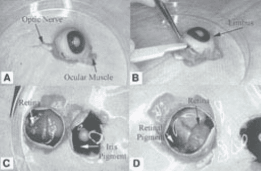
Fig. 1. Dissection of bovine eye to harvest retinae. (A) Bovine eye showing optic
nerve and partially trimmed ocular muscles; (B) circumferential incision posterior to
the limbus; (C) eye cup with vitreous removed showing the retina. The retina has been
gently moved away from the underlying pigment epithelium on the left side. (D) The
retina has been totally separated from the underlying retinal pigment epithelium but
remains attached via the optic nerve head.
252 Nayak and Herman
sterile 10-cm Petri dish containing 2% PSF in PBS prewarmed to 37°C (2 mL/
retina).
5. Repeat the eyeball dissection and retina excision for all the eyes and place the
retinae together in PSF/PBS buffer.
6. Examine retinal tissue for contamination with retinal pigment epithelium (small
fragments of black tissue) with the aid of a dissecting microscope placed in the
hood. Clean dissected retinae of pigmented tissue with a fine scissors and for-
ceps. Remove small free-floating fragments of pigmented tissue with a sterile
Pasteur pipet already drawn to a fine caliber in a bunsen burner flame, sterilized
by immersion in 70% ethanol for 30 min, and attached to a vacuum source with
an inline fluid trap. Excize any large vessels that are observed with fine scissors
and forceps.
3.2. Isolation and Explant of Retinal Microvessel Fragments
1. Transfer the cleaned retinae to another 10-cm Petridish containing PSF/PBS,
rinse, and then transfer to a dry sterile Petri dish (5 retinae/dish) and mince into a
homogeneous slurry by cross-cutting the tissue using two sterile scalpels. The
slurry from each batch of 5 retinae is transferred to a sterile 15-mL conical tube
by adding 5 mL PSF/PBS and transferring the suspension with a sterile 10-mL
pipet.
2. Pellet the retinal slurry by low-speed centrifugation (540g
max
, 3,000 rpm @ r
max
= 160 mm) for 5 min in a tabletop centrifuge. Aspirate off the buffer and estimate
the pelleted tissue volume. Add 5 vol of 0.1% collagenase (Worthington Type II
in DMEM) per vol tissue and incubate in a 37°C incubator for 1 hr with constant
agitation on a horizontally rotating table shaker at 250 rpm.
3. Centrifuge the digested retinal slurry at 540g
max
( 3,000 rpm @ r
max
= 160 mm)
for 5 min and remove and discard the supernatant. Resuspend the digest in growth
medium.
4. Take up the digested slurry with a 10-mL sterile disposable pipet and expel
against the wall of the centrifuge tube ten times to break up large aggregates.
5. Take up the triturated digest from 5 retinae into a sterile disposable 20-mL syringe
and pass through a sterile 100 µm Nitex mesh in a sterile Swinnex membrane
holder (see Note 3).
6. Pass the 100-µm filtrate over a 30-µm Nitex mesh and collect the 30-µm filtrate
in a sterile centrifuge tube.
7. Scrape the material retained on the meshes separately into growth medium and
plate into two 10-cm Petri dishes per fraction per 5 retinae. Also plate the 30-µm
filtrate into two 10-cm Petri dishes per 5 retinae of starting tissue. Place the petri
dishes in a 37°C incubator with a humidified atmosphere of 5% CO
2
in air.
8. After 24 h remove the medium and floating debris by aspiration, wash the
attached cells with PBS, and add fresh growth medium to the plates. Add 7 mL of
PBS to each Petri dish and aspirate off, leaving 1.5–2.0 mL on the dish. Replace
the lid of the dish and lift the dish and bang down sharply onto the counter top
three times to release tightly attached debris which is then aspirated off. This step
Pericyte Isolation and Culture 253
is repeated twice more followed by addition of fresh growth medium to the dishes,
which are then returned to the incubator.
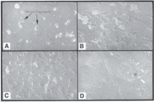
Immediately after plating, microvessel fragments can be observed by phase
contrast microscopy (Fig. 2A) and after one to several days cells can be seen to
have migrated out of the vessel fragments (Fig. 2B,C,D) and attached and
spread on the bottom of the Petri dish.
3.3. Weeding of Primary Cultures (see
Notes 4
and
13
)
1. Remove nonpericyte cell types from primary cultures by “weeding.” Nonpericyte
cells are identified by morphology (see Subheading 3.5.1.). Perform weeding
while the cells are still sparse.
2. Identify areas of the Petri dish that contain cells that do not exhibit pericyte mor-
phology by microscopy, ring with a marker pen on the bottom of the Petri dish,
and transfer to a sterile hood.
3. Remove and replace the medium with 2 mL of PBS and scrape the ringed areas
with a sterile Pasteur pipet or hypodermic needle. Follow by washing with PBS.
Check the scraped areas to ensure removal of nonpericyte cells by phase contrast
microscopy and rescrape if necessary. When the weeding is completed add
growth medium and return the plates to the incubator.
4. Continue to monitor the morphology of the cells, if cells of nonpericyte morphol-
ogy are seen repeat the weeding process.
Fig. 2. Explant culture of isolated retinal microvessels. (A) Appearance of vessel
fragments immediately after plating; (B,C) appearance of cells crawling out of the
vessel fragments after several days in culture; and (D) pericytes exhibiting typical
morphology, i.e., large flattened cells with multiple processes.
254 Nayak and Herman
3.4. Propagation of Pericyte Cultures
When the primary pericyte cultures are near confluence, they may be further
propagated by trypsinization and passage to additional plates (see Note 5).
1. Wash near-confluent primary pericyte cultures with PBS, then add 2 mL of
trypsin/EDTA solution per 10-cm Petri dish.
2. The pericytes will detach from the dish in 5–10 min at room temperature and
faster at 37°C. When the cells have detached (monitor by phase contrast micros-
copy) an equal volume of growth medium is added and the cell suspension is
transferred to a sterile centrifuge tube.
3. Pellet the cells by centrifugation at 540g
max
( 3,000 rpm @ r
max
= 160 mm) for 5 min.
4. Remove and discard the supernatant. Resuspend the pellet in growth medium for
replating.
5. Replate cells at a 1/3 ratio (each Petri dish of primary culture is replated on three
new Petri dishes or on a culture vessel with triple the surface area).
6. Remove the growth medium the following day and replace with fresh medium to
remove dead cells.
7. When these cultures are near confluence, the process is repeated (usually 3–4 d
later).
3.5. Identification of Pericytes (see
Notes 6–12
)
Pericytes may be identified by morphological criteria and also through
analysis of antigen expression detected by immunofluorescence with specific
antibodies (Figs. 3–5).
3.5.1. Cell Morphology
Pericytes in culture are large, well-spread cells with extremely irregular
edges (Fig. 2D) and prominent stress fibers (intracellular actin bundles). At
confluence, pericytes form multicellular nodules that may often be connected
by strands of cells. These morphological characteristics distinguish them from
ECs, which are polygonal and at confluence present a cobblestone-like appear-
ance (Fig. 3A). The subconfluent morphology of ECs is shown in Fig. 3E. At
sparse densities some ECs attempt to differentiate, which is seen morphologi-
cally as formation of processes that attempt to form a lumen (Fig. 3F). Smooth
muscle cells generally have a compact spindle morphology at sparse densities
and fibroblasts have an elongated spindle morphology, often with filopodial
extensions. Retinal pigment epithelial cells (RPE) contain dark pigment gran-
ules, which can be used to identify them, a feature that can be rapidly lost in
culture but is usually present in primary cultures from animals that are not
albinos. RPE may be seen as individual cells with a circular perimeter and
thick cables of intermediate filaments (cytokeratins) visible within the cell
(Fig. 3B,C,D). In small colonies the cell margins are indistinct or invisible by
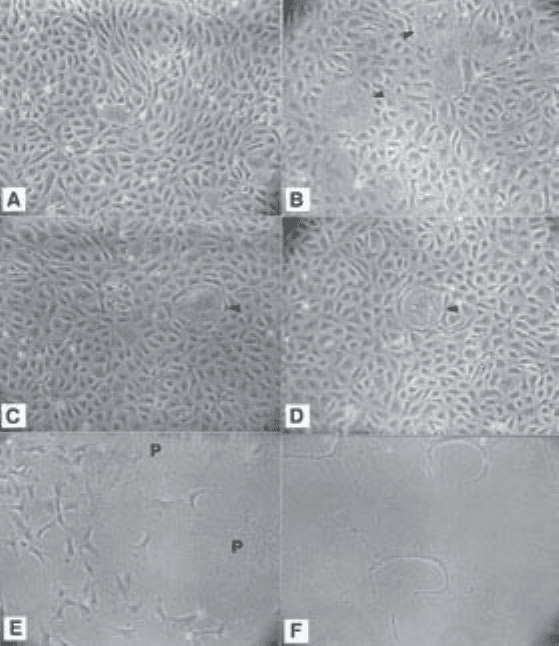
Pericyte Isolation and Culture 255
phase-contract microscopy but the nuclei are distinct. Large colonies may
detach and be seen as floating rafts of RPE cells. Colonies that remain attached
may undergo dome formation, which gives the appearance of blistering of the
monolayer. Good photographic examples of the various morphologies of RPE
cells in culture can also be seen in the literature (16). However, with careful
avoidance of pigmented tissue at the retina harvesting stage and diligent “weed-
ing” of the primary cell cultures we very rarely observe RPE contamination in
the cell cultures.
Fig. 3. Endothelial and epithelial cell morphology. (A) Typical cobblestone mor-
phology of confluent bovine retinal microvascular endothelial cells; (B,C,D) retinal
pigment epithelial cell contamination in confluent monolayers of ECs; and (E,F)
subconfluent EC morphology. At sparse cell densities ECs may attempt to differenti-
ate. This can be seen morphologically as an attempt to form a lumen (c-shaped cells).
P, pericyte.
256 Nayak and Herman
3.5.2. Immunophenotyping
In addition to morphological criteria, identification of pericytes and con-
taminating cells may be facilitated by immunological and biochemical mark-
ers (11,26–29). Table 1 shows some markers that are commonly employed in
the characterization of pericyte cultures.
For immunophenotyping studies cells must first be grown on multiple glass
cover slips, followed by fixation and permeabilization as required. Antibodies
to marker antigens are then bound and visualized by fluorescent secondary
antibodies (indirect immunofluorescence). The marker reagents listed above
are available commercially and should be used in accordance with the
manufacturer’s instructions. The following general protocol for immunofluo-
rescence can be adapted for use with each reagent.
Place sterile cover slips (autoclaved) in the bottom of 12-well cluster plates
and pipet 5,000 viable cells in growth medium into each well with a total
medium volume of 1 mL. Exchange with fresh medium the next day. On the
second day after plating, there should be sufficient cell numbers (check by
phase-contrast microscopy) for immunostaining.
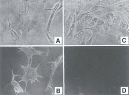
Fig. 4. Identification of pericytes by immunofluorescence with monoclonal anti-
body 3G5. (A) Phase-contrast photomicrograph of bovine retinal microvascular peri-
cyte culture; (B) Same field as A showing 3G5 immunofluorescence; (C)
phase-contrast photomicrograph of bovine microvascular ECs; and (D) same field as
C showing lack of 3G5 immunofluorescence. (Reproduced from: Nayak R.C. et al,
The J. Experiment. Med. (1988); 167, 1003–1015, by copyright permission of the
Rockefeller University Press).