Murray J. Clifford. Angiogenesis Protocols - Methods in Molecular Medicine, Vol. 46
Подождите немного. Документ загружается.

108 West et al.
(10–13). Variants of the shell-less model apply the sample to the yolk sac, or
vitelline membrane, which is superseded by the chorioallantoic membrane from
about d 7 of development. This form of assay has largely been employed to
study inhibitors, toxicity, granulation tissue formation, and early vascular
effects (14–19).
The CAM develops over the first 11 d of the 21-d gestation period. At about
24 h the development of blood islands around the embryo can be seen. Capil-
laries appear in the yolk sac of fertilized hens’ eggs at 48 hr and grow rapidly
over the next 6–8 d. The embryonic yolk sac itself consists of four layers:
ectoderm (bilayered vitelline membrane), somatic mesoderm, visceral meso-
derm (including blood vessels), and endoderm (16). The circulatory system
originates from the mesoderm and develops as three distinct divisions, in the
following sequence: the extraembryonic (for nutrition), the accessory or allan-
toic (for respiration and excretion), and the embryonic (for distribution within
the embryo). By d 4, the yolk sac membrane is well vascularized and continues
to develop until the chorioallantoic membrane supersedes it, about d 7. The
endothelial cells of this developing vasculature have an abundance of secre-
tory organelles, most likely related to the synthesis of the extracellular matrix
and basement membrane (20). After d 11, proliferation of endothelial cells in
existing capillaries is complete, and they appear to have acquired the morpho-
logical characteristics of differentiated cells (7,20).
During the first 10 d of incubation, the organs differentiate rapidly (21). The
heart undergoes a transition to the adult myosin phenotype on d 6 and is fully
chambered by d 8 (22). The spleen is recognizable by d 4 and of considerable
mass by d 6 (23). Between the d 4–5 of development, the mesonephric kidney
begins to secrete urine into the allantois, which arises as a diverticulum of the
hind gut (24). The liver develops between d 3 and 9. Detectable levels of pro-
teins concerned with blood clotting (fibrinogen and prothrombin) and globulin
(25) appear on d 12 or 13, but complement is not detected until at least d 17.
The chick embryo does not have a fully developed immune system until late
in its gestation. The thymus, which is first visible at d 5, is invaded by stem
cells on d 6 or 7 (26,27) and differentiates into cortical and medullary zones on
d 13. True lymphocytes are thought to be present by d 17 (28–30), but respon-
siveness to T-cell mitogens does not develop until d 20 (31). Cells bearing
B-determinants first appear in the cloacal bursa, about d 12. Yolk sac phago-
cytic cells, probably of endothelial origin, have been described (16) and mast
cells have been identified on d 3 of incubation (32), but inflammatory responses
are largely absent in the yolk sac membrane.
Given the immaturity of its immune system, it is surprising that one of the
main limitations of the CAM assay is that samples may induce nonspecific
inflammatory reactions, epithelial hyperplasia, or granulation tissue formation,
CAM Assay 109
all of which can stimulate a secondary angiogenesis reaction (16,33–35). How-
ever, similar indirect angiogenesis also occurs in most other in vivo assays.
Nonspecific reactions have been reported for a variety of carrier vehicles (e.g.,
Millipore filters, fiberglass discs, gelatin and polymer sponges, filter paper,
agarose or polyacrylamide discs, and Thermanox cover slips) and egg shell
itself. In addition, a high concentration of salt crystals can induce a
hyperosmotic damage to chorion epithelium, again inducing a nonspecific
response (35,36). The focal application of angiogenic stimulators or inhibitors
first described by Folkman (5) remains the most widely used method. How-
ever, in apparent angiogenic stimulation, the focal “spoke-wheel” vascular pat-
tern of the CAM may be the effect of local buckling and contraction of the
CAM, or a local distortion of supply vessels due to fibrosis, and may not result
from a local increase in capillary numbers (37,38). In response to the problems
of non-specific carrier and salt effects, and this potential localized physical
distortion of the CAM in focal application, several workers have developed
assay systems in which enough liquid sample is applied to cover the whole of
the CAM surface (39–43). However, in these methods the degree of angiogen-
esis can only be assessed by chemical means (protein/collagen synthesis,
through incorporation of radioactive amino acids, or DNA synthesis, using thy-
midine incorporation) or by image analysis, as the response affects the whole
area of the CAM.
Numerous methods of quantifying the CAM angiogenesis response have
been published in the last 25 years. Semiquantitative scoring methods depend
on a subjective grading scale for response, but with experience and blind
sample application this mode of assessment offers a convenient method to study
large numbers of samples and make serial observations (1,3,4,13,44–47). Use
of a numerical grading scale allows calculation of a coefficient of angiogenesis
(13,44,45), and statistical analysis using the Wilcoxon Matched-Pairs Signed-
Ranks test or Fisher’s test. Determination of the number of converging vessels
within a 1 mm radius of the application point (48,49), or the number of vessels
intersecting 3 concentric rings, at 4, 5 and 6 mm diameter from the sample
(50,51), have also been used as a measure of the angiogenic response. Several
laboratories have developed semiautomatic image analysis methods based on
these same principles (51–53).
Recently, a novel quantitative variant of the CAM assay has been proposed
(54). In this method the soluble test substance is prepared in a collagen solu-
tion, which is then allowed to gel at 37°C. Prior to application to the 8 d CAM,
the sample gel is placed between two parallel pieces of nylon mesh. New
vessels grow vertically into the collagen gel and the nylon meshes align the
vessels for rapid counting. It is claimed that new vessels can be quantified in
less than 1 min for each sample.
110 West et al.
Early methods were almost totally geared to assay of angiogenesis, but the
interest in negative regulators of angiogenesis and antiangiogenic drugs has
seen the development of assays for the inhibition of angiogenesis. The original
focal and liquid CAM assays have been, or can be, used in this context by
coapplication of both an angiogenic agent and the potential inhibitor. In
addition, several methods have been developed based on inhibition of the
developing vasculature of the yolk sac membrane and early CAM.
As the reader has no doubt gathered, almost every worker has his or her own
variation on the CAM assay. In the following subheadings, we outline four
major variants of the assay: (1) Focal sample application to the CAM in situ;
(2) liquid sample application to the CAM in situ; (3) an inhibition assay on the
yolk sac membrane/early CAM in situ, and (4) the shell-less yolk sac mem-
brane method for early vascular effects and inhibition. These have been used
routinely for at least the last five years, and in some cases for as long as 20 years.
2. Materials
2.1. Preparation of Eggs for In Situ Sample Application
1. Fertile hen eggs (see Notes 1, 2, and 3).
2. Betadine iodine scrub or 70% ethanol.
3. 37°C humidified incubator (fan assisted preferred) (see Note 4).
4. 2 mL syringe with G21 needle.
5. Modeler’s electric drill with selection of cutting tools (Zircon Drill, BDH
Cat No. 0190) or a diamond tipped glass-slide marker.
6. Household vacuum cleaner (necessary to remove shell dust) (see Note 5).
7. Fine pointed forceps and a microspatula.
8. Scotch “Magic” adhesive tape.
2.2. Sample Preparation for Focal Application
1. 1% aqueous methylcellulose (viscosity of 2% aqueous solution at 25°C, 4000
centipoises, Sigma).
2. Sample (sterile if possible) (see Note 6).
3. 10 µL automatic pipette.
4. 2-mm-diameter Teflon rods (flat ended) inserted vertically in a Teflon, or
Perspex, base. A 10 × 8 array of rods is suitable for most purposes (see Note 7).
5. Stainless steel dental needle, G30 or smaller.
6. Fine pointed forceps.
2.3. Collagen Synthesis Assay
1. [U-
3
H]- or [U-
14
C]- proline (Amersham/ ICN).
2. A sharp, sterile 1-cm diameter stainless steel cork-borer.
3. Ice-cold 5% and 20% aqueous trichloroacetic acid (TCA).
4. Refrigerated centrifuge capable of centrifuging 3 mL at 10,000g.
CAM Assay 111
5. 1 M NaCl in phosphate-buffered saline.
6. Filter sterilized (0.2 µm) sterile collagenase solution (Sigma Type II, 1 mg/mL in
Dulbecco’s Modified Essential Medium (DMEM), containing 200 U/mL penicil-
lin and 200 µg/mL streptomycin).
7. 37°C shaking waterbath or incubator.
8. Scintillation fluid and scintillation counter.
2.4. Visual Assessment of Angiogenesis
1. Stereomicroscope (Wild M32, continuously variable magnification). ×4–×8
objective and ×10 eyepiece most useful.
2. Fiberoptic light source (Schott K1500).
3. 35-mm camera mounted on microscope.
4. 4% paraformaldehyde in phosphate-buffered saline is prepared by dissolving
40 g of paraformaldehyde in 1 L of phosphate-buffered saline (PBS), by stirring
at 60°C in a fume hood until dissolved. This is stored frozen until required. Alter-
natively, commercial formalin solution (40% w/v) may be diluted with phosphate-
buffered saline or normal saline.
5. Syringe (5 mL) fitted with a 26-guage needle.
2.5. In Situ Inhibition Assay
1. 6-d-old hen eggs, prepared as in Subheading 3.1.
2. 1% aqueous methylcellulose (viscosity of 2% aqueous solution at 25°C, 4000
centipoises, Sigma).
3. Teflon slabs (50 × 100 × 3 mm; see Note 7).
4. 10 µL automatic pipette.
5. Sterile dissection forceps.
6. Liquid paraffin.
7. Tween 80 (Sigma).
8. Electric homogenizer.
9. Syringe (1 mL) fitted with a 26-guage needle.
10. Suitable dissecting microscope and camera system (optional).
2.6. Sample preparation for non- focal liquid application
1. Sterile (0.2 µm filtered) solution of sample in phosphate-buffered saline.
2. 1 mL syringe.
2.7. [
3
H]- Thymidine Incorporation Assay
1. Methyl-[
3
H]-thymidine (2 µCi/ml in phosphate-buffered saline).
2. Ice-cold normal saline.
3. Pointed scissors.
4. Homogenizer with a suitable head for a 5 mL volume.
5. Ice-cold 10% aqueous trichloroacetic acid.
6. Centrifuge to take “Universal” containers at 1000g.
112 West et al.
7. Instagel Plus (Packard Biosciences), a thixotropic scintillation fluid.
8. Scintillation counter.
2.8. Preparation of Shell-less Eggs
1. Fertile hen’s eggs (see Notes 1 and 2).
2. 70% ethanol.
3. 37°C humidified incubator (fan assisted preferred).
4. Laminar flow cabinet.
5. “Clingfilm hammocks,” formed by draping non-PVC clingfilm (Perfa-Cling, UK)
over a section of plastic drainpipe, about 7 cm diameter and 4 cm high, and secur-
ing with a rubber band.
6. Sterile petri dish.
7. Dissecting microscope.
8. Fiberoptic light source (Schott K1500).
2.9. Sample Application on Shell-less Eggs
1. 2 mm diameter methylcellulose discs (see Subheading 2.2.), filter paper discs
(Whatman no. 2) or nitrocellulose discs.
2. Fine forceps.
3. Methods
3.1. Preparation of Eggs for In Situ Sample Application
1. On d 0, bring the fertile hens’ eggs to room temperature, and wash with 2% v/v
aqueous Betadine, drain, and place in a fan-assisted humidified incubator/egg
incubator, at 37–37.5°C, preferably fitted with tilting shelves (see Note 8).
2. On d 3, remove the eggs from the incubator and swab with Betadine or 70%
alcohol. Make a small hole the shell at the pointed end of the egg and, using a
2-mL syringe with a 21G needle, remove 2 mL of albumen. Return the eggs to
the incubator and incubate horizontally with hole uppermost (see Note 9).
3. On d 4, swab the blunt end of the egg with Betadine or 70% alcohol and, using a
21G syringe-needle, or a fine-cutting drill bit, make a small hole/cut in the shell,
at the blunt end of the egg, to puncture the air sac. Then, using the same fine-
cutting drill bit, or a diamond-tipped glass slide marker, score a 1-cm square
window (size depends on further procedures) in the shell between the two previ-
ous cuts. After scoring, remove the square of shell with sharp pointed forceps and
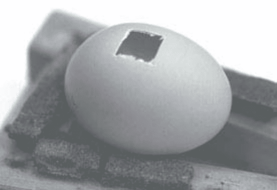
a small spatula, exposing the CAM (Fig. 1; see Note 10). This procedure causes
the membrane to drop as the egg contents fill the space left by collapse of the air
space at the end of the egg and the withdrawal of albumen (see Note 11). Dispose
of any nonviable eggs at this stage and, for each of the remainder, cover the
square opening in the shell with adhesive tape. Return the eggs to the incubator,
keeping them horizontally with the window uppermost, until sample application
on d 10 (d 8 may also be used; see Note 3).

CAM Assay 113
3.2. Focal Sample Application
1. Prepare a 1% w/v aqueous solution of methylcellulose and autoclave to sterilize.
Allow to cool whilst stirring and store sterile solution at 4°C (see Note 6).
2. Sterilize Teflon rods by autoclaving or 70% ethanol.
3. On day of sample application (see Note 15), in a laminar flow cabinet or clean
environment, mix equal volumes of sterile 1% methylcellulose and sample
(10 µL for each sample application). Vortex the mix thoroughly, because methyl-
cellulose is very viscous. Control discs of methylcellulose only should be pre-
pared in parallel (see Notes 12 and 13).
4. Pipette 10 µL of sample mix on to the top of a Teflon rod, or slab. After all the
samples have been applied to the Teflon rods, the samples are kept in the flow
cabinet until they have completely dried to a thin 2-mm diameter disc (30–60 min;
see Note 14).
5. On day of sample application, organize the surviving eggs into groups of at least
five eggs per sample. (See Note 15.) Apply samples to each egg in turn. Peel back
the adhesive tape to reveal the underlying CAM, and make a small puncture in
the CAM with a fine dental needle (see Notes 16, 17). Peel the sample disc from its
Teflon rod, using fine forceps, and place it on the CAM over the puncture. Reseal
the egg window and replace the eggs in the incubator for a further 3 d (see Note 15).
3.3. Collagen Synthesis Assay
1. Prepare eggs and samples as in Subheadings 3.1. and 3.2.
2. On d 10, immediately after application of the methylcellulose disc, apply 4 µL of
aqueous [U-
3
H]- proline (1 µCi) on to the sample pellet.
Fig. 1. Windowed egg showing exposed chorioallantoic membrane (CAM).

114 West et al.
3. After visual assessment of the angiogenic response on d 13 (Subheading 3.4.),
carefully excise the membrane using fine scissors and forceps.
4. With a sharp, sterile 1-cm-diameter cork-borer, cut a circle of each membrane
(1 cm diameter and approximately 5 mm radius from the point of sample applica-
tion) and transfer each separately to 3 mL of ice-cold 5% TCA.
5. After 4 hr at 4°C, recover the membranes by centrifugation at 10,000g for
10 min, at 4°C.
6. Wash the membranes twice with 3 mL of ice-cold 5% TCA, followed by three
washes with 3 mL 1 M NaCl in phosphate-buffered saline.
7. Digest washed membrane in 1 mL of sterile collagenase at 37°C, for 16 hr, with
shaking or mixing.
8. Terminate the digestion and precipitate undigested protein by the addition of
1 mL of ice-cold 20% TCA.
9. After 4 hr at 4°C, centrifuge the digest at 10,000g for 20 min and count an aliquot
of the supernatant, containing collagen peptides, in liquid scintillation counter to
determine [
3
H]- proline incorporation into collagen peptides. (See Note 18.)
3.4. Visual Assessment and Photography
1. On d 13 and/or d 14 (see Note 19), examine CAM for angiogenesis at site of
sample application. Part, or all, of the methylcellulose disc remains to indicate
the area.
2. Note the general condition of the CAM and embryo. Do not score dead or dying
eggs (membrane appears opaque and it is evident that there is a poor blood sup-
ply to the CAM vessels).
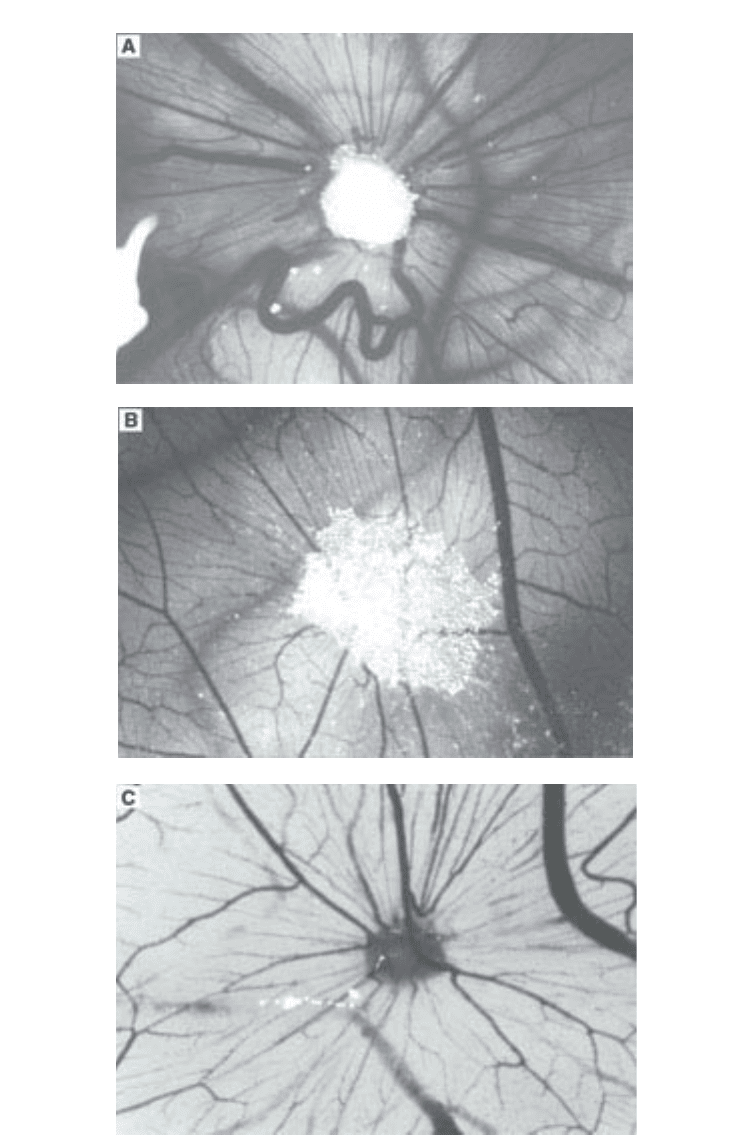
3. Score the degree of angiogenesis: 0, no change; 1, changes in vessels (disorga-
nized or bending), but not directed to the point of application; 2, a few
microvessels converge; moderate microvascular and large vessel convergence;
and 3, classical spoke wheel (Fig. 2; see Note 20).
4. The CAM can be photographed to provide a permanent record. A total magnification
(objective × ocular lens) of ×40 to ×80 is generally most suitable (see Note 21).
5. In addition, or alternately, fix the CAM and excise it for photography. Inject ice-
cold 4% paraformaldehyde–PBS under the CAM (approx 2 mL) and also flood
the upper surface of the CAM. Leave at 4°C for at least 4 hr (overnight fixation is
generally more practical). Excise the CAM, using small dissection scissors and
forceps (see Note 22), into PBS and place the CAM on a clean dry microscope
slide. Photograph under the dissecting microscope, as above.
Fig. 2. (see opposite page) Positive focal reactions: (A) Grade 4 reaction showing
“spoke wheel” pattern, with bending of large vessels (in situ, magnification ×16); (B)
weaker Grade 2 reaction, converging vessels on upper side of sample, largely
microvascular, typical of vascular endothelial growth factor (VEGF; in situ, mag-
nification ×32); and (C) paraformaldehyde-fixed CAM, showing a Grade 4 reaction
(magnification ×32).

CAM Assay 115
116 West et al.
3.5. In situ Inhibition assay
1. Prepare a 1% w/v aqueous solution of methylcellulose and autoclave to sterilise.
Allow to cool whilst stirring (see Note 6).
2. Sterilize Teflon rods by autoclaving or 70% ethanol.
3. Dissolve compounds to be tested in suitable solvents at 20 mM (see Notes 6 and 23).
4. Mix equal volumes of compound solution and 1% methylcellulose, and place
10 µl drops onto a Teflon slab (see Note 7). Empty discs (negative control) should
be prepared in parallel. Allow to dry.
5. Using sterile forceps, gently peel the methylcellulose discs from the Teflon slabs,
and place in the central area of the 15 mm diameter CAM of 6-d-old eggs (see
Notes 24, 25, and 26).
6. Return eggs to the incubator for a further 48 hr.
7. In a homogenizer, mix equal volumes of liquid paraffin and 4% (v/v) aqueous
Tween 80 to obtain a fine white emulsion (see Note 27).
8. Using a 1 mL syringe fitted with a 26-gauge needle, inject 1 mL of this emulsion
into the chorioallantois, so that the vascular network of the CAM stands out
against the white background.
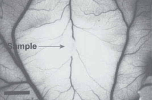
9. Observe with the naked eye. An area in which relatively large vessels (those
already present on d 6) cross an otherwise avascular zone of 3 mm or more in
diameter is scored as positive (Fig. 3; see Note 28).
10. The antiangiogenic activity of each compound is given as the percentage of tested
eggs presenting a positive response (see Notes 24 and 29).
3.6. Nonfocal (Liquid) Sample Application Technique
1. Prepare hen eggs as described in Subheading 3.1.
2. On d 10, organize the surviving eggs into groups of preferably no less than 10.
Prepare samples and inject 0.3 mL of test, or control, sample through the adhe-
sive tape onto the upper surface of the CAM (see Note 30).
3. On d 11, 18 hr after sample application (see Note 31), fix the CAM as outlined in
Subheading 3.4. Dehydrate and mount en face on large slides, using DPX resin
(see Note 32).
Visual analysis, ×30 magnification, shows general increase in vessel num-
ber and tortuosity with positive samples (Fig. 4). Subsequent image analysis of
vessel number, branching, and length can be used to quantify these changes.
Alternatively, measurement of the incorporation of methyl-[
3
H]-thymidine into
DNA may be used.
3.7. Assay of DNA Synthesis as Methyl-[
3
H]-Thymidine
Incorporation
1. On d 11 apply 0.5ml of methyl-[
3
H]-thymidine (2µCi/ mL of normal) onto the
CAM surface of each egg. Return to incubator for 20 min (see Notes 33 and 34).

CAM Assay 117
2. After 20 min, remove the eggs to a radioactive type bench and rapidly inject all
the eggs through the adhesive tape window, with 5 mL of ice-cold normal saline
to halt metabolism (see Notes 35 and 36).
3. Remove each CAM by holding the egg in the palm of one hand, window face
down, and piercing the side with pointed scissors above the level of the dropped
CAM. Cut around the shell with scissors, to separate the CAM from the rest of
the embryo. After cutting the umbilical vessels connecting the embryo to the
CAM, decant the residual yolk and embryo into a disposal container.
4. Cut around the edge of the CAM. Generally the dropped CAM remains inflated
by air above the window sealed by adhesive tape and a red ring is present at the
edge of the dropped CAM where it adheres to the shell (see Note 37).
5. Rinse each CAM briefly in saline, blot dry, and place in a 20 mL Sterilin dispos-
able vial, containing 5 mL of distilled water. These may be stored at 4°C over-
night if desired.
6. Homogenize each CAM briefly in the 5 mL of H
2
O using a powered homog-
enizer with a suitable head.
7. Add 5 mL of 10% trichloroacetic acid (TCA) to each and vortex.
8. Centrifuge the samples at approximately 1000g for about 8 min. Discard the
supernatant and resuspend the pellet in 10 mL TCA. Vortex thoroughly and cen-
trifuge 1000g for 8 min. Repeat the TCA wash to ensure adequate removal of
free isotope.
9. Resuspend the final precipitate in 2.5 mL of H
2
O, with vortexing, and immedi-
ately pour into scintillation vials containing 5 mL of Instagel Plus (a thixotropic
Fig. 3. In situ inhibition of angiogenesis in the CAM, d 8. The inhibitor was placed
in the methylcellulose disc (center) on d 6. Note the absence of small vessels around
the disc, the larger preexisting vessels being unaffected (bar, 4 mm).
