Messerschmidt U. Dislocation Dynamics During Plastic Deformation
Подождите немного. Документ загружается.

20 2 Experimental Methods
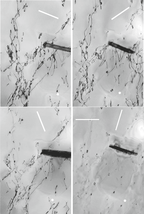
dislocations. An example is given in Fig. 2.6. In Fig. 2.6a, all dislocations in
the specimen area are visible. In Fig. 2.6b, the set marked A in Fig. 2.6a is
extinguished, in Fig. 2.6c it is set B. Finally, in Fig. 2.6d all dislocations except
some small loops are invisible. Considering the indexing of the g vectors in the
figure and the extinction rule (2.9), it may be concluded that the dislocations
marked A have Burgers vectors parallel to [010] and those marked B have such
parallel to [100]. For many TEM micrographs in this book, the g vectors are
indicated. If it is written, for example, g = (110) means that planes parallel
to the (110) plane are reflecting. In most cases, a reflection of higher order is
excited to obtain more narrow (sharper) images of the dislocations.
2.4 In Situ Straining Experiments in the Transmission
Electron Microscope
Performing complete plastic deformation experiments in situ in the specimen
chamber of a transmission electron microscope provides the unique opportu-
nity to observe the moving dislocations under stress at a resolution level that
allows conclusions to be drawn on many relevant mechanisms. Such experi-
ments were greatly facilitated by the commercial availability of high-voltage
electron microscopes (HVEM) with acceleration voltages of 1 MV, mainly in
Japan at the end of the 1960s. These microscopes allow the penetration of
specimens of thicknesses up to about 1–2 μm compared to some 100 nm in con-
ventional microscopes with an acceleration voltage of 200 kV. In addition, the
specimen chambers of most HVEMs offer sufficient room to insert elaborate
in situ stages between the pole pieces of the objective lenses. As will be men-
tioned below, the high voltage leads to the formation of radiation damage in
most materials, which may impair the reliability of these experiments. There-
fore, in situ experiments are also performed in conventional microscopes where
this problem usually does not occur, for example, [27]. A number of strain-
ing stages had been designed by the pioneering work of the group of Imura
and others, for example, [28–32], using different types of drive mechanisms.
The adjustment of proper imaging (diffraction) conditions requires the tilting
of the specimen. Most straining devices were designed for side entry specimen
stages and allow tilting only around a single axis. The present author designed
two straining stages for a 1 MV top entry HVEM with double tilting facilities,
one for room temperature [33] and another one for temperatures up to more
than 1,250
◦
C to be able to study also ceramic materials [34]. The stages will
be briefly outlined in the following.
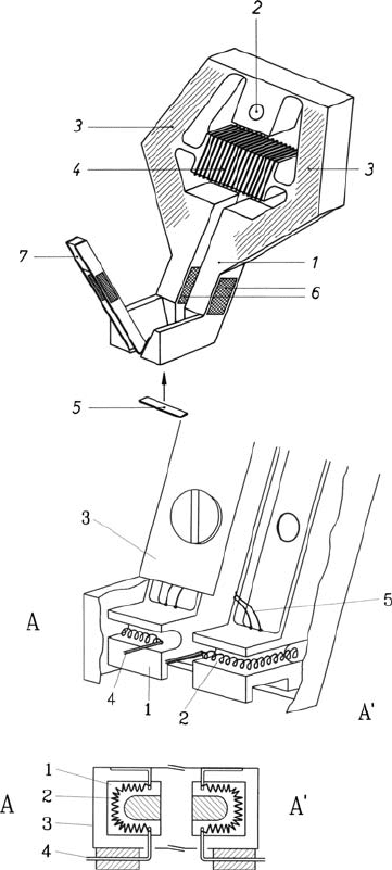
The room temperature stage shown in Fig. 2.7a consists essentially of a
single aluminium plate (1). It is supported by a screw at (2) and forms two
levers (3), which are connected with the support by thin parts acting as leaf
springs. The central bar (4) carries a heating coil. If the bar is heated, it pushes
the levers away from each other, resulting in a deformation of the specimen
(5), which is fixed to the levers from below. The hatched areas of the levers
2.4 In Situ Straining Experiments in the Transmission Electron Microscope 21
g
(110)
[1–10]
A
B
B
A
(a)
g
(–103)
[010]
(b)
g
(0–13)
[531]
(c)
1µm
g
(002)
[010]
(d)
Fig. 2.6. Analysis of the direction of Burgers vectors using the g times b rule. MoSi
2
single crystal deformed along a 011 direction at 380
◦
C. The thick dark contrast
near the center is a precipitate that can be used as a marker to identify individual
dislocations. The directions of the respective g vectors are indicated by the indexed
bars in the upper corners, the poles that are close to the viewing directions are
marked by the indexed white dots in the lower edges of the images. From the work
in [15]
22 2 Experimental Methods
Fig. 2.7. Outline of in situ straining stages for a top entry HVEM. These stages
are placed on double-tilting goniometer stages. (a) Room temperature deformation
stage. (b) Hot zone of high-temperature deformation stage. From [33,34]. Copyright
Elsevier Science (1976, 1994)
are water cooled. The whole equipment is inclined with respect to the electron
beam so that the latter can transmit the specimen. Four semiconducting strain
gauges forming a full bridge are glued to the lower parts of the levers for
measuring the load. Another set of strain gauges is fixed to a pair of leaf
springs (7) for measuring the specimen elongation. Thus, the stage allows the
recording of the load-elongation curve during the in situ experiment.
2.4 In Situ Straining Experiments in the Transmission Electron Microscope 23
Usually, high-temperature straining stages are heated by electrical resis-
tance heating. However, it is difficult to generate the high heating power
necessary to reach very high temperatures by this method because of the high
radiation losses. A solution to the problem is heating by electron bombard-
ment, which was introduced in the design of electron microscopy stages in [35].
The environment of the specimen in the high-temperature stage is outlined
in Fig. 2.7b in perspective and in cross-section views. Two double T-shaped
bars carry the specimen grips (1) at their lower ends. They are made from a
W-27at% Re alloy having a high strength at high temperatures. The specimen
is mounted between these grips from below by two tungsten pins fitting into
bores of the specimen. As seen in the cross-sectional view, the grips have a
U-shaped notch each. Tungsten filament coils (2) are situated in each notch.
They are attached to the thermal shields (3) via tungsten wires (4). As the
shields are at negative filament potential, they are electrically (and thermally)
insulated by small Al
2
O
3
spacers. For measuring the temperature, W-Re ther-
mocouple wires with 3 and 25% Re were welded individually to the top sides
of both grips. The T-shaped bars are connected to the drive mechanism. It
is made of copper and stainless steel and is similar to that of the room tem-
perature stage. For improving the heat transfer to the cooling water, heat
exchangers with copper lamellae are used. Outside, they carry the semicon-
ducting strain gauges for measuring the force acting on the specimen. The
whole system is digitally controlled by a personal computer equipped with
AD and DA converters. It provides the measurement of the temperatures of
both grips, of the electron beam current, and of the specimen load. The sys-
tem controls the electron beam current, the average temperature, the balance
between both grips, and the drive voltage.
The high-temperature straining stage allows a maximum specimen elon-
gation of 1 mm and a maximum load of 15 N. The thermal drive against water
cooling allows steady state behavior and ensures a very smooth and stable
operation. A number of experiments were performed at a grip temperature of
1,250
◦
C, corresponding to a specimen temperature of 1,150
◦
C, using a beam
current of about 60 mA at 700 V. The temperature is stable within about 3 K.
Duringtheinsituexperiments,theimages were recorded either as images
on photographic film with a high resolution or by a video system mounted in
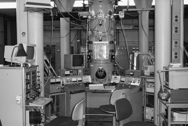
the base of the microscope with the usual TV resolution. Figure 2.8 shows the
HVEM equipped for performing straining experiments.
The specimens for in situ straining experiments have to meet the require-
ments of both transmission electron microscopy and of a tensile experiment.
In the case of these deformation stages, they are made from thin plates about
8 mm long, 1–2 mm wide, and 0.1 mm thick. For being fixed to the stage by
pins, they have two bores 5 mm apart. The thickness at the center must be
small enough for electron transparency, that is, about 1 μm. Besides, the edges
of the central thin area have to be thin enough to cope with the maximum
load of the stage, that is, about 10 μm. Depending on the material, this shape
can be produced by grinding, dimpling, and final ion milling, or by chemical
24 2 Experimental Methods
Fig. 2.8. View of the high-voltage electron microscope equipped for in situ straining
experiments. In the center, the microscope column and the control desk. At the top
right of the column, an extra turbo pump for improving the vacuum in the specimen
chamber. Left of the column, the video screen for the operator of the microscope.
In front, on the right side, the video recorder with another screen. On the left side,
the PC for controlling all functions of the straining stage
or electrolytical polishing. In the latter case, the thinning may be performed
in two steps using masks. In the first step, a shallow dimple is thinned from
one side through a mask with a hole smaller than the sample width. In the
second step, the whole central area is thinned, including also the edges of the
sample. For brittle materials like ceramics, semiconductors, or quasicrystals,
it is advantageous if there is no hole in the center of the specimen. For opti-
cally transparent materials, the thickness can be estimated by watching the
occurrence of Newton fringes so that the thinning can be stopped before the
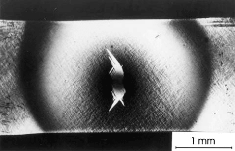
sample is perforated. Figure 2.9 presents an example of the central part of
aZrO
2
specimen without a hole after in situ straining. The bright contrasts
near the center show regions deformed by ferroelastic deformation. Metal spec-
imens usually have a hole in the center of the transparent area. Because of
the inhomogeneous cross section of in situ specimens, the stress distribution is
not uniform. According to calculations by the finite element method [37], the
stress state is complicated at the edges of the perforation lying in the tensile
direction. At the transverse edges, an almost uniaxial tensile stress occurs with
a stress concentration of three times the average stress in this cross section,
as results from technical elasticity calculations of a plate with a cylindrical
hole. Mainly these areas should be observed during in situ straining.
2.4 In Situ Straining Experiments in the Transmission Electron Microscope 25
Fig. 2.9. Micro-tensile sample of a ZrO
2
single crystal deformed during an in situ
straining experiment at 1,150
◦
C. The specimen was prepared by grinding, dimpling,
and ion milling. It does not have a perforation. The bright contrasts near the center
result from ferroelastic deformation. From the work in [36]
There are two problems limiting the reliability of in situ straining experi-
ments in an HVEM as discussed in [38,39]. The first one regards surface effects
due to the relatively low specimen thickness, even in the HVEM. The surfaces
close to the dislocations cause so-called image forces, which superimpose on
the forces from the applied stress. These forces are estimated in Sect. 3.2.3,
showing that the image forces are not of great importance in materials with
high flow stresses like many contemporary materials, but they may produce
artifacts in pure f.c.c. metals, which have very low flow stresses. Another
effect may be called the interruption of the dislocation kinetics. During defor-
mation, new dislocations are generated in the interior of the specimen, which
may move out of the foil, but no dislocations move into the foil from outside.
This results in differences in the dislocation density between the interior of the
crystal and the regions near the surfaces. Therefore, in situ straining experi-
ments are well suited to study the dynamics of individual dislocations rather
than the development of complex dislocation structures.
The second problem is the occurrence of radiation damage. The imag-
ing electrons of high energy may displace atoms from their regular places
forming vacancies and interstitial atoms. The threshold voltage for this pro-
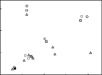
cess depends mainly on the atomic number of the material. As shown in
Fig. 2.10, the acceleration voltage of 1 MV exceeds the threshold voltage for
most materials. Even at 200 kV, displacement damage can occur in light mate-
rials. Whether the radiation damage disturbs the deformation experiment, or
not, depends on the temperature. At low temperatures, the generated pri-
mary defects are immobile. At intermediate temperatures, the defects diffuse,
at first forming small clusters and later on dislocation loops. This process is
described for ZrO
2
single crystals in [40]. In this temperature range, straining
experiments cannot be performed. At high temperatures, the defects anneal
26 2 Experimental Methods
1500
1000
500
U (kV)
Z
W
Au
Ta
Pb
Cd
Sn
Mo
Nb
f.c.c.
b.c.c.
h.c.p.
V
Co
Ni
Cu
Zn
Ti
Fe
Al
Mg
Si
20 40 60 800
0
Fig. 2.10. Dependence of the threshold voltage for displacement damage by electron
irradiation on the atomic number. Data from [38]
out at the near surfaces. How severe the radiation damage affects the dislo-
cation motion in a particular experiment has to be checked individually. In
most experiments described in this book, the effect is not strong so that the
results should be reliable.
As mentioned in the Introduction, video sequences of dislocation motion in
a number of materials can be downloaded from http://extras.springer.com/
2010/978-3-642-03176-2. These video clips will be described in some detail
in Part II in the chapters of the respective materials. Some videos showing
general features of dislocation motion will be referred to in the following chap-
ters. For a first glance at dislocation motion and generation, please look at
Video 9.17. The video presents a rotating source which, at each revolution,
produces two dislocations afterwards moving in opposite directions. They are
mostly straight and aligned along crystallographic orientations. In addition to
qualitative information on the dislocation behavior, in situ straining experi-
ments yield a wealth of quantitative data, including the indexing of the planes
of motion, the preferred orientations of dislocations, the density and strength
of obstacles to the dislocation motion, or kinematic data like waiting times
and jump distances. The evaluation of such data is discussed in the respective
chapters.
2.5 Other Methods
The methods described in this section supplement the main techniques pre-
sented above to study dislocation dynamics and the processes controlling
it. They are mostly limited either to particular materials or to particular
2.5 Other Methods 27
processes. The contents of this section is better understood after reading Part
Iofthisbook.
2.5.1 X-Ray Topography In Situ Deformation Experiments
Using X-ray topography, dislocations can be imaged in a similar way as in the
electron microscopy diffraction contrast. A recent review of the topography
techniques is given in [41]. Accordingly, in situ straining experiments can also
be performed in an X-ray topography arrangement to directly observe the
processes controlling the dislocation motion. This requires dedicated strain-
ing stages to be positioned on the goniometer head of the X-ray topographic
device. However, there are two differences with respect to TEM. The high-
energy photons have a much higher penetration power compared to the
electrons of usual energies, and so crystals of bulk dimensions can be observed.
Besides, the width of the dislocation images is considerably greater than in
TEM diffraction contrast, resulting in a poor resolution power for disloca-
tions. This limits the application to the observation of single dislocations in
crystals of a very high perfection with zero or very low dislocation densities as
semiconductor single crystals, or to the observation of groups of dislocations,
so-called slip bands, in otherwise perfect crystals. To record the processes by
means of video systems, X-ray beams of a high intensity are necessary. This
can be achieved by conventional high intensity rotating anode X-ray sources
or, more recently, by synchrotron radiation.
In the first type of experiments, mainly the multiplication of dislocations
in semiconductor crystals was studied [42, 43]. Microhardness indents were
placed on the surface to introduce nucleation sites. An example of a dislocation
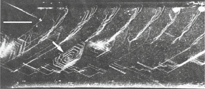
source, which had emitted a number of hexagonal dislocation loops in an Si
single crystal, is presented in Fig. 2.11.
The first occurrence and propagation of slip bands was studied in MgO sin-
gle crystals of thicknesses between about 60 and 320 μm[44]inanequipment
similar to that in [42]. First slip bands occur at about half the macroscopic
yield stress. During plastic instabilities causing decreases in the applied stress,
the bands propagate at high velocities of about 150 mm s
−1
. There exists a size
effect of an increasing yield stress with decreasing specimen thickness. In this
respect, the X-ray topography experiments are a link between macroscopic
tests and in situ straining experiments in the TEM. In optically transparent
crystals, similar studies on the propagation of slip bands can be performed by
optical birefringence microscopy.
2.5.2 Surface Studies of Slip Lines
The occurrence and propagation of slip bands can also be observed on the
crystal surface by imaging the steps resulting from the motion of disloca-
tions with Burgers vectors pointing out of the surface. These experiments can
be performed on very different levels of spatial and temporal resolution. In
28 2 Experimental Methods
1 mm
Fig. 2.11. Localized dislocation source in an Si single crystal observed during an
X-ray topography in situ straining experiment. The topography device included a
30 kW X-ray generator, the in situ straining stage, and a vidicon X-ray camera. From
[42]. Copyright Taylor & Francis Ltd. (1981). (http://www.informaworld.com)
the 1950s and 1960s, the surfaces of deformed crystals were imaged by opti-
cal microscopy or by surface replicas imaged in the TEM. These methods
do not resolve the steps produced by moving individual dislocations. Never-
theless, important information on the distribution and length of slip bands
formed by many dislocations has been obtained (e.g., the work by Seeger and
coworkers [45]).
To follow the temporal development of the slip bands, the optical micro-
scope was supplemented by a cine camera, first applied by Schwink and
Neuh¨auser [46], and reviewed in [47]. Later on, the cine camera was replaced
by a video camera. Different methods have been used to improve the time
resolution of specific processes, for example, a photo-diode was placed in the
intermediate plane of the microscope to record the intensity changes of an
imaged slip band (e.g., [48]). Recently, optical extensometers have been devel-
oped (e.g., [49]) where the gauge length of the specimen is coated with a
pattern of dark and bright stripes, which are scanned via a special line-scan
camera typically operating at a frequency of 250 Hz. The PC based evaluation
of the data allows the determination of the place and the time of a deformation
event. These data can be plotted in a correlation diagram.
The cinegraphic methods are mainly used to investigate the evolution of
slip bands at the heterogeneous deformation as it occurs at plastic instabilities
described in Sect. 5.3. The occurrence of slip bands can be correlated or un-
correlated. The heads of slip bands may move at velocities up to the m s
−1
range. In one study [48], a second video camera was placed on the opposite
side of the specimen to monitor the passage of slip bands through the crystal.
A method which is able to resolve slip steps of atomic height trailed
by moving individual dislocations is the so-called heavy metal decoration
2.5 Other Methods 29
technique [50,51]. It requires very smooth starting surfaces with a low density
of steps. Such surfaces can be obtained by cleaving ionic crystals in a very dry
atmosphere or in vacuum or by producing growth surfaces on metals. After
deforming the crystals and creating the slip steps in vacuum, a small amount
of gold or another heavy metal is evaporated on the surface. The gold forms
small nuclei distributed randomly on the atomically smooth surfaces. Along
monatomic steps, however, the nuclei are linearly arranged, thus decorating
the surface step. This method allows the paths of individual dislocations with
a Burgers vector component out of the surface to be imaged. The technique is
well suited to study cross slip as described in Sect. 4.3 and shown in Figs. 4.9
and 4.12.
In recent years, slip steps have also been imaged by atomic force
microscopy. Mostly, the resolution power is sufficient only to observe slip bands
(e.g., [52]). In some studies, also the steps of a few individual dislocations were
imaged showing cross slip events of superdislocations (e.g., [53]).
In general, the observations of slip bands on the crystal surfaces provide
a link between the behavior of individual dislocations and the macroscopic
plastic properties, in particular in the case of inhomogeneous deformation.
2.5.3 Internal Friction
If a material is deformed, the part W
el
of the total work W
t
expended is of
elastic nature, that is, it is re-gained after unloading. The rest is partly stored
in the material in the form of crystal defects produced by the deformation,
the so-called stored energy W
stor
, and partly transferred as friction W
fr
into
heat
W
t
= W
el
+ W
stor
+ W
fr
.
The stored energy is mostly only a small fraction (in the order of magnitude of
10%) of the nonelastic energy. Several methods have been designed to measure
the stored energy, mostly calorimetric ones. On principle, the nonelastic part
of the energy can be determined as the area enclosed in a stress–strain curve
of a specimen loaded and unloaded cyclically. Frequently, the driving stress is
harmonic
σ = σ
a
exp(iωt),
where σ
a
is the oscillation amplitude, ω the circular frequency, and t the time.
If the amplitudes are small so that no energy is stored, the behavior is anelastic
and the relative energy loss per cycle is called internal friction. An early
thorough review is given by DeBatist [54], and a later one in the proceedings
[55]. In most techniques, the specimens are excited in resonance modes. Two
types of measurement can be performed, either the oscillation amplitude is
maintained on a constant level and the necessary power is measured, or the
excitation is switched off and the decrease in the amplitude of the freely
vibrating specimen is recorded. One measure of the internal friction is the
ratio between the anelastic work ΔW and the total work W
t
during one cycle.
