Hughes M.P., Hoettges K.F. (Eds.) Microengineering in Biotechnology
Подождите немного. Документ загружается.


on, Hodgkin worked together with Katz (4) to demonstrate that
the sodium permeability causes the overshooting action potential.
Finally, Hodgkin and Huxley (5) developed a voltage-clamp circuit
to measure the ionic currents from the axon of a squid. These
experiments, using single electrodes, permitted the study of the
behaviour of single neurons. However, to understand the nervous
system, and the complex processing that relies on vast neural con-
nections, it is necessary to obtain information from many neurons at
the same time. The purpose of modern neural probes is to record
from as many individual neurons as possible without causing
damage to the cells. Some of the essential characteristics for these
modern devices are as follows:
l
Size: Since it is required to record from single neurons, the size
of the electrode has to be similar to that of the neurons, of the
order of micrometres.
l
Material: As the body’s immune system will reject any foreign
object, the electrode has to be of a material that does not
damage or cause rejection and that does not become affected
by the internal conditions within the body, i.e. it must be
biocompatible.
l
Insertion and attachment: The way the probe is inserted and
attached to the nerve has to be of a manner that the nerve
remains as intact as possible and the probe does not change
position with movement.
To obtain a neural probe with these characteristics, many
approaches have been investigated. Cuff electrodes, regeneration
probes and penetrating microelectrodes are the main approaches
to date. This chapter describes the development of implantable
neural probes for recording from neurons in vivo.
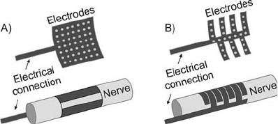
2. Cuff Electrodes
A cuff electrode is a device that encircles the whole nerve
(Fig. 6.1). It consists of an insulating sleeve placed around a
length of nerve, with electrodes around it. The advantages of this
technology are that the nerve remains intact during implantation
and surgical implantation is not difficult (44). Although the
implantation is not difficult, after surgery the nerve will swell and
connective tissue will form where surgery was performed. These
factors must be taken into consideration to avoid any neural
damage. Possible displacement can be avoided by mechanical
fixation. The main disadvantage of this technique is that the
recordings are made of the sum of superficial neurons, as the
electrode encloses the whole nerve. Signals from neurons inside
136 Bustamante Valles
the centre of the nerve have very low amplitude at the surface, and
may be lost in noise. Signal processing can be used to separate
information from different neurons and to obtain information
about neural coding; however, this is difficult to achieve. Other
important restrictions are the material and the geometry, which
have to be careful chosen since bending torques, compression to
the nerves or disruption to nerves blood supplies can provoke
severe nerve damage.
As described by Heiduschka and Thanos (44), the first elec-
trodes were made of rigid materials and with very few electrodes.
The electrodes were made of platinum foil (6) or platinum wire
electrodes (7). These first models presented major problems, such
as damaging the nerve due to mechanical displacement or causing
ischemia due to pressure on the nerves. The foil electrode also had
a problem with weak joints, since a weld is required between the
joint and the lead-out cable. Platinum wire electrodes were usually
sewn into the cuff wall. However, these electrodes were not likely
to stay inside the wall of the cuff, and connective tissue would grow
around them making then impossible to remove without damage
to the nerves. To overcome these problems, new geometries and
materials were developed. In the early 1980s, the use of platinum
wire for electrodes and leads was often substituted by Teflon
coated, multi-stranded stainless steel wires embedded into the
cuff wall, which have been widely used for the remaining 20
years (e.g. (8)). The electrode is made from the same lead-out
wire and as it is embedded into the cuff wall provides a mechani-
cally flexibility. The introduction of a self-spiralling cuff design by
Naples et al. (9) helped reducing the need for rigidity in the cuff
wall and minimising the wall thickness. Klepinski (10) patented a
chronically implantable cuff electrode with semi-rigid fingers
extending orthogonally from the central spine, which are bent
around the nerve. Sahin and Durand (11) reported an improved
nerve cuff electrode using electric currents to slow the velocity of
the action potentials, which creates an analogous effect of increas-
ing the cuff length without longer intercontact delays. Their data
suggested that this method could be used to improve the SNR of
Fig. 6.1. Cuff electrodes: (a) half cuff electrode (43). (b) flexible interdigitating cuff
electrode.
Microengineered Neural Probes for In Vivo Recording 137

nerve recordings. Takeuchi and Shimoyama (12) developed a
shape memory alloy cuff electrode, which has demonstrated to
be effective for chronic recordings in a wireless system on insects.
The biocompatibility of this material is still under investigation.
Analysis of the properties of cuff electrodes has been extensively
researched by Jezernik and Sinkjaer (13) from Aalborg University,
who described the statistical properties of the whole nerve cuff
recordings. Nakatani et al. (14) have developed methods to detect
action potentials using cuff electrodes with a low signal to noise
ratio, trying to address one of the main disadvantages of this type
of electrodes compared to intrafascicular electrodes. These cuff
electrodes were made of 75-mm-diameter stainless-steel wire and
they were designed for bipolar recording. The processing of the
data was not done in real-time, which would be necessary for real
clinical applications. Jensen and co-workers are currently investi-
gating the use of a triple electrode cuff electrode to provide motion
feedback for functional electrical stimulations system by detecting
joint rotation using recordings on sciatic nerves of rabbits (15).
Many types of cuff electrodes are already commercially avail-
able, but are mainly used for stimulation purposes. For example,
the Vagus Nerve Stimulation System to help treat patients with
epilepsy (Cyberonics Corp.); Bladder Control System from
Vocate; NeuroControl Corporation control bladder and bowel
voiding, or the Paralysed Hand Control System from FreeHand
NeuroControl Corporation to help activate grasp in high-level
quadriplegia. A more detailed review on cuff electrodes can be
found in Hoffer and Kallesoe (16).
Although there have been great advantages in the design of
cuff electrodes, they have many drawbacks when used for selective
neural recordings. Whilst cuff-based stimulator electrodes are
widely used, there are still no commercial devices which have
been used for recording neural activity. One of the main drawbacks
of the cuff type of sensor is selectivity.
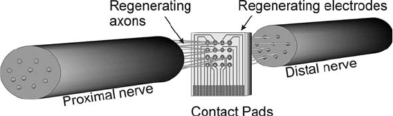
3. Regenerating
Electrodes
Regenerating electrodes are situated within the nerve. The probes
are sieve-shaped and contain holes, with electrodes on the side of
the holes or on the walls of these holes. The nerve is cut and the
probe is inserted in the gap (Fig. 6.2). The neurons are then
allowed to regenerate through the holes. Direct recordings can
be made from the neurons and the probe is mechanically stable as
the neurons grow through the holes and keep it in position. The
main drawback of this method is the need to cut the nerve, thereby
potentially causing irreparable damage, and the length of time it
138 Bustamante Valles
takes the nerve to regenerate. For example, Edell et al. (17)
reported nerve recovery after 332 days in New Zealand rabbits.
Akin et al. (18) achieved regeneration after 118 days on 21 out of
28 implants on rat nerves. These types of electrodes are used only
on peripheral nerves because the central nervous system does not
regenerate.
As described by Kovacs (19), the first work on constructing
regenerating electrodes was undertaken by Frishkoff (unpub-
lished) around 1965 with no successful results. Other authors
continued researching this technique and it was not until 1974
that Mannard et al. (20) reported the first recording using
nerve regeneration electrode units. The probes were made on
an epoxy wafer and the holes were mechanically drilled. The
first recordings using silicon-based regeneration electrodes were
not made until the beginning of the 1980s. Edell et al. (17)
reported the development of regenerating nerve electrodes
using silicon bars, 40 mm wide and 200 mm apart in a comb
configuration. These probes had 20 40 mm electrodes and
were implanted in rabbit peripheral nerves. The results showed
the ability of this design to record action potentials up to
600 mV in amplitude. Kovacs et al. (21) developed a new
process for the fabrication of regenerating microelectrode arrays
for peripheral and cranial nerve applications using BiCMOS
processes (CMOS technology not involving high temperature
process steps nor heavily doped etch stop layers). A typical
probe had 100 mm
2
microelectrodes and 90 90 mm via-
holes. These devices were implanted in the rat peroneal nerve
and the frog auditory nerve; action potential signals were
recorded from several microelectrodes in parallel. Another
design of regeneration probes was developed by Akin et al.
(18). This probe consisted of a silicon rim support, with a
thickness of 15 mm, and different sizes of via-holes. The elec-
trodes had areas from 100 to 2,000 mm
2
and were made of
iridium. A silicon ribbon cable was integrated to connect the
probe to the outside world. These electrodes were tested on
peripheral taste neurons of rats. Regeneration was successful
and electrophysiological recordings were obtained.
Fig. 6.2. Regenerating electrodes.
Microengineered Neural Probes for In Vivo Recording 139
Wallman et al. (22) created and tested regenerated electrodes
using via-holes with either 30 or 90 mm and varying the ratio of the
total via-hole cross-sectional area in relation to the actual surface
area of the electrode. Previously, recordings with 50 or 10 mm
were used with unsuccessful results. The best results were achieved
with 30 mm via-holes and a ratio of hole to surface area of 30%.
A hybrid system using sieve electrodes is being investigated by
Stieglitz et al. (23). This system consists of a microfabricated
flexible sieve polyamide device with microelectrodes and inte-
grated biological cells. The device has 19 electrodes for stimulation
and recording purposes and the adaptation of the implanted bio-
logical cells to the nerve stump has been demonstrated. Their
future work will focus on the integration of cell containments
with their microsystem.
This type of neural probes provides a close contact with the
neurons, allowing a selective recording and stimulation besides
mechanical stability. However, there is no guarantee of healing
and regeneration through the probe. The lesion to the nerve
caused in order to implant the probe will only heal under favour-
able conditions, which cannot always be controlled.
Cuff electrodes and regenerating-type electrode have been
developed for many years now. There have been great advances;
however, there are many drawbacks associated. Penetrating probes
offer good compromise between cuff and regenerating-type elec-
trodes. As described next, they allow a better selectivity than cuff
electrodes as they are in direct contact with neurons but they are
not as invasive as regenerative-type electrodes.
4. Penetrating
Electrodes
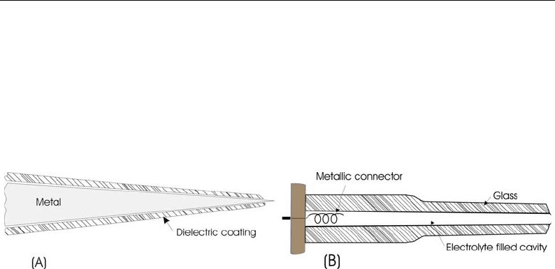
The third type of device is a ‘‘needle like’’ probe carrying one or
more electrodes. These probes penetrate inside the nerve and can
have direct contact with one or more neurons. The first types of
penetrating electrodes were simple insulated metal wires or glass
filled pipettes with a metal contact inside (Fig. 6.3).
Fig. 6.3. First microelectrodes, modified from Ferris (45) (a) Cross section of a metal wire electrode. (b) Cross section of a
glass electrode.
140 Bustamante Valles
These wire or glass microelectrodes were used for the early
neurophysiological experiments, for example, in the work of
Hodgkin and Huxley mentioned before, and they are still in use
today (24). Very fine metal tips could be created, sometimes up to
0.2 mm in diameter as reported by Grundfest et al. (25). A review
of the first type of electrodes can be found in work by Kruger (26).
These probes usually allow only single unit recordings; whilst this
was very useful for the understanding of the nerve system, simul-
taneous recordings from many neurons are needed as the nervous
system is made of large and complicated neural connections.
The use of photographic techniques was developed to help the
creation of multielectrode microprobes. Gross et al. (27) devel-
oped a 36-electrode array with photoetching techniques for in
vitro recordings. The contacts had 12 mm diameter and neural
activity from several neurons was recorded. Pine (28) introduced
two rows of electrodes 250 mm apart with electrodes surfaces of
8 10 mm produced by photoetching techniques. A review on
microetching and metal deposition techniques can be found in
Pickard et al. (29).
Another technique emerged in the early 1970s with the use of
the semiconductor industry for the fabrication of microelectrodes.
One of the first electrodes reported using silicon techniques was
that by Wise et al. (30). They produced an array of gold electrodes
supported on a silicon carrier. Interelectrode spacing was accu-
rately controlled and tip diameters as small as 2 mm were produced.
Successful recordings were obtained from cat cortex. Later, Wise
and Angell (31) reported a multielectrode structure containing
integrated junction-FET input stages. Prohaska et al. (32) obtained
an eightfold microelectrode using thin-film technology with elec-
trodes contact areas of 50 50 mm and separation of 300 mm.
Since then, many microprobes with different characteristics (such
as geometrical areas, materials and number of electrodes) have
been fabricated. Gold, platinum and iridium have been widely
used for recording neural activity. They all present good electrical
and biocompatibility characteristics; however, if the probe is to be
used for stimulation purposes, iridium has a higher current density
capability. The area of the electrodes reported varied from 10 up to
2,500 mm
2
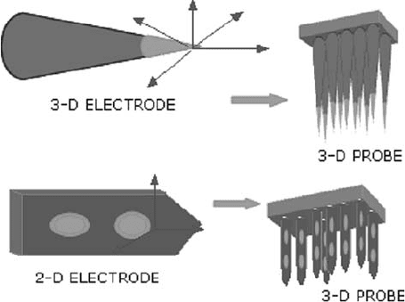
. The different types of design include planar electrodes
in a two-dimensional plane or electrodes with a three-dimensional
form. A simple wire penetrating electrode would usually have
three-dimensional recording capabilities, meaning it would be
able to record from all its surroundings. Usually the silicon probes,
will have electrodes in a two-dimensional plane, being less able to
record action potentials that occur behind the plane of the elec-
trode. To overcome this problem and still use the advantage of
silicon probes, three-dimensional arrays have been fabricated using
an array of planar shanks, or using electrodes needles in a brush-
like array (e.g. Fig. 6.4).
Microengineered Neural Probes for In Vivo Recording 141
The University of Surrey, in collaboration with the Uni-
versity of Southampton, has been working for more than 10
years in the development of 2D silicon microprobes (33). The
probes were fabricated using a bonded and etched-back silicon-
on-insulator wafer (BESOI), which permits the reduction of
boron doping for the future implementation of on-site elec-
tronics. The probes used different sizes of electrodes allowing
the investigation of the effects of diverse recording areas on
the neural recordings. The probes also had different sizes of
shank width, from 122 to 250 mm. These probes have success-
fully recorded neural activity from locust nervous system and
the different designs permit the study of the different record-
ing characteristics.
One of the groups that have worked for many years
developing penetrating silicon probes is at the Centre for
Neural Communication Technology under the direction of
Anderson and Wise. Their work has been directed to the
development of multichannel silicon substrate probes with
one to four shanks and up to 16 (177 mm
2
) iridium electro-
des per shank. A diagram of one of these probes is found
in Fig. 6.5. Some of the latest publications from this group
report single-unit recordings from three-dimensional
electrode arrays (Fig. 6.6) containing on-chip signal cir-
cuitry (34).
Another recent development is found in Xu et al. (35). They
reported a metal microelectrode array (Fig. 6.7), similar to tung-
sten-wire probes or tetrode probes, but made using metal shanks
and electrical insulation by conformal coating of Parylene-C film.
The recording sites have precise opening sizes defined by
Fig. 6.4. Two- and three-dimensional neural probes.
142 Bustamante Valles
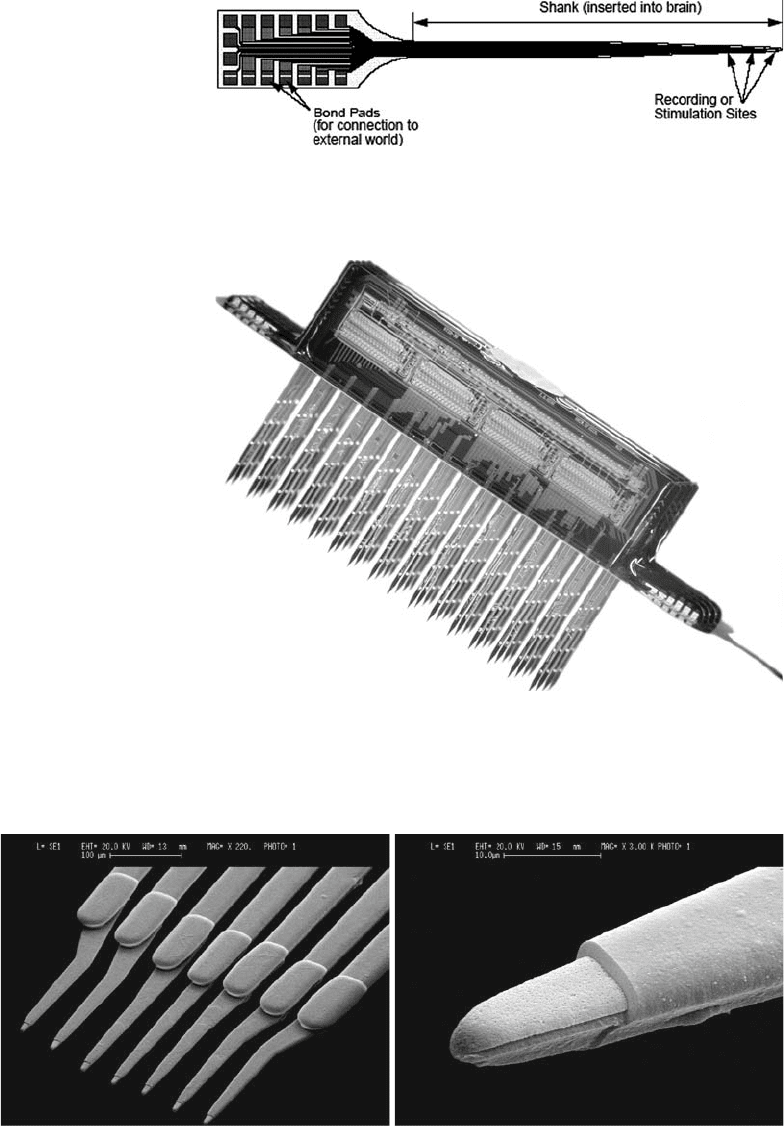
Fig. 6.6. 3-D probe array, courtesy Dr K. Wise. Reprinted with permission from Proceed-
ings of the International Conference on Solid State Sensors and Actuators (Transducers
01), Munich. ª 2001 IEEE.
ab
Fig. 6.7. SEM of metal microelectrode (a) View of the shank design; (b) close view of the recording tip. Pictures courtesy
Dr. C. Xu.
Fig. 6.5. CNCT, Schematic of a single shank acute probe. Picture courtesy
Dr. D. Anderson.
Microengineered Neural Probes for In Vivo Recording 143
photolithography and oxygen plasma etching. Recordings were
made from cockroaches and signals were acquired from all indivi-
dual electrodes in the probe.
The University of Utah, Department of Bioengineering,
under the direction of Professor Horch, have worked extensively
developing a neural-controlled prosthetic arm. This arm would be
controlled by neural signals from motor nerves in the amputee’
stump. To sense these nerve signals, flexible microwires were
developed. Ti/W, Au and Pt were used to produce mechanically
stable and highly flexible metallised fibres insulated with silicone
elastomer (36).
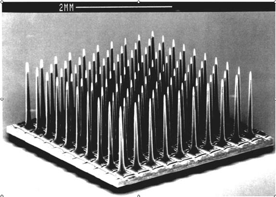
Within the same University, The John Moran Laboratories for
Applied Vision and Neural Sciences have worked for many years on
the development of three-dimensional electrode arrays. The Utah
Intracortical Electrode Array (UIEA) consists of 100 penetrating
silicon microelectrodes with a spacing of 400 mm(Fig. 6.8).
However, chronic recording applications are still limited. These
probes have been implanted in cat sensory cortex for up to 13
months. In some cases evidence of chronic injury response
was found and only 11 electrodes were connected to the exterior
(37–39).
Stieglitz and Gross (40) recently reported a process technol-
ogy for polyamide-based devices. These are flexible microprobes
with electrodes located on the top and back of the flexible device
(Fig. 6.9). The electrodes have a diameter of 10 mm. First proto-
types were built and electrically tested; however, they have not yet
Fig. 6.8. The Utah Intracortical Electrode Array, courtesy Dr. R. Normann.
144 Bustamante Valles
been tested in any animal model. This work is done at the Fraun-
hofer-Institute Biomedical Engineering, Neural Prosthetics Unit,
Ingbert Germany.
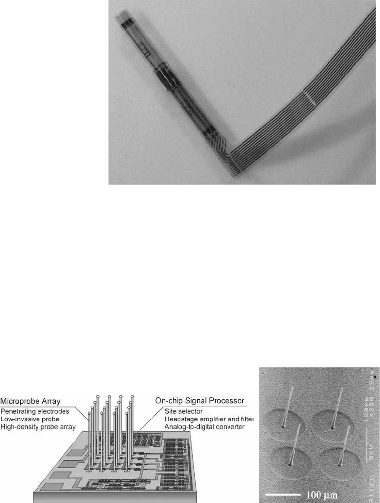
At the Toyohashi University of Technology, in Japan, Kawano
et al. (41) have developed a micro-Si wire probe array with on chip
circuits using a selective Au-Si
2
H
6
vapour–liquid–solid growth
method. Si probes with 160 mm in length and 3.5 mm in diameter
at the tip were grown (Fig. 6.10). Simulated stimulation of neural
activity was recorded to evaluate these prototypes, which could
mean that these probes could be useful for real neural recordings.
Fig. 6.9. Photograph of the flexible microprobe, courtesy of Dr. Koch.
ab
Fig. 6.10. (a) Schematic diagram illustrating an image of ‘‘smart neural probe chip,’’ comprising a multichannel
penetrating Si microprobe electrode array and with on-chip integrated circuit (IC) signal processors. The Si probe array
was fabricated by selective growth of Si probes, which realised low neural invasiveness probes and high-density probe
array with distribution similar to neurons. The greatest merit of this technology is that the microprobe array can be
combined with numerous CMOS-IC on the same substrate. (b) SEM image of Si microprobe array, each with 160 mmin
length and 3.5 mm in diameter at tips. Pictures courtesy Dr. T. Kawano.
Microengineered Neural Probes for In Vivo Recording 145