Hermanson G. Bioconjugate Techniques, Second Edition
Подождите немного. Документ загружается.


23
One of the most popular methods of noncovalent conjugation is to make use of the nat-
ural strong binding of (strept)avidin for the small molecule biotin. The strength of the
(strept)avidin–biotin interaction has made it a useful tool in specifi c targeting applications and
assay design. Since each (strept)avidin molecule contains a maximum of four biotin binding
sites, the interaction can be used to enhance the signal strength in immunoassay systems.
Modifi cation reagents that can add a functional biotin group to proteins, nucleic acids,
and other molecules now come in many shapes and reactivities (Chapter 11 and Chapter 18,
Section 3). Depending on the functionality present on the biotinylation compound, specifi c
reactive groups on antibodies or other proteins may be modifi ed to create a (strept)avidin bind-
ing site. Amines, carboxylates, sulfhydryls, and carbohydrate groups can be specifi cally targeted
for biotinylation through the appropriate choice of biotin derivative. In addition, photoreactive
biotinylation reagents (Chapter 11, Section 3.4) are used to add nonselectively a biotin group to
molecules containing no convenient functional groups for modifi cation. In this manner, oligo-
nucleotide probes often are modifi ed for detection with (strept)avidin conjugates (Chapter 27,
Section 2.3).
The following sections discuss the concept and use of the (strept)avidin–biotin interaction in
bioconjugate techniques. Preparation of biotinylated molecules and (strept)avidin conjugates
also are reviewed with suggested protocols. For a discussion of the major biotinylation rea-
gents, see Chapter 11 and Chapter 18, Section 3.
1. The Avidin–Biotin Interaction
Avidin is a glycoprotein found in egg whites that contains four identical subunits of 16,400 Da
each, giving an intact molecular weight of approximately 66,000 (Green, 1975). Each subunit
contains one binding site for biotin, or vitamin H, and one oligosaccharide modifi cation (Asn-
linked). The tetrameric protein is highly basic, having a pI of about 10. The biotin interaction
with avidin is among the strongest noncovalent affi nities known, exhibiting a dissociation con-
stant of about 1.3 10
15
M. Tryptophan and lysine residues in each subunit are known to be
involved in forming the binding pocket (Gitlin et al ., 1987, 1988).
Avidin–Biotin Systems
900
The tetrameric native structure of avidin is resistant to denaturation under extreme chao-
tropic conditions. Even in 8 M urea or 3 M guanidine hydrochloride the protein maintains
structural integrity and activity (Green, 1963). When biotin is bound to avidin, the interaction
promotes even greater stability to the complex. An avidin–biotin complex (ABC) is resistant
to break down in the presence of up to 8 M guanidine at pH 5.2. A minimum of 6–8 M guani-
dine at pH 1.5 is required for inducing complete dissociation of the avidin–biotin interaction
(Cuatrecasas and Wilchek, 1968; Bodanszky and Bodanszky, 1970). Since the subunits in avidin
are not held together by disulfi de bonds, conditions that cause denaturation also result in subu-
nit disassociation.
The strength of the noncovalent avidin–biotin interaction along with its resistance to break
down makes it extraordinarily useful in bioconjugate chemistry. Biotinylated molecules and
avidin conjugates can “ fi nd ” each other under the most extreme conditions to bind and com-
plex together. The biospecifi city of the interaction is similar to antibody–antigen or receptor–
ligand recognition, but on a much higher level with respect to affi nity constants. Variations in
buffer salt, pH, the presence of denaturants or detergents, and extremes of temperature will
not prevent the interaction from occurring (Ross et al ., 1986).
The only disadvantage to the use of avidin is its tendency to bind nonspecifi cally with
components other than biotin due to its high pI and carbohydrate content. The strong posi-
tive charge on the protein causes ionic interactions with more negatively charged molecules,
especially cell surfaces. In addition, carbohydrate binding proteins on cells can interact with
the polysaccharide portions on the avidin molecule to bind them in regions devoid of targeted
biotinylated molecules. These nonspecifi c interactions can lead to elevated background signals
in some assays, preventing the full potential of the avidin–biotin amplifi cation process to be
realized.
Streptavidin is a similar biotin binding protein to avidin, but it is of bacterial origin and
originates from Streptomyces avidinii. Due to streptavidin ’s structural differences, however, it
can overcome some of the nonspecifi c binding defi ciencies of avidin (Chaiet and Wolf, 1964).
Similar to avidin, streptavidin contains four subunits, each with a single biotin binding site.
After some post-secretory modifi cations, the intact tetrameric protein has a molecular mass of
about 60,000 Da, slightly less than that of avidin (Bayer et al ., 1986, 1989).
The primary structure of streptavidin is considerably different than that of avidin, despite
the fact that they both bind biotin with similar avidity. This variation in the amino acid
sequence results in a much lower isoelectric point for streptavidin (pI 5–6) compared to the
highly basic pI of 10 for avidin. Moderation in the overall charge of the protein substantially
reduces the amount of nonspecifi c binding due to ionic interaction with other molecules. Of
additional signifi cance is the fact that streptavidin is not a glycoprotein, thus there is no poten-
tial for binding to carbohydrate receptors. These factors lead to better signal-to-noise ratios in
assays using streptavidin–biotin interactions than those employing avidin–biotin.
Both avidin and streptavidin can be conjugated to other proteins or labeled with various detec-
tion reagents without loss of biotin binding activity. Streptavidin is slightly less soluble in water
than avidin, but both are extremely robust proteins that can tolerate a wide range of buffer con-
ditions, pH values, and chemical modifi cation processes. Bioconjugate techniques can utilize the
- or N-terminal amines on these proteins for direct conjugation or employ modifi cation reagents
to transform their existing functional groups into other reactive groups (Chapter 1, Section 4).
In the following sections, the use of the term “ (strept)avidin ” is meant to infer that either
avidin or streptavidin can be used in the associated protocols, conjugates, and applications.
1. The Avidin–Biotin Interaction 901

902 23. Avidin–Biotin Systems
2. Use of (Strept)avidin–Biotin Interactions in Assay Systems
The specifi city of biotin binding to (strept)avidin provides the basis for developing assay sys-
tems to detect or quantify analytes. Biotinylated molecules can be targeted in complex mix-
tures by using the appropriate (strept)avidin conjugates. If the biotinylated component has
affi nity for binding a particular antigen, then the antigen can be located through the use of
an (strept)avidin conjugate containing a detectable molecule. A series of (strept)avidin–biotin
interactions can be built upon each other—utilizing the multivalent nature of each tetrameric
(strept)avidin molecule—to further enhance the detection capability for the target.
A common application for (strept)avidin–biotin chemistry is in immunoassays. The spe-
cifi city of antibody molecules provides the targeting capability to recognize and bind particu-
lar antigen molecules. If there are biotin labels on the antibody, it creates multiple sites for
the binding of (strept)avidin. If (strept)avidin is in turn labeled with an enzyme, fl uorophore,
etc., then a very sensitive antigen detection system is created. The potential for more than one
labeled (strept)avidin to become attached to each antibody through its multiple biotinylation
sites is the key to dramatic increases in assay sensitivity over that obtained through the use of
antibodies directly labeled with a detectable tag.
There are several basic immunoassay designs that make use of the enhanced sensitiv-
ity afforded by the (strept)avidin–biotin interaction. Most of these assays use conjugates of
(strept)avidin with enzymes (such as horseradish peroxidase (HRP) or alkaline phosphatase),
although other labels (such as fl uorophores) can be used as well. In the simplest assay design,
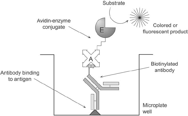
called the labeled avidin–biotin (LAB) system ( Figure 23.1 ), a biotinylated antibody is allowed
to incubate and bind with its target antigen. Next, a (strept)avidin–enzyme conjugate is intro-
duced and allowed to interact with the available biotin sites on the bound antibody. Just as in
Figure 23.1 The basic design of the LAB assay system.

other enzyme-linked immunosorbent assay (ELISA) tests, substrate development then provides
the chemical detectability necessary to quantify the antigen (Guesdon et al. , 1979).
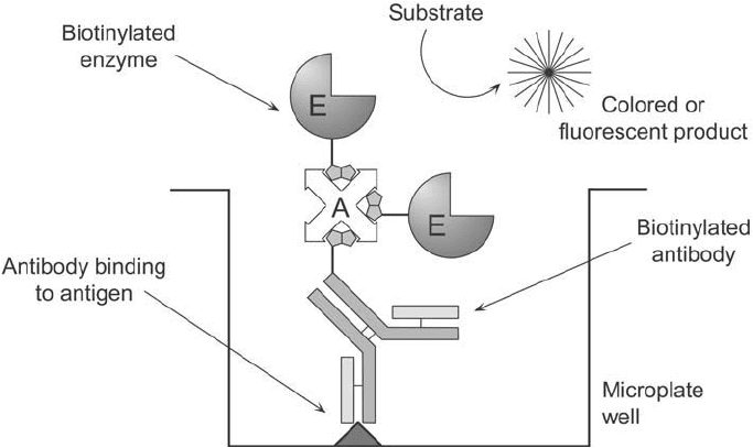
In a slightly more complex design, the bridged avidin–biotin (BRAB) system uses (strept)
avidin’s multiple biotin binding sites to create an assay of potentially higher sensitivity than
that of the LAB assay. Again the biotinylated antibody is allowed to bind to its target, but
in the next step an unmodifi ed (strept)avidin is introduced to bind with the biotin binding
sites on the antibody. Finally, a biotinylated enzyme is added to provide a detection vehicle
(Figure 23.2 ). Since the bound (strept)avidin still has additional biotin binding sites avail-
able, the potential exists for more than one biotinylated enzyme to interact with each bound
(strept)avidin. In some cases, sensitivity can be increased over that of the LAB technique by
using this bridging ability of (strept)avidin.
A modifi cation on this theme can be used to produce one of the most sensitive enzyme-
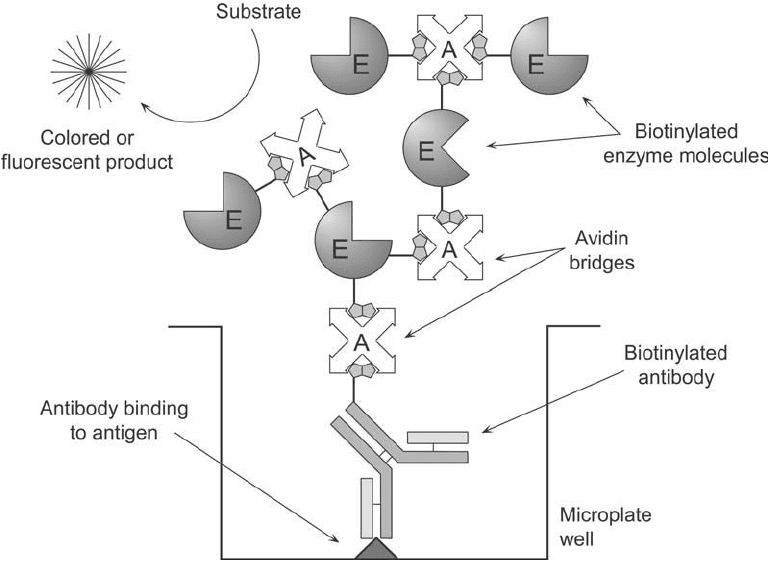
linked assay systems known. The ABC system (for avidin–biotin complex) increases the
detectability of antigen beyond that possible with either the LAB or BRAB designs by forming
a polymer of biotinylated enzyme and (strept)avidin before its addition to an antigen-bound,
biotinylated antibody (Bayer et al., 1988). When (strept)avidin and a biotinylated enzyme are
mixed together in solution in the proper proportion, the multiple binding sites on (strept)avidin
create a linking matrix to form a high-molecular-weight complex. If the biotinylated enzyme is
not in large enough excess to block all the binding sites on (strept)avidin, then additional sites
will still be available on this complex to bind a biotinylated antibody which is bound to its
complementary antigen. The large complex provides multiple enzyme molecules to enhance the
sensitivity of detecting antigen ( Figure 23.3 ). Thus, the ABC procedure is currently among the
highest-sensitivity methods available for immunoassay work.
Similar techniques can be used to devise (strept)avidin–biotin assay systems for detection
of nucleic acid hybridization. DNA probes labeled with biotin can be detected after they bind
Figure 23.2 The basic design of the BRAB assay system.
2. Use of (Strept)avidin–Biotin Interactions in Assay Systems 903

904 23. Avidin–Biotin Systems
their complementary DNA target through the use of (strept)avidin-labeled complexes (Bugawan
et al., 1990; Lloyd et al., 1990). Direct detection of hybridized probes can be accomplished,
similar to the LAB method, by incubating with an (strept)avidin–enzyme conjugate followed by
substrate development. BRAB-like and ABC-like assays also can be utilized to enhance further
a DNA probe signal (Chapter 27, Section 2.3).
Non-enzyme assay systems can be designed with the (strept)avidin–biotin interaction, as well.
Fluorescently labeled (strept)avidin molecules can be used to detect a biotinylated molecule after
it has bound its target. In fact, a single preparation of a fl uorescent (strept)avidin derivative can
be used as a universal detection reagent for any biotinylated targeting molecule. The main appli-
cation of this technique is in cytochemical staining wherein the fl uorescence signal is used to
localize antigen or receptor molecules in cells and tissue sections. In addition, detection of ana-
lytes on arrays commonly is done using fl uorescently labeled (strept)avidin conjugates to bind to
biotinylated primary antibodies interacting with specifi c targets on the array surface.
Other tags or probes can be coupled to (strept)avidin and used in a similar fashion. For
instance, radiolabeled (strept)avidin can be employed as a universal detection reagent in radio-
immunoassay designs (Wojchowski and Sytkowski, 1986) (Chapter 12). (Strept)avidin labeled
with
125
I can be used to localize biotinylated monoclonal antibodies directed against tumor
cells in vivo for imaging purposes (Paganelli et al., 1988). Chemical tags such as in hydrazide-
(strept)avidin derivatives can be made to site-direct (strept)avidin ’s interaction toward oxi-
dized carbohydrate residues for specifi c detection of glycoconjugates (Section 5, this chapter).
Figure 23.3 The assay design of the ABC system.
Colloidal gold-labeled (strept)avidin can be used as highly sensitive detection reagents for
microscopy techniques (Cubie and Norval, 1989) (Chapter 24). Finally, cytotoxic substances
coupled to (strept)avidin can be used to direct cell-killing activity toward a tumor-cell-bound,
biotinylated monoclonal antibody (or other targeting molecule) for cancer therapy (Hashimoto
et al ., 1984) (Chapter 21).
Universal detection reagents also can be constructed through biotinylation techniques.
Modifi cation of immunoglobulin binding proteins with biotin tags, for instance, creates a reagent
useful for the general assay of antibody molecules. In this sense, biotinylated protein A or bioti-
nylated protein G can be used to detect the binding of any primary IgG to its antigen target (pro-
vided there is no other antibody molecules presence to cause nonspecifi c binding of the protein
A component). Subsequent addition of a labeled (strept)avidin molecule binds to the biotinylated
protein A, completing the formation of a detection complex (Jagannath and Sehgal, 1989).
To develop assay systems using the (strept)avidin–biotin interaction, it is fi rst necessary to
produce the associated (strept)avidin conjugates and/or biotinylated components. When the
LAB technique is employed, the (strept)avidin conjugate is made using crosslinking agents, not
biotinylation reagents, in order to maintain the binding capacity of the (strept)avidin tetramer
toward other biotinylated molecules. In the BRAB assay system, (strept)avidin is left unconju-
gated and acts merely as the multivalent bridging molecule, while both the targeting molecule
and the detection molecule are biotinylated. The components for the ABC assay are identical to
the BRAB system.
The following sections discuss the main techniques used to make (strept)avidin conjugates
and various biotinylated components. Chapter 11 and Chapter 18, Section 3 should be con-
sulted for a complete overview of biotinylation reagents.
3. Preparation of (Strept)avidin Conjugates
Conjugates of (strept)avidin with other protein molecules must be prepared to design systems
using the LAB assay technique. Suitable protein molecules attached to (strept)avidin either
possess indigenous detectability, such as in the case of ferritin or phycobiliproteins, or possess
catalytic activity (enzymatic) that can be utilized to produce a detectable substrate product.
The majority of conjugation procedures for making (strept)avidin–protein conjugates use the
amines, sulfhydryls, or carbohydrates on each protein as functional groups for crosslinking.
Perhaps the most common conjugates of (strept)avidin involve attaching enzyme molecules
for use in ELISA systems. As in the case of antibody–enzyme conjugation schemes (Chapter 20),
by far the most commonly used enzymes for this purpose are HRP and alkaline phosphatase.
Other enzymes such as -galactosidase and glucose oxidase are used less often, especially with
regard to assay tests for clinically important analytes (Chapter 26).
Other proteins commonly crosslinked to (strept)avidin are chromogenic or fl uorescent mole-
cules, such as ferritin or phycobiliproteins (Chapter 9, Section 7). These conjugates can be used in
microscopy techniques to stain and localize certain antigens or receptors in cells or tissue sections.
The following sections discuss three main methods for preparing these types of
(strept)avidin–protein conjugates. They involve using an N-hydroxysuccinimide (NHS) ester–
maleimide heterobifunctional crosslinker, making use of the carbohydrate on glycoproteins for
reductive amination coupling, and employing the old technique of homobifunctional crosslink-
ing with glutaraldehyde.
3. Preparation of (Strept)avidin Conjugates 905
906 23. Avidin–Biotin Systems
3.1. NHS Ester–Maleimide-Mediated Conjugation Protocols
Heterobifunctional crosslinking agents can be used to control the degree of protein conjuga-
tion, thus limiting polymerization and controlling the molar ratio of each component in the fi nal
complex (Chapter 5). Particularly useful heterobifunctionals include the amine- and sulfhydryl-
reactive NHS ester–maleimide crosslinkers discussed in Chapter 5, Section 1. Chief among these
is succinimidyl-4-( N-maleimidomethyl)cyclohexane-1-carboxylate (SMCC) or sulfo-SMCC
(Chapter 5, Section 1.3), which contains a reasonably long spacer and an extremely stable male-
imide group due to the adjacent cyclohexane ring in its cross-bridge.
Conjugations done with SMCC usually involve up to three steps. In the fi rst stage, one of
the proteins is modifi ed at its amine groups via the NHS ester end of the crosslinker to form
amide linkages, which upon modifi cation then create derivatives that terminate in reactive
maleimide groups. If the other protein to be conjugated does not contain sulfhydryl residues
necessary to react with the maleimide-activated protein, it must be modifi ed to contain them
(Chapter 1, Section 4.1). Finally, the two reactive components are mixed together in the proper
ratio to effect the conjugation reaction.
For the preparation of (strept)avidin–enzyme conjugates, either protein may be fi rst modi-
fi ed with SMCC and the other one modifi ed to contain SH groups. Since (strept)avidin does
not possess any free sulfhydryls—and the disulfi des present in (strept)avidin are inaccessible
to easy reduction—it must be modifi ed with either a crosslinker or with a thiolating agent
before conjugation. If the enzyme employed contains free sulfhydryls in its native state, such as
-galactosidase, then it is convenient to activate (strept)avidin with SMCC and simply add the
sulfhydryl-containing protein to it for conjugation. If the enzyme does not contain free sulfhy-
dryls (as is the case with alkaline phosphatase or HRP), then the choice of which component
gets maleimide activated and which gets thiolated is up to the individual.
The following protocol describes the activation of (strept)avidin with sulfo-SMCC and its
subsequent conjugation with an enzyme modifi ed to contain sulfhydryls using N-succinimidyl-
S-acetylthioacetate (SATA) (Chapter 1, Section 4.1). A method for the opposite approach,
wherein the enzyme is activated with SMCC and the (strept)avidin component is thiolated, is
presented immediately after this protocol. This strategy may be the most common approach to
forming these conjugates ( Figure 23.4 ). In addition, since there are enzymes commercially avail-
able that are preactivated with SMCC (Thermo Fisher), their use may be the easiest solution to
forming conjugates.
Protocol for the Conjugation of SMCC-Activated (Strept)avidin with Thiolated Enzyme
Activation of (Strept)avidin with SMCC
1. Dissolve (strept)avidin (Thermo Fisher) in 0.1 M sodium phosphate, 0.15 M NaCl, pH
7.2, at a concentration of 10 mg/ml.
2. Add 1.0 mg of sulfo-SMCC (Thermo Fisher) to each ml of (strept)avidin solution. Mix to
dissolve.
3. React for 30–60 minutes at room temperature. Since maleimide groups are labile in aque-
ous solution, extended reaction times should be avoided.
4. Immediately purify the maleimide-activated (strept)avidin away from excess crosslinker and
reaction by-products by gel fi ltration on a desalting resin. A spin column will facilitate the

most rapid purifi cation. Use 0.1 M sodium phosphate, 0.15 M NaCl, pH 7.2, as the chro-
matography buffer. Pool the fractions containing protein (the fi rst peak eluting from the
column). After elution, adjust the protein concentration to 10 mg/ml for the conjugation
reaction (centrifugal concentrators work well for this step). At this point, the maleimide-
activated (strept)avidin may be frozen and lyophilized to preserve its maleimide activity.
The modifi ed protein is stable for at least 1 year in a freeze-dried state. If kept in solution,
the maleimide-activated (strept)avidin is labile and should be used immediately to conju-
gate with a thiolated enzyme following the procedure described below.
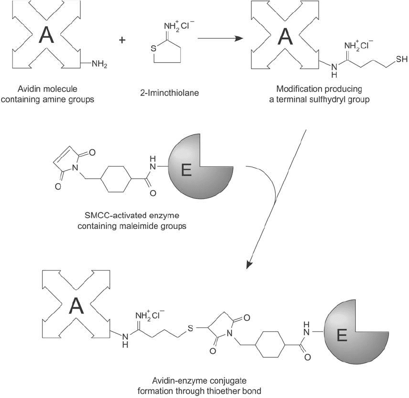
Figure 23.4 Avidin may be modifi ed with 2-iminothiolane to produce sulfhydryl groups. Subsequent reaction
with a maleimide-activated enzyme produces a thioether-linked conjugate.
3. Preparation of (Strept)avidin Conjugates 907
908 23. Avidin–Biotin Systems
Modifi cation of Enzyme with SATA
If -galactosidase is used to conjugate with an SMCC-activated (strept)avidin, then there is
no need to thiolate the enzyme, since it contains sulfhydryls in its native state (Fujiwara et al. ,
1988; Sivakoff and Janes, 1988). For conjugations using HRP, alkaline phosphatase, or glucose
oxidase, however, thiolation is necessary to add the requisite sulfhydryls.
1. Dissolve the enzyme to be modifi ed in 0.1 M sodium phosphate, 0.15 M NaCl, pH 7.2, at
a concentration of 10 mg/ml.
2. Prepare a stock solution of SATA (Thermo Fisher) by dissolving it in DMSO at a concen-
tration of 13 mg/ml. Use a fume hood to handle the organic solvent.
3. Add 25 l of the SATA stock solution to each ml of 10 mg/ml enzyme solution. For differ-
ent concentrations of enzyme in the reaction medium, proportionally adjust the amount
of SATA addition; however do not exceed 10 percent DMSO in the aqueous reaction
medium.
4. React for 30 minutes at room temperature.
5. To purify the SATA-modifi ed enzyme perform a gel fi ltration separation using a desalt-
ing resin or dialyze against 0.1 M sodium phosphate, 0.15 M NaCl, pH 7.2, containing
10 mM EDTA. Purifi cation is not absolutely required, since the following deprotection
step is done using hydroxylamine at a signifi cant molar excess over the initial amount of
SATA added. Whether a purifi cation step is done or not, at this point, the derivative is
stable and may be stored under conditions which favor long-term enzyme activity.
6. Deprotect the acetylated sulfhydryl groups on the SATA-modifi ed enzyme according to
the following protocol:
a. Prepare a 0.5 M hydroxylamine solution in 0.1 M sodium phosphate, pH 7.2, contain-
ing 10 mM EDTA.
b. Add 100 l of the hydroxylamine stock solution to each ml of the SATA-modifi ed
enzyme. Final concentration of hydroxylamine in the enzyme solution is 50 mM.
c. React for 2 hours at room temperature.
d. Purify the thiolated enzyme by gel fi ltration on a desalting resin using 0.1 M sodium phos-
phate, 0.1 M NaCl, pH 7.2, containing 10 mM EDTA as the chromatography buffer. To
obtain effi cient separation between the thiolated protein and excess hydroxylamine and
reaction by-products, the sample size applied to the column should be at a ratio of no
more than 5 percent sample volume to the total column volume. Collect 0.5 ml fractions.
Pool the fractions containing protein by measuring the absorbance of each fraction at
280 nm.
Production of Conjugate
1. Immediately mix the thiolated enzyme with an amount of maleimide-activated (strept)
avidin to obtain the desired molar ratio of enzyme-to-(strept)avidin in the conjugate.
Use of a 4:1 (enzyme:avidin) molar ratio in the conjugation reaction usually results in
high-activity conjugates suitable for use in many enzyme-linked immunoassay procedures
employing the LAB approach.
2. React for 30–60 minutes at 37 °C or 2 hours at room temperature. The conjugation reac-
tion also may be done at 4 °C overnight.
A variation of the above method can be used, wherein the enzyme is fi rst activated with
SMCC and conjugated to a thiolated (strept)avidin molecule. This approach probably is the
most common way of preparing (strept)avidin–enzyme conjugates, and since the preactivated
enzymes are readily available (Thermo Fisher), it also may be the easiest.
Protocol for the Conjugation of SMCC-Activated Enzymes with Thiolated (Strept)avidin
Activation of Enzyme with Sulfo-SMCC
The following protocol describes the activation of HRP with sulfo-SMCC. Other enzymes may
be activated in a similar manner. The activated enzyme possesses maleimide groups that are
relatively unstable in aqueous solution. Therefore, the thiolation reaction should be coordi-
nated with the activation process so that the fi nal conjugation can be done immediately. Note :
If preactivated enzymes are obtained (Thermo Fisher), this step may be eliminated.
1. Dissolve HRP in 0.1 M sodium phosphate, 0.15 M NaCl, pH 7.2, at a concentration of
10 mg/ml.
2. Add 3.3 mg of sulfo-SMCC (Thermo Fisher) to each ml of the HRP solution. Mix to dis-
solve and react for 30 minutes at room temperature. Alternatively, two equal additions
of crosslinker may be done—the second one after 15 minutes of incubation—to obtain
even more effi cient modifi cation.
3. Immediately purify the maleimide-activated HRP away from excess crosslinker and reac-
tion by-products by gel fi ltration on a desalting column. Use 0.1 M sodium phosphate,
0.15 M NaCl, pH 7.2, as the chromatography buffer. HRP can be observed visually as it
fl ows through the column due to the color of its heme ring. Pool the fractions containing
the HRP peak. After elution, adjust the HRP concentration to 10 mg/ml for the conjuga-
tion reaction. At this point, the maleimide-activated enzyme may be frozen and lyophi-
lized to preserve its maleimide activity. The modifi ed enzyme is stable for at least 1 year
in a freeze-dried state. If kept in solution, the maleimide-activated HRP should be used
immediately to conjugate with thiolated (strept)avidin following the protocols outlined
below.
Thiolation of (Strept)avidin
1. Dissolve (strept)avidin in 0.1 M sodium phosphate, 0.15 M NaCl, pH 7.2, at a concen-
tration of 10 mg/ml.
2. Prepare a stock solution of SATA by dissolving it in DMSO at a concentration of 13 mg/ml.
Use a fume hood to handle the organic solvent.
3. Add 25 l of the SATA stock solution to each ml of 10 mg/ml (strept)avidin solution. For
different concentrations of protein in the reaction medium, proportionally adjust the
amount of SATA addition; however do not exceed 10 percent DMSO in the aqueous reac-
tion medium.
4. React for 30 minutes at room temperature.
5. To purify the SATA-modifi ed (strept)avidin use gel fi ltration on a desalting column or
dialyze against 0.1 M sodium phosphate, 0.15 M NaCl, pH 7.2, containing 10 mM
EDTA. At this point, the derivative is stable and may be stored under conditions which
favor long-term (strept)avidin activity.
3. Preparation of (Strept)avidin Conjugates 909
