Hawkes P.W., Spence J.C.H. (Eds.) Science of Microscopy. V.1 and 2
Подождите немного. Документ загружается.

Chapter 4 Analytical Electron Microscopy 295
high-energy X-rays, transmission through the detector is possible.
Measurement of the current is carried out from the back electrode,
which is connected to a fi eld effect transistor (FET) that acts as a fi rst
stage of amplifi cation.
The detector and FET are cooled to low temperature by a Cu rod
connected to a liquid nitrogen reservoir (Figure 4–19) to prevent the
diffusion of Li in the electric fi eld and to reduce the thermal noise of
the e–h generation and the FET electronics. The mechanical design of
the detector assembly is crucial to reduce the transfer of mechanical
vibrations into noise in the spectra. For example, bubbling in the Dewar
arising from fl oating ice crystals and vibrations from the microscope
frame or other components (such as fans) touching the detector can
result in additional noise. The detector should therefore be supported
by the same support mechanism as the frame of the microscope column
and be isolated from the rest of the microscope components.
2.3.2 Detector Windows
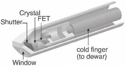
Due to the low operating temperature, the detector can act as a cold
trap attracting contaminants to the detector front. For this reason,
detector windows are used to isolate the detector from the vacuum of
the microscope (Figure 4–21). The window technology has evolved in
recent decades with the initial technology based on Be windows (7–
8 µm in thickness) allowing X-rays for elements down to Na to be
detected due to the absorption of lower energy photons in this mate-
rial. This limitation for the detection of light elements has led to the
development of polymer-based thin windows and some implementa-
tion of systems without windows at all (called windowless detectors).
Various polymer-based window materials are available in ultrathin
windows (UTW). These use proprietary technology combining light
element composites (polymers, diamond, nitrides, etc.) some of which
are strong enough to withstand atmospheric pressure (labeled as atmo-
spheric thin windows—ATW). Due to the combination of various X-ray
absorbing materials the sensitivity of the detector system for light ele-
ments strongly varies at low energy due to the absorption the “soft”
X-rays into the window material. UTWs, for example, allow detection
of elements down to B but with reduced sensitivity as compared to
Figure 4–21. Schematics of the detector front of a TEM with components
inserted into the TEM column detector window. (Courtesy of N. Rowlands
and A. Kirk, Oxford Instruments.)
296 G. Botton
windowless detectors that are actually able to detect X-rays down to
the BeKα line. The latter technology, however, normally requires the
use of ultrahigh vacuum in the microscope to prevent rapid contamina-
tion of the crystal by the microscope environment and the related
absorption of soft X-rays by the ice contamination layer. Whether win-
dowless or UTW, most detectors are equipped with crystal heaters that
gently warm up the crystal surface causing desorption of the ice layer
built up on the crystal surface. Ice contamination is revealed by a
reduction of the intensity of soft X-ray lines as compared to higher-
energy lines such as demonstrated by the NiL/NiK ratios measured as
a function of time. Since the Ni-L line at 0.85 keV is much more strongly
absorbed than the NiK (at about 7 keV) set of lines the ratio is an
effective means to appreciate the contamination effect (L’Espérance
et al., 1990).
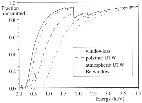
The detector effi ciency (Figure 4–22) accounts for all these effects
and represents the fraction of X-rays that is transmitted through the
window system as compared to the incident intensity as a function of
energy. Windowless detectors still show an important drop in effi -
ciency at low energy due to the presence of the dead layer and metal-
lization in front of the intrinsic active portion of the detector. Even for
UTW and ATW detectors, the effi ciency drops signifi cantly for low-
energy X-rays resulting in diffi culties for the analysis of light elements
in low concentration. Discontinuities in the detector effi ciency are
visible at the energies corresponding to the absorption edges of the
elements contained in the window material (e.g., C, O, B, N, for example),
the detector, and metallization. As discussed above, metallization is
required for the application of a bias on the semiconductor crystal but
it is also necessary on the window material to prevent light (generated
by some samples by cathodoluminescence) from entering the detector
system and resetting the signal amplifi cation system.
Figure 4–22. Detector effi ciency curves for various materials used as detector
windows. (From Williams and Carter, © 1996, with permission from Springer
Science+Business Media.)
Chapter 4 Analytical Electron Microscopy 297
In addition to the fact that high-purity Ge (HPGe) crystals can be pro-
duced and no Li additions are required, Ge-based detectors offer the
advantage that the absorption of X-ray is stronger (1 e–h pair/2.9 eV) and
higher-energy lines can be analyzed as the related high-energy X-rays
are not transmitted through the crystal. The width of the X-ray peaks is
narrower for Ge detectors than Si(Li) detectors leading to better sensitiv-
ity and less overlap in measurements. Some AEM systems are therefore
equipped with a combination of both Si(Li) and HPGe detectors for a
more effi cient analysis of X-rays from a larger range of elements.
2.3.3 Signal Processing
The e–h pair-generated current is detected by the FET as a pulse signal
that is subsequently fed into a main amplifi er system as a voltage. The
sequence of pulses, separated by a time interval, generates a staircase
signal where each step represents a photon arrival and the height is
linked to the energy of the photon. After the integrated signal reaches
a threshold level, the FET must be reset to a base value by the means of
a light pulse generated by a light-emitting diode in a “optoelectronic
feedback” system. This is necessary to avoid saturation of the signals.
Each step rise lasts in the order of 150 ns. Pulses can be amplifi ed and
shaped for subsequent analysis (to determine exact height and thus
photon energy) with analog technology. Analog systems give the user
fl exibility on the process time of the pulse and thus accuracy in the
signal analysis. High processing speeds of pulses (in the order of few
microseconds process time per pulse) result in low energy resolution of
the peaks due to the uncertainty in the pulse height and thus the energy
of the photon. Low processing speed (about 50 µs/pulse) results in more
accurate determination of the pulse height and more accurate determi-
nation of the X-ray energy. During analysis of the pulses, the detector is
effectively not able to process more photons entering the detector result-
ing in analysis dead time, which represents the time the detector is not
processing signals. Due to this limitation, the output count rate is not
linear with input count rate at high X-ray fl uxes. Count rates imposing
detector dead times in the order of 60% are acceptable for modern
systems and exhibit a nearly linear response. Above 60% dead time,
there is a drop in the output rate with an increase in the input rate.
Therefore, with thick samples and thus large photon fl uxes, high pro-
cessing speeds are required to reduce the process time and the resulting
dead time. Recent developments have allowed much faster pulse pro-
cessing with digital technology resulting in higher throughputs of
signals and linear response of the system with respect to the input
count rates. With digital technology, the voltage rise output from the
FET is directly digitized and can subsequently be processed with
numerical pulse processing techniques leading to reduced noise and
better high-count rate responses. Details of the various tests and proce-
dures to determine linearity of the system response and examples are
given in Williams and Carter (1996) and Goldstein et al. (2003).
2.3.4 Peak Shapes
The intrinsic width of an X-ray emission line is in the order of 1–2 eV.
The width of the X-ray peaks as processed by the detector system,

298 G. Botton
however, depends on the generation of e-hole pairs and the noise intro-
duced during the measurement process. A peak in the spectrum rep-
resent the distribution of X-rays “detected” at a given energy with each
incident photon generating a variable number of e–h pairs due to sta-
tistical fl uctuations in the generation process. The standard deviation
of the number of e–h pairs produced is one of the factors affecting the
width of the peaks. The noise of the detector system and detector
collection artifacts, however, also contribute to the broadening. The
broadening due to the statistical generation of e–h pairs is given by
∆EFE
s
= 235. ε
with ε representing the energy for e–h pair creation
(3.8 eV in Si, 2.9 eV in Ge), E the energy of the X-ray peak, and F a
parameter representing the statistical correlation in the e–h pair gen-
eration process known as the Fano factor, which varies between 0 and
1. F = 1 if there is no correlation between the e–h generation events and
F = 0 if the processes are completely deterministic (the process is com-
pletely reproducible and yields the same result time after time). Noise
and artifacts also contribute to the broadening ∆E
N
yielding a total
broadening of a Gaussian peak distribution ∆E = FWHM (full width
at half maximum) of the X-ray peaks as
∆E
2
= (∆E
s
)
2
+ (∆E
N
)
2
= (2.35)
2
εFE + (∆E
N
)
2
(4)
For Si(Li), F = 0.12, and the FWHM of the MnKα line (used as a refer-
ence for resolution because of availability of radioactive standards
producing MnKα peaks) is around 138–140 eV. If the noise contribu-
tions were completely removed, the theoretical resolution is solely
based on the e–h generation process and would be around 110 eV for
the MnKα peak. The energy width decreases at lower energy with
FWHM below 100 eV for light elements. Since the noise term is not
linear with energy and depends on the processing time of the amplifi -
cation system, a full prediction of the peak resolution depends on the
operating conditions and energy. Reference values given for the MnKα
lines are used to compare the electronics and detector performance
and are usually given at optimum process time with typical values
around 140 eV FWHM at the MnKα line. Improved energy resolution
is achieved even for high count rates on digital detectors and with
HPGe detectors due to the lower ε values (FWHM for MnKα is around
120 eV). Noise introduced by vibrations (e.g., mechanical coupling with
environment and/or ice crystals fl oating in the Dewar) can also con-
tribute to peak broadening, hence lowering the spectral resolution, and
should be minimized.
2.3.5 Detector and Signal Processing Artifacts
A summary of detection artifacts is presented here with further details
given in more extended reviews (e.g., Goldstein et al., 2003; Williams
and Carter, 1996). In perfect detector conditions, the peak shape is
expected to be Gaussian but small distortions can arise if the genera-
tion of e–h pairs is perturbed. For example, recombination of e–h pairs
in the dead layer or at lattice defects generated by high-energy incident
electrons accidentally entering the crystal can give rise to a phenome-
non known as incomplete charge collection. This effect gives rise to
Chapter 4 Analytical Electron Microscopy 299
low-energy tails in the peak distribution as not all the e–h pairs are
collected.
Incident X-rays can cause fl uorescence of the SiKα line (or a Ge line).
If SiKα photons are not absorbed within the detector and exit the active
area, incident photons will have lost a fraction of their energy in this
process equivalent to the SiKα ionization energy (1.74 keV). This will
cause an “escape peak” in the spectrum at an energy E
es
= E − E
SiKα
. This
effect is particularly important when small trace elements are investi-
gated since there is potential overlap between E
es
and X-ray lines (for
example Fe overlaps with the escape peak of the CuKα line).
If the count rate is high, there is the possibility that two incident
photons of energy E will be perceived by the pulse counter as one
single photon of energy 2E. This effect, known as a “sum peak,” is visible
when count rates are above the reliable limit of the system (which of
course varies depending on the processing technology). High count
rates, leading to dead times greater than around 60%, are likely to lead
to sum peaks.
Internal fl uorescence peaks can be also detected if the incident photons
generate a Si (or Ge) Kα peak in the dead layer of the detector, which
is subsequently detected in the active area of the detector. This effect
is small but can, once again, be signifi cant for trace analysis.
High-energy incident electrons can also generate spurious signals
and damage the semiconductor crystals. The location of the detector
and the operation of the microscope should be such that these contribu-
tions are minimized (for example, objective apertures must be removed
during acquisition).
2.3.6 Geometry of the EDXS Detector in the AEM
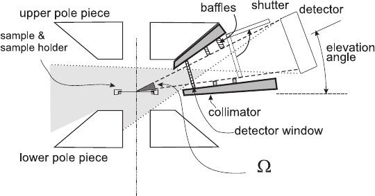
To optimize the solid angle and thus the collection of the X-ray radia-
tion generated by the incident electron, the detector is placed as close
as possible to the sample area (Figure 4–23). The Si(Li) detector active
area A is typically 10 mm
2
with recent systems as large as 30 mm
2
. For
a detector positioned at a distance R with respect to the optic axis (and
the origin point of the emission) the solid angle Ω = A/R
2
(measured
Figure 4–23. Interface of the detector with the microscope sample area.
(Adapted from Otten, 1996.)
300 G. Botton
in steradians) is the key parameter determining how effective the
system collects emitted X-rays. For optimal solid angle, the detector
normal is in direct line of sight to the emission point and not tilted
away from it. Typical solid angles in current AEM are 0.13 sr but with
combinations of large detector areas and effective coupling with the
microscope specimen area, solid angles in the order of 0.3 sr have been
achieved. For Ω = 0.13 sr the fraction of collected X-rays with respect to
the full emission solid angle is only 1%! The detection of X-rays is
therefore a very ineffi cient process considering that the X-ray emission
is fully isotropic.
The elevation angle (also known as the “take-off” angle in the litera-
ture) is an important parameter affecting the quantifi cation of data
through the absorption correction and the quality of the spectra. A
large elevation angle minimizes the path length of X-rays into the
sample (see the quantifi cation section) and also reduces the continuum
background emission, which is forward peaked. High detector eleva-
tion angles, however, are impractical in the TEM due to the fact that
the detector would need to be above or within the objective lens at a
large distance from the sample, resulting in even lower collection effi -
ciency. In addition, backscattered electrons have direct sight to the
detector and can cause signifi cant contributions and potential damage
to the detector. Lower elevation angles (0–20°) allow larger solid angles
and lead to an effective shielding of the backscattered electrons by the
objective lens magnetic fi elds. This shielding is not as effective for high
elevation angles.
The interest in large solid angles and the proximity of the detector
to the sample lead to signifi cant drawbacks in terms of spurious signal
collection. The fi eld of view of the detector is much larger than the
sample area and X-rays generated by backscattered electrons or by
fl uorescence of hard X-rays generated in upper parts of the illumina-
tion area of the microscope easily enter into the detector (see Section
7.2). High-e nergy backscattered electrons can also enter the detector
and generate additional secondary electrons/X-rays while low-energy
electrons would spiral away from the detector due to the high magnetic
fi eld of the objective lens or the presence of a magnetic trap in the
detector system (Figure 4–19). To reduce these effects, detectors are
equipped with collimators that limit the fi eld of view to the smallest
possible area, thus preventing hard X-rays generated in the illumina-
tion system from directly hitting the detector, and contain baffl es that
reduce the effects of potential incident backscattered electrons that
might enter the collimation system.
Many other contributions arising from stray electrons hitting the
microscope components such as apertures, cold traps, the polepieces,
etc. lead to increased noncharacteristic signals resulting in weak detec-
tion limits. As demonstrated in the work of Nicholson et al. (1982),
many of these contributions can be reduced by improving the micro-
scope and detector chamber using coatings to cover the microscope
components with low atomic number materials and by improving the
collimation system. These effects can be minimized in systems using
the precautions discussed in Section 7.2.
Chapter 4 Analytical Electron Microscopy 301
2.4 EELS
2.4.1 Spectrometers
The measurement of energy losses suffered by the incident electrons
as they exit the sample is carried out with energy loss spectrometers.
These devices also make it possible to select electrons with a particular
energy loss (or no loss at all) with the use of energy-selecting slits. The
ability to select electrons with a particular energy loss is called energy-
fi ltered microscopy. The technique also allows the operator to obtain
images and diffraction patterns where parts of the inelastically scat-
tered electrons are fi ltered out so that information deriving from the
elastically scattered electrons only is used.
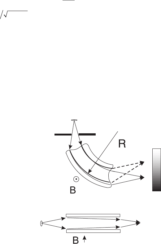
These instruments are based on the use of a magnetic fi eld that
modifi es the trajectory of the electron according to the electron energy.
The radius of curvature R
e
of the electron trajectory is related to their
velocity ν and magnetic fi eld strength B
f
as
R
m
eB
v
e
f
=
γ
0
(5)
where
γ=
−
1
1
22
vc
is the relativistic factor and m
0
is the rest mass
of the electron. Slower electrons will follow trajectories with a smaller
radius of curvature and will be dispersed on a detector plane located
after the spectrometer. The dispersion refers to the separation of elec-
tron energies in space and is typically in the order of 1–2 µm/eV at
100 keV. The exact location of this detector depends on the implementa-
tion of the spectrometer and its coupling with the microscope column
(see below). The most common electron optical component generating
the magnetic fi eld is a magnetic sector (used in various confi gurations,
whether the spectrometer is implemented within the microscope
column or after the viewing chamber of standard TEMs). Current fl ow
in the prism generates the required fi eld B
f
that disperses the electrons
(Figure 4–24). Other approaches to fi ltering have been implemented in
Figure 4–24. Prism spec-
trometer system showing
the bending of electrons
as they travel through the
spectrometer. Dispersion
of electrons according to
their energy is achieved
by the spectrometer in one
direction and focusing is
achieved in the other
direction of travel.
302 G. Botton
prism-mirror spectrometers (see below) and Wien spectrometers,
which combine magnetic and electrostatic fi elds in different confi gura-
tions (Egerton, 1996).
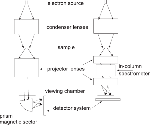
Two types of energy fi ltering spectroscopy approaches should be
distinguished (Figure 4–25). The so-called in-column fi lter/spectrom-
eters are located within the projector lens system/postspecimen area
of the microscope column. These spectrometers generate electron tra-
jectories and dispersion that will result in the transfer of the electrons
into the projector lens system and viewing chamber of the microscope.
The alternative approach is realized with the postcolumn spectrome-
ters/fi lters attached at the bottom of the microscope column. In the case
of in-column fi lters, energy loss spectra and/or energy fi ltered images
(obtained by selection of electrons of a particular value of energy loss
using a slit) are realized. For postcolumn spectrometers, dedicated
imaging lenses are required to generate energy-fi ltered images after
selection of electrons of a particular energy loss. Two implementations
of postcolumn spectrometers therefore exist. For acquisition of spectra
only, the magnetic prism is followed by a series of optical components
dedicated to increase/vary the dispersion at the detector system. For
energy-fi ltered imaging, a more elaborated series of nonround lenses
(i.e., based on multipoles) and a removable energy selecting slit are used
to provide both spectroscopy and imaging capabilities.
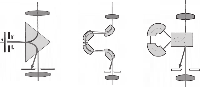
There are various implementations of in-column fi lters. The earliest
commercial applications were based on the electrostatic mirror-prism
system initially proposed by Castaing and Henry (1962) implemented
in the Zeiss microscope (Figure 4–26a). These instruments were devel-
oped on 80 keV microscopes and thus remained very popular for bio-
logical applications, although excellent fundamental electron scattering
experiments were carried out on such instruments (Mayer et al., 1995).
The fi rst portion of the prism is used for an initial dispersion and the
Figure 4–25. Schematic diagrams
of energy-fi ltered electron micro-
scopes. The postcolumn confi gura-
tion (left) is based on the simple
prism attached at the bottom of the
microscope while the in-column
confi guration (right) is achieved by
various components inserted in the
projector lens system.
Chapter 4 Analytical Electron Microscopy 303
electrostatic mirror’s function is to defl ect the electrons back on the
second section of the prism and then the optic axis. The mirror voltage
must be close to the accelerating voltage of the microscope. After the
mirror-prism, electrons are dispersed and continue to travel down the
optic axis of the microscope. Energy-fi ltered images are obtained by
allowing electrons to pass through the slit and the projector lens
system. Energy loss spectra, angular resolved energy-scattering dia-
grams (showing the energy loss distribution as a function of scattering
angle), and fi ltered images and diffraction patterns can be obtained by
careful selection of the operating conditions of the microscope and
crossover points. This can be achieved by selecting, with the micro-
scope postspecimen lenses (objective, intermediate), the object point
entering the spectrometer and the transfer of the crossover points on
the viewing screen. Subsequent implementations of the in-column
fi lters in higher voltage instruments (100, 200 keV microscopes) are
based on OMEGA-type spectrometers that use four magnetic sectors
(Figure 4–26b) generating the dispersion and transfer of electrons back
onto the optic axis of the microscope. Aberrations of the spectrometer
that lead to loss of resolution in spectra and generate nonuniformities
in the energy distribution of electrons in images are reduced by a
combination of design of the magnetic sectors entrance and exit faces,
the symmetry of the confi guration (the fact that the aberration of the
fi rst two sectors is compensated by the aberrations in the opposite
direction of the third and fourth sectors), and the use of a series of
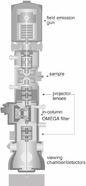
multipoles within the path of the electrons. These fi lters are introduced
within the projector lens system (Figure 4–27). The last implementation
of in-column spectrometers is the Mandolin fi lter recently developed
(Essers and Benners, 2006) (Figure 4–26c). This fi lter generates larger
dispersions (a factor 3 larger than OMEGA) and is optimized for lower
aberrations. This system is ideally suited for energy fi ltering with very
narrow energy windows and large fi elds of view.
abc
Figure 4–26. Various in-column spectrometer confi gurations. (a) Mirror-prism spectrometer, (b)
OMEGA fi lter, and (c) Mandolin fi lter. (Courtesy of P. Schlossmacher, Zeiss SMT.) (See color plate.)
304 G. Botton
Images, diffraction patterns (fi ltered/nonfi ltered), and spectra for
in-column fi lters can be observed directly on the viewing screen of
the microscope and recorded with analog techniques (on negatives),
imaging plates, or a digital camera following conversion of the
incident electrons to photons using scintillator materials (YAG,
phosphor, etc.).
For postcolumn spectrometers/imaging fi lters, the technology is
based on the magnetic sector (Figure 4–24). Aberrations of the prism
can be minimized through design of the spectrometer entrance and
exit faces and the use of multipole correcting elements before the prism.
Only one prism generates the required dispersion to form a spectrum
in the dispersion plane. The early spectrometers were used to generate
energy loss spectra by making use of a serial detection system where
the spectrum is scanned, using an electrostatic fi eld, in front of an
energy slit. The number of electrons (or the current) entering the slit
is subsequently measured with a scintillator detector and photomulti-
plier with pulse counting or current measurement methods. This serial
detection process (one energy recorded at a time) is extremely ineffi -
cient for recording large energy ranges (several seconds/minutes) and
Figure 4–27. Schematic diagram of the
in-column energy-fi ltered microscope
(Zeiss-Libra 200). (Courtesy of P.
Schlossmacher, Zeiss SMT.) (See color
plate.)
