Hammond C. The Basics of Crystallography and Diffraction
Подождите немного. Документ загружается.

274 Electron diffraction and its applications
diffraction, no simple analogy to the structure factor equation for X-ray diffraction. (i.e.
no simple way of relating atomic positions to diffracted beam intensities.)
The scattering described above is the dominant scattering process in the ‘thin foil’
(∼ 1 nm–100 nm) crystalline specimens used in transmission electron microscopy
(TEM). It is described as an elastic scattering process because the scattered electrons
lose no energy and therefore suffer no change in wavelength. It is the physical basis of
the ‘spot’ electron diffraction patterns described in Sections 11.2 and 11.3.
There are in addition a number of inelastic scattering processes in which the incident
electrons lose energy and therefore suffer a change in wavelength. They are of increasing
importance in ‘thicker thin-foil’ specimens in TEM and the ‘bulk’ specimens used in
scanning electron microscopy (SEM). We have already described some such inelastic
scattering processes in Section 9.8; the large energy losses suffered by electrons as they
enter the anode of an X-ray tube give rise to the continuous and characteristic X-radiation
(Fig. 9.24). There are in addition several inelastic scattering processes which involve
much smaller energy losses—thermal diffuse or phonon scattering, plasmon scattering,
single valence electron scattering—the physics of which are described in the books on
electron microscopy listed in Further Reading. The important point for us is that, since
the energy losses are small, the wavelength changes are also small and such inelastically
scattered electrons can then be elastically scattered, giving rise to Kikuchi patterns in the
TEM and electron backscattered diffraction (EBSD) patterns in the SEM. The geometry
of these diffraction patterns and their importance in the precise determination of crystal
orientation are described in Section 11.5.
Finally, we should note, as with light microscopy, the importance of diffraction in
relation to image formation and resolution. This is a very large topic which is introduced
very briefly in Section 11.6.
11.2 The Ewald reflecting sphere construction for
electron diffraction
Since electron wavelengths are very much smaller than the lattice parameters of crystals,
which are normally in the region of 0.3 nm upwards, the diameter of the reflecting sphere
is very large in comparison with the size of the unit cell of the reciprocal lattice. Referring
to the reciprocal lattice sections in Figs 8.6 and 9.l4(a) and (b) for example, for the case
of electron diffraction, the centre of the reflecting sphere would be situated along the
direction of the incident beam at a point far to the left of the origin of the reciprocal
lattice, well beyond the left-hand edge of the paper.
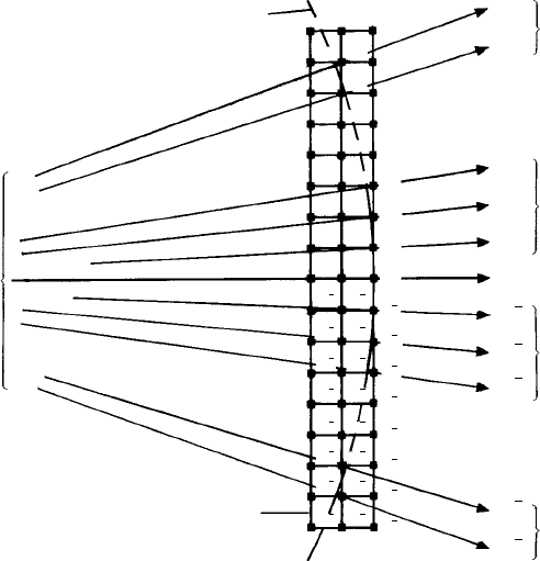
The situation for a simple cubic crystal (as for Fig. 9.l4(a)) is illustrated in Fig. 11.1,
which shows again that part of the reciprocal lattice section perpendicular to the b
∗
reciprocal lattice vector or y -axis (out of the plane of the paper) which lies close to the
surface of the reflecting sphere, only part of which near the origin can be shown since its
radius is so large. The incident electron beam is directed along the a
∗
reciprocal lattice
vector or x-axis and the centre of the reflecting sphere is far to the left of the diagram.
Close to the origin, 000, the curvature of the sphere is so small that it approximates
to a plane perpendicular to the direction of the electron beam and hence passes (in this
section) very close to the 001, 002, 003 and 00
¯
1, 00
¯
2, 00
¯
3, reciprocal lattice points.

11.2 The Ewald reflecting sphere construction 275
208
Surface of reflecting
sphere
Section of the
reciprocal lattice close
to the reflecting sphere
Reflected
beams from
centre of
the
reflecting
sphere
Incident
beam
108
008
107
Reflections
from FOLZ
Reflections
from ZOLZ
Reflections
from ZOLZ
Reflections
from FOLZ
Direct beam
106
003
002
001
000
007
006
005
004
003
002
001
000
209 107
206 106
205 105
204 104
203 103
202 102
201 101
200 100
201
101
001
001
002
003
106
107
002
003
004
005
006
007
008
202 102
203 103
204 104
205 105
206 106
207 107
208 108
Fig. 11.1. The Ewald reflecting sphere construction for a simple cubic crystal in which the electron
beam is incident along the a
∗
reciprocal lattice vector (x-axis). The b
∗
reciprocal lattice vector (y-axis)
is perpendicular to the plane of the page. A section of the sphere (centre beyond the left-hand edge of
the page) and the reflected beams passing at small angles from the centre of the sphere through the
reciprocal lattice points in ZOLZ (giving spots close to the direct beam) and FOLZ (giving spots further
away from the direct beam) are indicated.
Now in order for Bragg’s law to be satisfied exactly, a reciprocal lattice point must
intersect the Ewald reflecting sphere. However, since reciprocal lattice points are broad-
ened into nodes (Section 9.3) they may be extended sufficiently to intersect the sphere
and hence give rise to diffracted beams. This situation also arises in X-ray diffraction
but to a lesser extent than in electron diffraction (a) because the reciprocal lattice points
of the thin foil crystal sections used in electron microscopy are extended perpendicular
to the foil surface and are therefore approximately parallel to the electron beam and
(b) because thin foils are often slightly bent or ‘buckled’ such that the orientation of
the electron beam varies slightly over the surface of the foil—i.e. it is never incident
precisely along one crystallographic direction as indicated in Fig. 11.1.
Hence (referring again to Fig. 11.1), the reciprocal lattice points 001, 002, 003, 00
¯
1,
00
¯
2, 00
¯
3 which lie in a reciprocal lattice row perpendicular to the electron beam direction
all give rise to reflected beams, which are directed from the centre of the sphere (to the
far left of the page) as indicated. Similar considerations apply to the reciprocal lattice
row in the ‘next layer above the page’, i.e. the row through 010 (above 000), 011, 012,

276 Electron diffraction and its applications
013, etc. and also the next layer below, i.e. through 0
¯
10 (below 000), 0
¯
11, 0
¯
12, 0
¯
13,
etc. All of these reciprocal lattice points lie in the (0kl) plane or section of the reciprocal
lattice which is perpendicular to the electron beam direction. In summary, therefore,
the reciprocal lattice points close to the origin, in the (0kl) reciprocal lattice section
perpendicular to the electron beam direction, give rise to reflected beams.
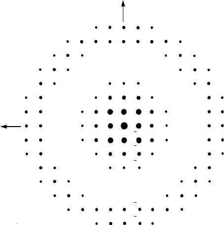
Since the diffraction angles are small (Fig. 11.1), the pattern of spots on the screen per-
pendicular to the beam corresponds to the pattern of reciprocal lattice points. Figure 11.2
shows this pattern of spots from the (0kl) reciprocal lattice section grouped around the
direct beam. For simplicity and clarity, only those reflected beams of type 00l (in the
section perpendicular to the y-axis, Fig. 11.1) are indexed.
These diffraction spots are said to belong to the zero order Laue zone (ZOLZ for
short) since they all arise from reciprocal lattice points in a section through the origin
or, equivalently, they all arise from planes in a zone—the zone axis being parallel (or
nearly parallel) to the electron beam direction.
The pattern of electron diffraction spots (for a particular incident beam direction) in
the zero order Laue zone is identical to that in the zero level X-ray precession photograph
(when the crystal is precessed by the appropriate angle about the same incident beam
direction). This is seen by comparing Fig. 9.18(c) with Fig. 11.2. The far simpler set-
up (a stationary crystal) in electron diffraction ‘works’ because of the ‘flatter’ (large
107
z-axis
y-axis
106
003
002
001
000
001
002
003
106
107
Fig. 11.2. The diffraction pattern on a screen perpendicular to the electron beam direction. The 0kl
ZOLZ spots are grouped around the direct beam (centre spot) and the 1kl FOLZ spots are grouped
roughly in a ring outside. Only the h0l spots in the section perpendicular to the y-axis (Fig. 11.1) are
indexed (compare with Fig. 9.18(c)).
11.3 The analysis of electron diffraction patterns 277
diameter) Ewald reflecting sphere, the extension of the reciprocal lattice nodes and the
slight buckling of the thin foil specimens—all of which serve to provide the requisite
diffraction conditions without the necessity of precessing the specimen over a very much
smaller angular range.
Referring again to Fig. 11.1, as we proceed along the surface of the reflecting sphere
away from the origin, we begin to intersect the next row of reciprocal lattice points
which does not pass through the origin. The reciprocal lattice points in this row which
lie close to the sphere, i.e. 106, 107, 10
¯
6 and 10
¯
7 (and in the corresponding rows above
and below the plane of the diagram, all of which lie in the (1kl) section of the reciprocal
lattice) will also give rise to diffraction spots as indicated. These spots, which lie outside
those from the zero order Laue zone away from the centre of the pattern (Fig. 11.2)
are said to belong to the first order Laue zone (FOLZ for short). Continuing outwards
towards the edge of Fig. 11.1 we come to reciprocal lattice points in the second order
Laue zone (SOLZ for short). These are just the first two in a series of higher order Laue
zones (HOLZ for short).
11.3 The analysis of electron diffraction patterns
The analysis of electron diffraction patterns is essentially a process of identifying the
reciprocal lattice sections which give rise to them. Several computer programs are now
available (seeAppendix 1) which generate reciprocal lattice sections, given as input data
the lattice parameters, type of unit cell (and in some cases the space group and atomic
positions) and the electron beam (zone axis) direction. Several examples showing the
ZOLZ only are given in Fig. 11.3. Care must be exercised in using such data because a
number of different electron diffraction patterns from different types of high-symmetry
crystals have identical symmetry and only differ in scale. For example the
111
fcc
and
[000l]
hcp
patterns shown in Figs 11.3(b) and (d) both consist of hexagonal patterns of
points.
The first step in the analysis of electron diffraction patterns is measurement of the
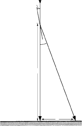
d-spacings. This is an easy matter. Figure 11.4 shows a set of reflecting planes at the
Bragg angle θ (much exaggerated) to the electron beam. The diffracted beam, at angle
θ +θ = 2θ, to the direct beam, falls on the screen at a distance R from the centre spot.
Figure 11.4 does not represent the actual ray paths in the electron microscope, which
are of course determined and controlled by the lens settings: hence L, the camera length,
is not the actual distance between the specimen and screen, but is a ‘projected’ distance
which varies with the lens settings.
From Fig. 11.4, tan 2θ = R/L. Since θ is small, tan 2θ ≈ 2θ (2θ of course being
measured in radians). Similarly for Bragg’s law, sinθ ≈ θ henceλ ≈ 2d
hkl
θ. Eliminating
θ from these equations we have:
d
hkl
= λL/R
where λL is known as the camera constant and which, like L, varies with lens settings
in the microscope. The units of λL are best expressed in terms of the product of R, the
distance measured on the screen (e.g. mm) and d
hkl
(e.g. nm, Å or pm). Although the

278 Electron diffraction and its applications
(004)
(222)
(404)
(422)
(440)
(220)
(242)
(022)
(044)
(224)
(202)
(220)
(242)
(440)
(312)
(222)
(132)
(110)
(220)
(312)
(222)
(132)
(220)
(2020)
(2110)
(1010)
(2200)
(1210)
(1100)
(1120)
(0220)
(0110)
(1210)
(1100)
(2200)
(0110)
(0220)
(1010)
(2110) (1120)
(2020)
(0000)
(110)
(000)
(422)
(404)
(202)
(224)
(044)
(022)
(000)
(113)
(111)
(220)
(002)
(113)
(111)
(220)
(111)
(222)
(113)
(002)
(004)
(111)
(222)
(a) (b)
(c) (d)
(113)
(222)
(000)
Fig. 11.3. Examples of the computer-generated diffraction patterns, drawn to the same scale (λL =
16.1 Å mm). (a) Aluminium (fcc), zone axis =
110
; (b) aluminium (fcc), zone axis =
111
; (c) iron
(bcc), zone axis =
112
; (d) titanium (hcp), zone axis =[0001].
ångström (Å) is not an SI (Système International) unit, it is coherent with the SI system
(1 Å = 10
−10
m) and still remains in widespread use in crystallography (a) because
it is roughly equal to atomic diameters and (b) because X-ray d
hkl
-spacing data in the
Powder Diffraction File (see Section 10.3) is recorded in ångströms.
The accuracy with which the d
hkl
-spacings can be measured is very limited because
of the size or ‘diffuseness’ of the diffraction spots. Moreover the camera constant, λL,
will only be known to a high degree of accuracy if a ‘standard’ specimen is used which
gives a diffraction pattern with spots of accurately known d
hkl
-spacings. Otherwise the
small variations in lens settings from specimen to specimen and from day-to-day will
limit the accuracy of λL values at best to three significant figures. Hence, as pointed out
above, patterns which have identical symmetry but which only differ slightly in scale
with respect to the d
hkl
-spacings of the diffraction spots are not readily distinguished.
The spots may then be indexed by reference to tables of data such as are contained
in the Powder Diffraction File, which show the (Laue) indices, hkl, corresponding to

11.3 The analysis of electron diffraction patterns 279
Trace of
reflecting
planes
Centre
spot
Diffracted
spot
R
u
2u
L
Fig. 11.4. Idealized representation of the direct and diffracted ray paths in the electron microscope
between the specimen and the screen. L is the camera length and R is the distance between the centre
spot (direct beam) and the diffracted spot.
the measured d
hkl
-spacings. In doing so, a further step is required: the indexing must be
such that the addition rule (Section 6.5.2) is satisfied. A given set or family of planes
{hkl} (which are invariably listed in data files simply as hkl—no brackets) will consist
of a number of variants of identical d
hkl
-spacings. For example, in the cubic system (see
Section 5.4), there are six variants of the ‘plane of the form’ {100}, all of which have
identical d
hkl
-spacings. The procedure consists of choosing the appropriate variants such
that the addition rule is satisfied. Consider, for example, Fig. 11.3(a). The four spots of
identical d
hkl
-spacings closest to the centre spot are all reflections from planes of the form
{111}. The particular indices have been chosen such that the indices of the remaining
spots are determined correctly. For example, d
∗
1
¯
11
+ d
∗
¯
111
= d
∗
002
and the zone axis,
[110], is found by cross-multiplication of any pair of these indices (see Sections 5.6.2
and 6.5.7), the ‘choice’ only needs to be made once, and having been made, all the other
spots in the pattern can be indexed consistently by repeated application of the addition
rule; again, with reference to Fig. 11.3(a), d
∗
1
¯
11
+ d
∗
002
= d
∗
1
¯
13
and so on. The ‘choice’
is of course arbitrary: the number of different choices which can be made, all leading
to a self-consistently indexed diffraction pattern, depends on the number of equivalent
variants of the zone axis. In a cubic crystal there are twelve variants of the
110
direction,
hence there are twelve ways in which a pattern like that of Fig. 11.3(a) can be indexed.
The availability of computer-generated diffraction patterns has greatly facilitated
the indexing and verification of electron diffraction pattern analysis. However, it must

280 Electron diffraction and its applications
be said that many ‘useful’ diffraction patterns—i.e. those which may be of potential
scientific interest, which have been obtained under conditions of experimental difficulty
and which may consistofmany spots from several crystals of varying degrees of visibility
on the film—still require for their solution careful observation and measurement and an
awareness of the possible occurrence of additional complicating factors such as double
diffraction (see Appendix 6). Those books on electron diffraction techniques in Further
reading provide a more detailed and thorough coverage of these topics.
11.4 Applications of electron diffraction
11.4.1 Determining orientation relationships between crystals
One of the greatest advantages of electron diffraction is the facility in the electron
microscope of being able to select a particular area of the specimen and to vary the
diameter of the beam to obtain diffraction patterns simultaneously from those phases
of interest. Moreover, the reflections from the phases present may be distinguished by
the technique of dark field imaging. Each electron diffraction pattern may be indexed
according to the procedures described in Section 11.3. The orientation relationship(s)
may then be given in terms of parallelisms between zone axes and planes.
Figure 11.5(a) shows an electron diffraction pattern of α-iron a = 2.866 Å (bcc—
strong reflections) in which are precipitated (or exsolved) particles of Fe
2
TiSi, a =
5.732 Å (fcc—weak reflections) in a single crystallographic orientation. It is obvious,
simply by inspection or looking, that there is a clearly defined orientation relationship
between the iron matrix and theprecipitateand, because of the coincidence of many ofthe
(a)
(b)
211
011
211
111
111
200
200
000
200
200
Fig. 11.5. (a) An electron diffraction pattern of α-iron (bcc, a = 2.866 Å: strong reflections) and
Fe
2
TiSi (fcc, a = 5.732 Å: weak reflections) in a single crystallographic orientation. The camera
constant λL = 31.5 Å mm. (b) The patterns indexed according to one (arbitrary) variant of the ‘cube–
cube’orientation relationship: (200)
Fe
2
TiSi
(200)
α−Fe
; [0
¯
11]
Fe
2
TiSi
[0
¯
11]
α−Fe
. (Photographby courtesy
of Dr. D. H. Jack.)
11.4 Applications of electron diffraction 281
reflections, that the lattice parameters are related in some rational ratio. The d -spacings
of the reflections may be measured (Section 11.3) and, given the lattice parameters and
the conditions for reflection for the cubic F (fcc) and cubic I (bcc) lattices (Table A6.2),
the reflections may be indexed. Figure 11.5(b) shows the patterns indexed according to
one (arbitrary) variant of the orientation relationship. Note that the 200 Fe
2
TiSi spot is
half the distance (and therefore twice the d -spacing) of the 200 α-Fe spotfrom the centre;
these planes are therefore parallel and the lattice parameter of Fe
2
TiSi is twice that of α-
Fe. Similarly, the 022 Fe
2
TiSi reflection (by vector addition of
¯
111 and 111) is coincident
with 011 α-Ti. The zone axes, obtained by cross-multiplication (Section 11.3), are both
[0
¯
11].
1
The orientation relationship may be specified by quoting the parallelisms:
(200)
Fe
2
TiSi
(200)
α-Fe
[0
¯
11]
Fe
2
TiSi
[0
¯
11]
α-Fe
.
This is known as the ‘cube-cube’ orientation relationship because it is equivalent to the
statement that the x, y, z axes of both crystals are mutually parallel.
Other, more complicated, orientation relationships may be determined by the same
simple approach, but to go from the parallelisms between the planes and zone axes
observed for the particular patterns to the establishment of possible parallelisms between
planes not observed in the patterns (i.e. those that are not (nearly) parallel to the electron
beam direction), requires a knowledge of the stereographic projection (see Chapter 12).
11.4.2 Identification of polycrystalline materials
The Powder Diffraction File may be used to identify polycrystalline specimens from
their electron diffraction patterns. However, its use is much more limited owing to the
fact that the d-spacings of the diffraction rings can be measured to a much lower level of
accuracy and only qualitative estimates can be made of their relative intensities. Hence
the use of search procedures, as described in Section l0.3.2, is uncommon. Usually we
have a fair idea as to what the specimen might be and make direct comparisons between
the experimental data and that of ‘possible’ patterns in the File. Exercise 11.4 gives an
example of this in the case of a single-crystal electron diffraction pattern.
Figure 11.6 gives an example of an electron diffraction ‘ring’ pattern from a fine-
grained polycrystalline thin foil specimen of aluminium. It is, in effect, the superposition
of many single-crystal patterns. Notice the variations in intensity and the sequence of
spacings of the rings. Aluminium can immediately be recognized as having the fcc struc-
ture since the rings occur in the characteristic sequence ‘two-together’, ‘one-on-its-own’,
‘two-together’, etc. (see Note 3, Table A6.2). Hence the rings may simply be indexed
‘by inspection’, i.e.
111,200, 220, 311,222, 400, 331,420.
1
The order of cross-multiplication is important; one should read anticlockwise. E.g. for the α-iron diffrac-
tion spots write down (211) as the first line in the memogram and then (011) as the second line. This gives (for
the usual right-handed axial system) the zone axis direction upwards (i.e. anti-parallel to the electron beam
which is much easier to deal with when plotting the data on a stereographic projection (Chapter 12)).

282 Electron diffraction and its applications
Fig. 11.6. An electron diffraction pattern of polycrystalline aluminium, showing varying intensities
of the diffraction rings and the sequence of ring spacings characteristic of the fcc structure.
11.4.3 Identification of quasiperiodic crystals
In Section 4.8 we showed how quasiperiodic crystals or crystalloids could nucleate and
grow without any long range translational symmetry and that their existence was first
recognised from the ten-fold symmetry of their electron diffraction patterns. Figure 11.7
shows the first observed such electron diffraction pattern of a nodule-like phase in a
rapidly quenchedAl-25 wt.% Mn alloy. Such a ten-fold pattern does not however consti-
tute sufficient evidence for quasiperiodicity since multiply-twinned crystals (consisting
of 10 twin-related segments radiating out from a central point rather like the slices of
a cake) can also give rise to such ten-fold patterns. Nor is this a remote possibility and
some alloy systems may exhibit quasiperiodic crystals or multiply-twinned crystals (the
microcrystalline state) depending upon heat treatment. For example the Al
65
Cu
20
Fe
15
alloy gives twinned microcrystals when slowly cooled from the melt which on heating
transform to quasiperiodic crystals.
2
There are two ways in which these quasiperiodic and multiply-twinned states may
be distinguished. First the evidence from microscopy: twin boundaries and the changes
in orientation of lattice fringes at twin boundaries may be observed by high resolution
electron microscopy—which constitutes firm evidence for the microcrystalline phase.
Second, the fine detail of the spots in the electron diffraction patterns: the segments
or ‘slices of cake’ in multiply-twinned crystals are not perfectly related by five-fold
2
Quasicrystals: A Primer by C. Janot, Clarendon Press, Oxford, 1992.

11.5 Kikuchi and electron backscattered diffraction patterns 283
Fig. 11.7. Electron diffraction pattern of the first observed icosahedral particle in an Al-25 wt.% Mn
alloy, April 1982. (Photograph by courtesy of Prof. D. Shechtman.)
symmetry; there is some misalignment which results in spots of a triangular ‘shape’ (i.e.
made up of a group of little sub-spots) rather than the clear and distinct single spots of
Fig. 11.7. Other, more complicated five-fold patterns of spots may also arise. It is not
surprising that the first reported occurrences of quasiperiodic crystals were greeted with
considerable scepticism throughout much of the crystallographic community.
11.5 Kikuchi and electron backscattered diffraction
(EBSD) patterns
11.5.1 Kikuchi patterns in the TEM
We first consider the trajectories, or paths, of the inelastically scattered electrons within
the specimen (Section 11.1). These occur over a range of angles, the scattering being
most intense at small angles to the incident beam and decreasing at larger angles, giving
in effect a ‘pear-shaped’ distribution of intensity as shown schematically in the upper
part of Fig. 11.8. This distribution of intensity around the incident electron beam (the
centre spot) is shown in the lower part of Fig. 11.8. Such a distribution is only observed in
practice in ‘thicker’ specimens (in the range 200 nm upwards) and in which the amount
of inelastic scattering is significant. It is almost entirely absent in the thin foil specimens
from which the electron diffraction patterns shown in Figs 11.5(a), 11.17, 11.18 and
11.19 were obtained.
Now we describe the elastic scattering, or Bragg reflection, of the weakly inelastically
scattered electrons that have suffered negligible changes in wavelength. Consider a
specimen in which a set of hkl planes is at an angle to the incident beam slightly smaller
