Hammond C. The Basics of Crystallography and Diffraction
Подождите немного. Документ загружается.


254 X-ray diffraction of polycrystalline materials
SiO
2
46-1045
dÅ
4.2550 16 100 1.1530 1 311
3.3435 100 101 1.1407 <1 204
2.4569 9 110 1.1145 <1 303
2.2815 8 102 1.0816 2 312
2.2361 4 111 1.0638 <1 400
2.1277 6 200 1.0477 1 105
1.9799 4 201 1.0438 <1 401
1.8180 13 112 1.0346 1 214
1.8017 <1 003 1.0149 1 233
1.6717 4 202 0.9896 <1 115
1.6592 2 103 0.9872 <1 313
1.6083 <1 210 0.9783 <1 304
1.5415 9 211 0.9762 <1 320
1.4529 2 113 0.9608 <1 321
1.4184 <1 300 0.9285 <1 410
1.3821 6 212 0.9182 <1 322
1.3750 7 203 0.9161 2 403
1.3719 5 301 0.9152 2 411
1.2879 2 104 0.9089 <1 224
1.2559 3 302 0.9009 <1 006
1.2283 1 220 0.8972 <1 215
1.1998 2 213 0.8889 1 314
1.1978 <1 221 0.8814 <1 106
1.1840 2 114 0.8782 <1 412
1.1802 2 310 0.8598 <1 305
Int hkl hkldÅ Int
Silicon Oxide
Quartz, syn
Rad. CuK
1
Sys. Hexagonal
a 4.91344(4) b
Ref. Ibid.
D
x
2.65
Ref. Swanson, Fuyat, Natl. Bur. Stand. (U.S.), Circ. 539,324(1954)
Color White
See following card.
Integrated intensities. Pattern taken at 23(1) C.Low tempertature
quartz. 2 determination based on profile fit method. O
2
Si type.
Quartz group. Silicon used as internal standard. PSC: hP9. TO
replace 33-1161. Structure reference: Z.Kristallogr., 198 177(1992)
n 1.544 1.553 Sign + 2V
D
m
2.66 SS/FOM F
30
= 539(.002,31)
Z 3 mp
c 5.40524(8) A C 1.1001
S.G. P3
2
21 (154)
Cut off
Ref. Kern, A., Eysel, W., Mineralogisch-Petrograph.Inst., Univ.
Heidelberg, Germany, ICDD Grant-in-Aid, (1993)
Int. Diffractometer I/I
cor.
3.41
1.540598 Filter Ge Mono.d-sp Diff.
Fig. 10.8. The Powder Diffraction File Card No. 46-1045, for quartz. The left-hand side gives crys-
tallographic optical and physical information and the source of the data. The right-hand side lists the
d-spacings, in descending order, with the strongest line assigned an intensity of 100. Formerly (up to set
24) the cards included along the top left edge the d-spacings of the three strongest lines and the largest
d-spacing recorded in the pattern. (International Centre for Diffraction Data.)
than observed (a ‘c’ mark). Figure 10.8 shows an example of a Powder Diffraction File
card, that for quartz, set number 46, card number 1045.
A comparison of an unknown phase with so many others is potentially a daunting
task! It is greatly facilitated by a search procedure based on that first devised by J. D.
Hanawalt in 1936 and now refined and speeded up using computer search procedures.
The phases aregroupedtogether (into what is known asHanawaltgroups) according to
the d-spacing of their strongest reflection. There are forty Hanawalt groups covering the
whole range of d -spacings. For example, the strongest line of quartz (relative intensity
100) is that at 3.34 Å (Figs 10.4 and 10.8); quartz therefore lies in the Hanawalt group
with d -spacings in the range from 3.39 to 3.32 Å (Fig. 10.9—first column). Within
each Hanawalt group the patterns are listed in the second column in order of decreasing
d-spacings of the second strongest line which for quartz is that at 4.26 Å; on reading
down the list of patterns in the second column of the Hanawalt group a potential ‘match’
can be made with the second strongest line and then (from the number of possibilities
available) final confirmation made with the d-spacings and intensities of the third, fourth
etc. strongest lines in thesubsequent columns. Figure10.9 is fromtheHanawalt Inorganic
Search Manual showing some entries in the Hanawalt group 3.39–3.32 Å which includes
that for quartz, File No. 46-1045.
In practice the procedure is by no means as straightforward as this. First, many
minerals and metallic alloys give very nearly the same diffraction pattern because of the
existence of solid solution ranges, which result in very small shifts in lattice parameter.

10.3 Applications of X-ray diffraction techniques 255
3.40
x
4.29
9
4.09
9
6.63
7
4.82
6
3.31
6
2.67
6
2.33
6
o*144 CIF
2
OBF
4
26- 235
3.34
8
i
4.29
x
10.5
6
2.64
6
2.71
5
5.27
4
5.25
4
3.79
3
oI32 Na
2
Ta
3
O
6
F
5
25- 871
3.32
x
4.29
6
2.99
6
2.87
6
2.52
6
4.70
5
2.38
5
2.30
5
Cs
2
Al(NO
3
)
5
28- 274
3.31
x
4.29
x
3.68
x
2.91
x
2.34
x
2.06
x
1.99
x
1.93
8
Pu(OH)CO
3
•xH
2
O 33- 982
3.37
x
4.28
3
1.84
2
1.55
1
2.47
1
2.31
1
1.39
1
2.14
1
hP18 AlPO
4
10- 423Berlinite, syn
3.35
8
4.28
x
3.71
6
2.85
6
2.17
6
4.07
4
3.03
4
2.73
4
K
2
N
2
V
6
O
21
20-1379
3.34
x
4.28
3
4.27
3
3.38
3
2.77
3
2.33
2
2.79
2
2.32
2
mP28 CsLiSeO
4
29- 411 2.60
3.33
x
4.28
x
3.24
x
8.58
8
2.91
8
2.37
8
1.87
8
4.95
6
hP20.70 Cs
0.35
V
3
O
7
30- 385
3.36
x
4.27
3
1.82
2
2.28
2
1.54
2
2.46
1
2.13
1
1.37
1
hP9 SiS
2
47-1376
3.34
x
4.27
4
3.19
2
2.70
2
7.28
2
4.91
2
1.82
2
3.13
1
mP100 CaAl
2
Si
2
O
8
•4H
2
O 20- 452Gismondine
3.30
x
4.27
7
3.71
7
3.45
7
3.92
5
3.34
5
3.27
5
2.96
5
aP26 KFeSi
3
O
8
16- 153
3.38
x
4.26
x
4.16
x
1.68
x
6.70
8
2.97
8
2.86
8
2.56
8
Cs
2
VCl
5
•4H
2
O 20- 301
3.37
x
4.26
8
4.18
8
2.98
8
8.56
6
6.76
6
3.49
6
3.09
6
mC40 Cs
2
VCl
5
•4H
2
O 34- 322
3.36
x
4.26
x
4.21
x
2.86
x
5.90
8
5.55
8
3.26
8
2.94
8
KAlH(P
3
O
10
) 38- 55
3.36
9
4.26
x
2.98
x
5.95
8
5.55
8
4.02
6
3.26
6
2.87
6
KCrHP
3
O
10
41- 442
3.34
x
4.26
7
4.33
5
3.40
5
3.38
4
2.05
3
4.89
2
2.02
2
oP36 KNaSiF
6
43-1313
3.34
x
4.26
9
3.66
9
2.61
9
2.48
9
1.80
9
3.97
8
3.05
8
hP168 3PbCO
3
•2Pb(OH)
5
•H
5
O 9- 356
3.34
x
4.26
7
2.13
6
7.40
5
3.49
5
2.58
5
2.24
5
2.21
5
hP70 Ca
3
Ge(SO
4
)
2
(OH)
6
•3H
2
O 19- 225Schaurteite
3.34
x
4.26
8
2.13
8
7.40
6
2.57
6
2.03
6
3.49
4
2.24
4
hP70 Ca
3
Mn
+4
(SO
4
)
2
(OH)
6
•3H
2
O 20- 226Despujolsite
3.33
x
4.26
4
3.27
4
4.31
3
2.89
3
2.75
3
3.88
2
3.19
2
K
2
Ca(VO
3
)
4
37- 180
3.30
x
4.26
8
4.24
7
3.66
7
2.89
6
2.30
6
2.63
5
2.32
5
Sm
2
O(CO
3
)
2
•xH
2
O 28- 994
3.30
8
4.26
x
2.89
x
4.45
5
3.14
5
2.56
4
3.89
2
2.66
2
mP16 CsO
3
45- 947cesium ozonide
3.34
x
4.26
2
1.82
1
2.46
1
1.54
1
2.28
1
1.38
1
2.13
1
hP9 SiO
2
46-1045
3.41
Quartz, syn
3.31
9
4.25
x
4.09
9
3.72
7
2.98
7
2.20
7
1.93
7
5.47
6
o*66 ClPtOF
8
26- 415
3.30
9
4.25
x
4.09
9
3.67
9
7.42
6
5.47
6
2.96
6
1.91
6
o*66 (ClF
2
O)AsF
6
26- 117
3.40
x
4.24
6
3.00
4
5.72
3
2.65
3
2.85
2
2.37
2
0.00
1
tI24 KAuF
4
27- 395
3.37
x
4.24
3
2.32
2
2.12
2
2.45
1
1.84
1
1.83
1
1.54
1
hP18 GaPO
4
8- 497
3.36
x
4.24
x
2.86
x
5.90
8
5.55
8
3.26
8
2.94
8
6.60
6
KAlH(P
3
O
10
) 38- 55
3.33
x
4.24
x
3.39
4
2.64
3
3.25
3
2.60
2
4.38
2
3.44
2
mP44 SrUMo
4
O
16
38-1360
3.41
8
4.23
x
2.87
x
2.49
x
2.06
8
1.99
8
1.95
8
1.93
8
mC12 -WP
2
35-1467
3.39
9
4.23
x
5.06
9
3.45
9
3.18
9
3.05
9
5.25
7
3.76
7
mC* RbAl
2
(H
2
P
3
O
10
)(P
4
O
12
) 37- 261
3.39
8
4.23
x
2.99
9
2.89
8
2.77
7
5.93
6
3.24
5
4.75
4
RbGaHP
3
O
10
34-1347
3.38
x
4.23
9
3.83
4
2.50
3
3.10
2
1.71
2
3.31
2
2.21
2
mP62 K
2
Mn(SeO
4
)
2
•6H
2
O 53-1151
3.37
x
4.23
x
3.46
x
3.00
x
2.13
x
6.00
8
2.69
6
2.45
6
CsSnFBrI 30- 369
3.36
x
4.23
x
2.37
x
2.29
x
2.15
x
1.90
x
9.50
8
3.95
5
Al
8
La
2
S
15
41- 734
3.36
x
4.23
5
1.64
5
2.72
4
2.44
3
2.22
3
1.93
3
3.14
2
Mo
3
O
8
•xH
2
O 21- 574Ilsemannite
3.35
8
4.23
x
3.12
x
3.43
8
3.84
6
3.75
6
3.39
6
3.37
6
-Cs
2
Cu(SO
4
)
2
28- 288
3.32
x
4.23
x
1.88
7
3.45
7
2.76
3
2.25
3
2.12
3
2.64
3
oP36 -NaSrGaF
2
44-1352
3.40
x
4.22
8
3.20
7
3.14
7
5.81
6
3.62
5
3.03
4
2.64
4
Na
3
VO
2
(SO
4
)
2
41- 175
3.41
8
4.21
x
3.11
x
2.75
8
7.58
7
5.60
7
4.46
7
3.31
7
Na
2
SbF
3
SO
4
28-1038
3.41
9
4.21
x
2.99
9
3.24
8
2.91
8
2.77
7
5.95
6
4.77
6
RbCrHP
3
O
10
41- 443
3.40
8
4.21
x
3.30
8
3.22
8
6.97
5
4.76
5
3.11
5
2.58
5
a*15.54 UO
3
•0.393H
2
O 41- 443
3.39
x
4.21
9
3.92
9
2.78
8
1.89
8
3.78
5
2.96
5
2.08
5
Pb
2
Sb
2
S
5
36- 94
3.34
x
4.21
4
3.15
4
2.11
4
2.71
3
2.05
2
4.73
2
2.56
1
Cs
3
Zr
1.5
(PO
4
)
3
52-1181
3.33
8
4.21
x
3.82
7
2.96
7
2.47
7
2.34
7
2.01
7
1.89
7
CsXeOF
5
35-1267
3.30
8
4.21
x
3.40
8
3.22
8
6.97
5
4.76
5
3.11
5
2.58
5
a*15.54 UO
3
•0.393H
2
O 43- 346
3.41
9
4.20
x
3.65
7
2.77
7
2.71
7
2.96
5
2.11
5
4.00
4
Tl
2
Si
2
S
5
38- 914
3.39
x
4.29
9
5.88
8
3.12
8
2.98
8
2.30
8
1.96
8
1.78
8
hP15 Li
2.33
Fe
0.67
S
2
36-1089
3.39
x
4.29
4
2.47
1
2.31
1
2.23
1
2.14
1
1.99
1
1.83
1
h** BPO
4
11- 237
3.36
9
O
QM Strongest Reflections PSC Chemical Formula Mineral Name;Common Name
3.39 – 3.32 (±.02)
PDF#
I/I
c
i
i
i
i
i
i
i
i
i
i
O
O
O
O
O
O
O
O
O
O
O
O
O
O
O
O
4.29
x
2.85
9
5.60
5
4.02
5
2.58
5
1.98
5
1.90
5
V
0.87
Cr
0.13
O
2.17
35- 332
Fig. 10.9. Part of the Hanawalt Search Manual for Inorganic Phases, showing some entries in the
Hanawalt Group 3.39–3.32 Å. The intensities of the reflections are indicated by subscripts x = 100,
9 = 90, 8 = 80, and so on. Notice that some patterns are entered in this Hanawalt Group according to
their second -strongest or third -strongest lines; e.g. K
2
N
2
V
6
0
21
(20-1379) or RbGaHP
3
O
10
(34-1347)
respectively. This is because they have intensities over 75% of that of the strongest line and may
experimentally be identified as the strongest lines. (International Centre for Diffraction Data.)
Second, the crystals in the powder specimen may not be randomly orientated—the
specimen may be textured or may show preferred orientation, which implies that the
relative intensities of the reflections may be altered as compared with those in the File.
Third, the unknown specimen may consist of more than one phase—and if they are in
roughly equal proportions the strongest lines may be ‘mixed up’.
These problems are partly (but not wholly) circumvented by a series of ‘entry rules’,
the details of which are given in the Search Manual. First, those patterns in which the
d-spacing of the strongest reflection lies close to one of the boundary values of the
Hanawalt group are entered in both groups—the one above and the one below. Second,
those patterns in which the intensity of the second strongest reflection is greater than 75%
of the intensity of the strongest (and in which there is therefore a strong possibility of
the mis-identification of the strongest reflection) are entered twice; the second strongest
reflection determining the Hanawalt group and the position in the group listed (as before)
in accordance with the (decreasing) d-spacing of the second strongest reflection. Similar

256 X-ray diffraction of polycrystalline materials
rules apply to the third and fourth strongest reflections such that a pattern may be listed
several times. Examples of such permutations are shown in Fig. 10.9.
10.3.3 Measurement of crystal (grain) size
This is based on the determination of β, the line broadening in the Scherrer equation
(Section 9.3) and an estimate of K, the ‘shape’ correction factor (which does not differ
significantly from unity). However, this is not a straightforward business because the
observed broadening, β
obs
, also includes ‘instrumental’factors (see Section 9.3) such as
detector slit width, area of specimen irradiated, possible presence of a Kα
2
component
in the X-ray beam, etc. The contributions from these factors have to be ‘subtracted’ from
the observed peak breadth (or, strictly speaking, they have to be deconvoluted from
the peak). The usual procedure is to measure the instrumental broadening, β
inst
,ina
large-grained material in which grain size broadening is assumed to be negligible, and
to determine β either by a simple subtraction, due to Scherrer, i.e. β = β
obs
– β
inst
,or
by a subtraction of squares, due to Warren, i.e. β
2
= β
2
obs
− β
2
inst
. In practice, attempts
to measure ‘absolute’ grain sizes by X-ray diffraction are fraught with difficulty because
of the presence of imperfections (particularly sub-grain boundaries), lattice strains, etc.
which contribute to the broadening of the X-ray peaks.
10.3.4 Measurement of internal elastic strains
The elastic strains to be measured can be distinguished into those occurring on a ‘macro’
scale and those occurring on a ‘micro’ scale. Macroscale refers to the situation where
the whole material is subject to some directional residual tension or compression; the
resultant strains, which are manifested in terms of increases (tension) or decreases (com-
pression) in the d
hkl
-spacings are therefore also directional and the measured values vary
with the orientation of the reflecting planes—those parallel to the stress axis for example
suffering no change.
In order to measure such strains (and to determine the stress axis) in the diffracto-
meter, the specimen needs to be rotated from the symmetrical (Bragg–Brentano)
geometry as described above. In practice, measurements of peak shift are made for
various settings of the specimen and from which the direction and magnitude of the
stress can be extrapolated.
Microscale refers to the situation in which the directions (and magnitudes) of the
internal strains vary from crystal to crystal. The subsequent elastic strains give rise, not
to a shift, but to a broadening of the diffraction peaks from which an estimate of the
micro-stress can be made. The relevant equation is that derived for the limit of resolution
(Section 10.3.1), in which e, the elastic strain, is substituted for δd/d , i.e.
=−cot θδθ.
Hence the broadening, β, expressed as the half-height peak width 2δθ, is:
β =−
2
cot θ
=−2 tan θ.

10.4 Preferred orientation and its measurement 257
Notice that this equation is very similar to the Scherrer equation for broadening
arising from crystal size; the important difference being that in this case β varies as 1/cot
θ or tan θ and in the Scherrer equation β varies as 1/cos θ or sec θ . As in the case of
grain size broadening (Section 10.3.3), the instrumental broadening, β
inst
, measured for
a stress-free material, must be subtracted from β
obs
, the observed broadening for the
micro-strained material. Having measured the strain , the stress may be determined by
substituting the appropriate value of the Young modulus, E.
Finally, the elastic strain ε may be expressed in terms of the extension of reciprocal
lattice points into ‘nodes’ just as we did for a crystal of finite thickness t (Section 9.3.1).
Since
ε =
δ
(
d
hkl
)
(
d
hkl
)
=
δ
1
d
∗
hkl
1
d
∗
hkl
=−
δ
d
∗
hkl
d
∗
hkl
remembering the differential d
1
x
=−
dx
x
2
then the extension is
δ
d
∗
hkl
=−εd
∗
hkl
=−
ε
d
hkl
i.e. unlike the extension arising from crystal size in which every reciprocal lattice point
is extended the same amount, 1/t, in this case the reciprocal lattice points are more
extended the further we go from the origin in reciprocal space.
10.4 Preferred orientation (texture, fabric) and its
measurement
There are essentially two methods of recording preferred orientation: film methods which
make use of flat-plate film cameras, and counter methods which make use of a special
design of diffractometer called a texture goniometer. Film methods have the advantage
that the cones of reflected beams from planes of different d
hkl
spacings can be recorded
‘all at once’ so that you can see at a glance, from the varying intensities around the
diffraction rings, whether the crystals are in random orientation or not. In practice the
film may be set ‘behind’ the specimen as in Fig. 10.5 such that the cones of high-angle
reflections are recorded (the back reflection X-ray method; or the film may be set ‘in
front of the specimen such that the cones of low-angle reflections are recorded) the
transmission X-ray method. In the latter case the specimen must be thin enough for the
reflected beams to pass through with sufficient intensity to be recorded—which sets a
practical limitation in the case of ‘bulk’specimens which cannot be cut into thin sections.
However, film methods have the disadvantage that, except for the case of fibres, the
data cannot easily be represented in a quantitative way—the varying intensities around
the diffraction rings depend also on the orientation of the specimen with respect to the
incident beam which means that several exposures need to be made and the results
collated and the intensities analysed in terms of the ‘blackening’ of the films—a tedious
business. In the case of fibres only one orientation and hence only one photograph is
258 X-ray diffraction of polycrystalline materials
needed—the fibre axis normally being set vertical and perpendicular to the direction of
the (horizontal) incident beam.
In the texture goniometer the detector is set at a fixed 2θ angle to record the diffracted
intensity from just one set of d
hkl
planes. All the movement takes place at the specimen
which can be rotated about two axes as described below such that the detector in effect
scans a wide range of orientations of the selected d
hkl
planes. The detector may then be
re-set at another 2θ angle to record another set of d
hkl
planes and the process repeated.
It is therefore, in contrast to film methods, a sequential procedure but has the enormous
advantage that the output from the detector can be represented in the form of pole figures
or processed numerically to give what is known as an orientation distribution function
(ODF) which provides a complete description of the texture or fabric of the material.
10.4.1 Fibre textures
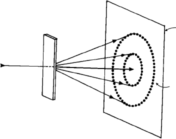
Figure 10.10 shows the simple experimental set-up for the transmission X-ray method
to record the low 2θ angle (highest d
hkl
) reflections. The fibre, or reference direction is
set vertical and perpendicular to the incident X-ray beam. Figure 10.11 is an example
of the application of the technique in the study of polymers. Figure 10.11(a) shows the
diffraction pattern of a thin section of an injection-moulded specimen of polypropene
n(CH
2
–CHCH
3
) which partially crystallizes on cooling from the melt; the diffraction
rings are of uniform intensity which shows that the crystallites are in random orientation.
Figure 10.11(b) is of the same material which has been plastically deformed in tension
to a strain of about 500%. The diffraction rings are no longer continuous but consist
of peaks of intensity which show that the crystallites now lie in a particular orientation
with respect to the tensile axis. The diffraction pattern approaches that of a rotation
or oscillation photograph of a single crystal (Fig. 9.17), the peaks of intensity lying in
layer-lines perpendicular to the axis of rotation/oscillation. Given the structure and unit
cell of polypropene and the {hkl} reflections which occur, we can index the reflections
Incident
beam
Specimen
Diffraction
rings
Film
Fig. 10.10. Transmission X-ray powder method. The diffraction rings consist of reflections from many
crystallites as indicated by the dots. Each ring corresponds to a cone of reflected beams of semi-angle 2θ.

10.4 Preferred orientation and its measurement 259
(a)
(b)
Fig. 10.11. Transmission X-rayphotographs ofpolypropene n(CH
2
–CHCH
3
): (a)as-crystallized from
the melt (crystallites in random orientation); (b) after 500
◦
/a plastic deformation in tension (crystal-
lites orientated with the polymer chains along the (vertical) tensile axis). (Photographs by courtesy of
Dr. J. Way.)
and hence determine which crystallographic direction is orientated along the tensile axis.
Intuitively we may guess that the crystallites are orientated such that the polymer chains
lie in the direction of the tensile axis and the demonstration that this is indeed the case
is given in Exercise 10.4.
10.4.2 Sheet textures
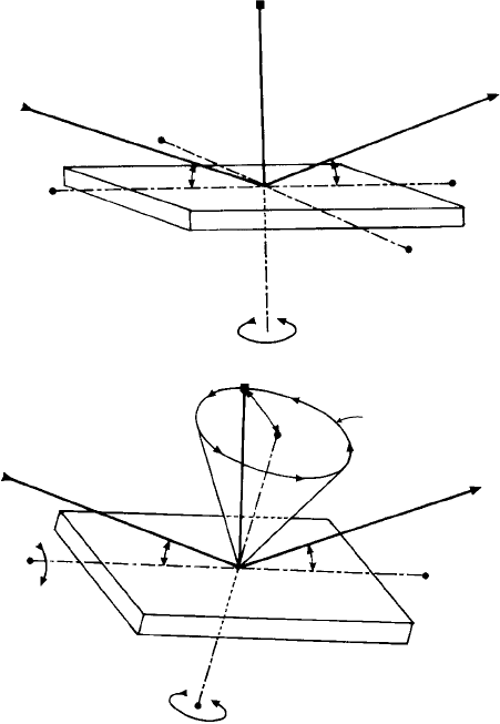
The texture goniometer consists essentially of a cradle in which a flat-surface specimen
(as in a diffractometer) can be rotated about two axes as indicated in Fig. 10.12(a), one of
which, O … O is normal to the specimen surface and the other B … B lies in the specimen
surface and in the plane containing the incident and diffracted beams (i.e. perpendicular
to the direction A…A). In the ‘starting position’ the specimen is set in the symmetrical
or Bragg-Brentano focusing arrangement as shown in Fig. 9.20(a) and all the reflected
intensity received by the detector arises from the crystal planes lying parallel to the
specimen surface. The specimen is then tilted a small angle β (say 5
◦
) about the axis
B … B. The reflected intensity received by the detector now arises from the crystal planes
also tilted 5
◦
to one side of the specimen surface. Note that this angle of tilt is not the
same as the angle α in Fig. 9.20(b) which is in effect a tilt about the other directionA…A
in Fig. 10.12(a). The specimen is then rotated about the normal O … O; in a complete
revolution of 360
◦
the detector records the reflected intensity from the planes tilted in
all directions at 5
◦
to the specimen surface. Now the specimen is tilted about the axis
B … B say another 5
◦
and again rotated 360
◦
about its normal; the recorded intensity
now arises from all the planes tilted 10
◦
from the specimen surface—and so on.
Figure 10.12(b) shows the geometry involved for an angle of tilt β; the diffracted
intensity arises from the planes whose normals lie in a cone normal to the specimen
surface of semi-apex angle β. The ‘vertical’edge of the cone (in the plane of the incident
260 X-ray diffraction of polycrystalline materials
Incident
beam
B
A
θ
O
θ
Reflected
beam
B
A
O
(a)
Reflecting plane normals
lying in a cone of
semi-angle
Incident
beam
Reflected
beam
θ
θ
β
β
B
B
O
O
(b)
β
Fig. 10.12. The geometry of the texture goniometer, the specimen is rotated about the axis O … O
normal to the specimen surface and tilted about the axis B … B lying in the specimen surface and in
the plane containing the incident and diffracted beams, (a) indicates ‘starting position’(reflecting plane
normals perpendicular to specimen surface) and (b) at angle of tilt β (reflecting plane normals lying in
a cone at angle β to specimen surface).
and diffracted beams) corresponds to the normals of the planes which are reflecting at
any point during the 360
◦
rotation.
Tilting about the axis B … B maintains the symmetrical Bragg–Brentano focusing
geometry (unlike, as in Fig. 9.20(b), tilting about the other axis A…A) but clearly as β
increases the incident and diffracted beams lie at an increasingly small ‘glancing’ angle
to the specimen surface such that the practical limit of tilt is about 80
◦
. Hence the planes
which lie perpendicular to the specimen surface (normals lying in the specimen surface)
cannot be recorded. This problem is overcome either by using a transmission method or
by sectioning the specimen in another orientation.
10.4 Preferred orientation and its measurement 261
(a)
(b)
(002)
Fibre axis
10.000
5.00
15.000
20.000
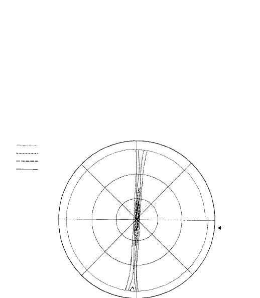
Fig. 10.13. (a) Graphitized carbon tape showing the orientations of the graphite layers at a fracture
surface (left) at the edge of the tape and (right) in the centre (photograph by courtesy of Dr S. Lu and
Professor B. Rand). (b) The corresponding pole figure, the plane of projection is the plane of the tape
and the fibre or tape direction is as indicated. The pole figure extends to ∼ 80
◦
and the limit of the spiral
is shown by the thin line. The intensity-contours are indicated in the legend.
In practice the mechanics of the texture goniometer are such that the specimen is
tilted and rotated at the same time—say a 5
◦
tilt for 360
◦
rotation, such that the recorded
intensity arises from planes lying in a spiral of 5
◦
pitch. The data is then plotted in
a stereographic projection—a pole figure (Section 12.5.3) and presented in terms of
contours of intensity data. Finally, in order that the X-ray beam may sample as large
an area as possible, the specimen is oscillated in its cradle, or shunted to and fro, by a
mechanical device rather like the connecting-rod to the wheels in a steam engine. It is
the one feature of the texture goniometer which students always notice first!
Figure 10.13(a) is a scanning electron micrograph of a graphitized carbon tape show-
ing clearly the graphite layers lying parallel to the surface of the tape except at the
edges where they ‘curl over’—but always lying parallel to the long direction of the tape.
Figure 10.13(b) is the corresponding pole figure, the 2θ angle being set to record the
262 X-ray diffraction of polycrystalline materials
0002 graphite planes. They peak in the centre (the planes parallel to the surface of the
tape) and also towards the transverse directions (the planes curled over at the edges). This
is a nice example because the relationship between the pole figure and the micrograph
can be visualized very easily. In most other materials the micrographs do not reveal the
orientations of the reflecting planes at all and their orientations are only revealed in the
pole figure.
10.5 X-ray diffraction of DNA: simulation by light
diffraction
The 1952 X-ray photograph of deoxyribonucleic acid (DNA) is of such importance in
the history of science that I have placed it, along with the 1912 X-ray photograph of
zinc blende, at the very beginning of this book (p. xv). The caption indicates how some
of the features of this pattern relate to the structure of DNA. We are, of course, unable
to attempt a complete analysis of the positions and intensities of the diffraction spots
from a structure of such complexity, but we can make some progress towards that goal
by making use of the simple ideas about diffraction that we covered earlier in this book
together with a simplified (two-dimensional) model of DNA itself.
1
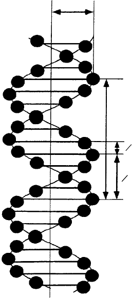
Figure 10.14 shows a projection of a simplified DNA model showing schematically
the two helical backbones of the phosphate groups (indicated by dots along the sine
waves) and the equidistant planar bases (indicated by horizontal bars), p = 3.4 nm is the
helical repeat distance; the two helices are separated by 3/8 p and the distance between
the bases is 1/10 p. The diameter of the helices is 0.6 p ≈ 2.0 nm.
Wewill now represent this structure inthe simplest possible way—as arow of horizon-
tal lines of length 0.6 p and separation p (Fig. 10.15(a)). The corresponding diffraction
pattern (or optical transform) is shown in Fig. 10.16(a). It is produced in the same way
as the patterns shown in Fig. 7.3 except that, in practice, the diffracting mask is made
up not of one, but of many sets of such lines, irregularly spaced, so as to give sufficient
diffracted light intensity. It is also made to such a scale that the distance p between the
lines and the wavelength of laser light is in the same proportion to the actual helix repeat
distance (3.4 nm) and typical X-ray wavelengths (∼0.15 nm).
The vertical separation between the laterally-streaked spots is given by the simple
diffraction grating equation (Section 7.4): sin a
n
= nλ/p and since λ p, sin α
n
≈ α
n
and the spots are nearly equally spaced.
Now we will improve our model by inclining the lines at (approximately) the same
angle as the ‘arms’ or the ‘zigs’ and ‘zags’of the helices (Fig. 10.15(b)). The diffraction
pattern from one of these again shows laterally-streaked spots with the same vertical
1
The analysis which follows is largely based on the work of Prof. Amand Lucas and his colleagues of the
University of Namur, Belgium, to whom I am very grateful. Their analysis entitled ‘Revealing the backbone
structure of B-DNAfrom laser optical simulations of its X-ray diffraction diagram’ is published in the Journal
of Chemical Education 76, 378–83, 1999. It is also available from the authors as a videotape (together with
a diffraction mask) entitled DNA—from light to life, and is also included in the Vega Science Film Series
(Videotape VRS-7 ‘How X-rays cracked the structure of DNA’), available from the Vega Science Trust,
University of Sussex, UK, BN1 3QJ and via the vega website; www.vega.org.uk.
10.5 X-ray diffraction of DNA: simulation by light diffraction 263
p
p
0.3
1
10
3
8
p
p
p
Fig. 10.14. Amodel of DNAshowing schematically the two helical backbones ofthe phosphate groups
(indicated by dots) and the equidistant planar bases (indicated by horizontal bars), p = 3.4 nm is the
helical repeat distance, the two helices are separated by 3/8 p and the distance between the bases is
l/10 p. The diameter of the helices is 0.6 p = 2.0 nm (from A. A. Lucas, Ph Lambin, R. Mairesse and
M. Mathot. J. Chem. Educ.76, 378, 1999).
separation but with maximum intensities in a direction perpendicular to the inclined
lines (Fig. 10.16(b)). Clearly, for the arms of the helices inclined in the opposite sense
the diffraction pattern will be mirror-related to that in Fig. 10.16(b) in a vertical line.
Now we combine the two sets of lines to give a zig-zag pattern (Fig. 10.15(c)) which
roughly represents the projection of a single helix; the resulting pattern (Fig. 10.16(c))
now shows (as expected) the two characteristic rows or arms of spots radiating out from
the centre and the angle between them, which is the angle between the arms of the helices
and can, together with the repeat distance p, be used to estimate the width of the helices,
i.e. the diameter of the DNA structure. We may refine our model by representing the
helix more properly as a sine wave but the resulting pattern is only changed in detail.
2
2
Examples of diffractionpatterns of sine waves of various forms are given in the Atlas of Optical Transforms
(see Chapter 7).
