Cohen I.M., Kundu P.K. Fluid Mechanics
Подождите немного. Документ загружается.

762 Compressible Flow
Show that the loss of stagnation pressure is nearly 34.2 kPa. What is the entropy
increase?
5. A shock wave generated by an explosion propagates through a still
atmosphere. If the pressure downstream of the shock wave is 700 kPa, estimate the
shock speed and the flow velocity downstream of the shock.
6. A wedge has a half-angle of 50
◦
. Moving through air, can it ever have an
attached shock? What if the half-angle were 40
◦
?[Hint: The argument is based entirely
on Figure 16.20.]
7. Air at standard atmospheric conditions is flowing over a surface at a Mach
number of M
1
= 2. At a downstream location, the surface takes a sharp inward turn
by an angle of 20
◦
. Find the wave angle σ and the downstream Mach number. Repeat
the calculation by using the weak shock assumption and determine its accuracy by
comparison with the first method.
8. A flat plate is inclined at 10
◦
to an airstream moving at M
∞
= 2. If the chord
length is b = 3 m, find the lift and wave drag per unit span.
9. A perfect gas is stored in a large tank at the conditions specified by p
o
,
T
o
. Calculate the maximum mass flow rate that can exhaust through a duct of
cross-sectional area A. Assume that A is small enough that during the time of interest
p
o
and T
o
do not change significantly and that the flow is adiabatic.
10. For flow of a perfect gas entering a constant area duct at Mach number M
1
,
calculate the maximum admissable values of f, q for the same mass flow rate. Case (a)
f = 0; Case (b) q = 0.
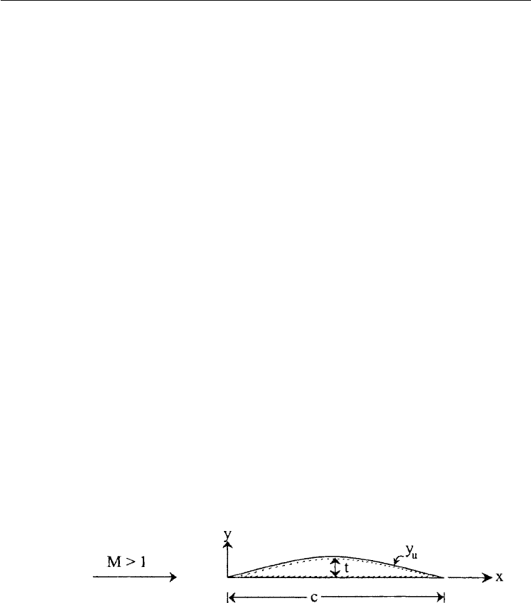
11. Using thin airfoil theory calculate the lift and drag on the airfoil shape given
by y
u
= t sin(π x/c) for the upper surface and y
l
= 0 for the lower surface. Assume
a supersonic stream parallel to the x-axis. The thickness ratio t/c 1.
12. Write momentum conservation for the volume of the small “pill box” shown
in Figure 4.22 (p. 121) where the interface is a shock with flow from side 1 to side 2.
Let the two end faces approach each other as the shock thickness → 0 and assume
viscous stresses may be neglected on these end faces (outside the structure). Show
that the n component of momentum conservation yields (16.29) and the t component
gives u · t is conserved or v is continuous across the shock.

Supplemental Reading 763
Literature Cited
Ames Research Staff (1953). NACA Report 1135: “Equations, Tables, and Charts for Compressible Flow.”
Becker, R. (1922). “Stosswelle und Detonation.” Z. Physik 8: 321–362.
Cohen, I. M. and C. A. Moraff (1971). “Viscous inner structure of zero Prandtl number shocks.” Phys.
Fluids 14: 1279–1280.
Cramer, M. S. and R. N. Fry (1993). “Nozzle flows of dense gases.” The Physics of Fluids A 5: 1246–1259.
Fergason, S. H., T. L. Ho, B. M. Argrow, and G. Emanuel (2001). “Theory for producing a single-phase
rarefaction shock wave in a shock tube.” Journal of Fluid Mechanics 445: 37–54.
Hayes, W. D. (1958). “The basic theory of gasdynamic discontinuities,” Sect. D of Fundamentals of
Gasdynamics, Edited by H. W. Emmons, Vol. III of High Speed Aerodynamics and Jet Propulsion,
Princeton, NJ: Princeton University Press.
Liepmann, H. W. and A. Roshko (1957). Elements of Gas Dynamics, New York: Wiley.
Shapiro, A. H. (1953). The Dynamics and Thermodynamics of Compressible Fluid Flow, 2 volumes.
New York: Ronald.
von Karman, T. (1954). Aerodynamics, New York: McGraw-Hill.
Supplemental Reading
Courant, R. and K. O. Friedrichs (1977). Supersonic Flow and Shock Waves, New York: Springer-Verlag.
Yahya, S. M. (1982). Fundamentals of Compressible Flow, New Delhi: Wiley Eastern.
This page intentionally left blank

Chapter 17
Introduction to Biofluid
Mechanics
Portonovo S. Ayyaswamy
University of Pennsylvania
Philadelphia, PA
1. Introduction ..................... 765
2. The Circulatory System in the
Human Body .................... 766
The Heart as a Pump ............ 769
Nature of Blood .................. 773
Nature of the Blood Vessels ....... 779
3. Modelling of Flow in Blood Vessels 782
General Introduction ............. 782
Hagen-Poiseuille Flow ........... 783
Pulsatile Flow Theory............ 791
Blood Vessel Bifurcation: An
Application of Poiseuille’s Formula
and Murray’s Law............. 807
Flow in a Rigid Walled Curved
Tube.......................... 812
Flow in Collapsible Tubes ........ 818
Laminar Flow of a Casson Fluid
in a Rigid Walled Tube ........ 826
Pulmonary Circulation ........... 829
The Pressure Pulse Curve in the Right
Ventricle ...................... 830
Effect of Pulmonary Arterial Pressure
on Pulmonary Resistance ...... 830
4. Introduction to the Fluid Mechanics
of Plants ........................ 831
Xylem ........................... 833
Xylem Flow ..................... 835
Phloem .......................... 835
Phloem Flow .................... 836
Exercises ........................ 837
Acknowledgment ................ 838
Literature Cited ................. 838
1. Introduction
This chapter is intended to be of an introductory nature to the vast field of biofluid
mechanics. Here, we shall consider the ideas and principles of the preceding chapters
in the context of fluid motion in biological systems. First we will learn about some
aspects of the fluid motion in the human body, and later we will learn about some
aspects of fluid mechanics of plants.
The human body is a complex system that requires materials such as air, water,
minerals and nutrients for survival and function. Upon intake, these materials have to
be transported and distributed around the body as required. The associated biotrans-
port and distribution processes involve interactions with membranes, cells, tissues, and
organs comprising the body. Subsequent to cellular metabolism in the tissues, waste
765
©2010 Elsevier Inc. All rights reserved.
DOI: 10.1016/B978-0-12-381399-2.50017-4

766 Introduction to Biofluid Mechanics
by products have to be transported to the excretory organs for synthesis and removal.
In addition to these functions, biotransport systems and processes are required for
homeostasis (physiological regulation–for example, maintenance of pH and of body
temperature), and for enabling the movement of immune substances to aid in body’s
defense and recovery from infection and injury. Furthermore, in certain other special-
ized systems such as the cochlea in the ear, fluid transport enables hearing and motion
sensing. Evidently, in the human body, there are multiple types of fluid dynamic sys-
tems that operate at multiple and widely disparate scales. These scales are at various
levels such as macro, micro, nano, pico and so on. Systems at the micro and macro
levels, for example, include cells (micro), tissue (micro–macro), and organs (macro).
Transport at the micro, nano and pico levels would include ion channeling, binding,
signaling, endocytosis, and so on. Tissues constitute organs, and organs as systems
perform various functions. For example, the cardiovascular system consists of the
heart, blood vessels (arteries, arterioles, venules, veins, capillaries), lymphatic ves-
sels, and the lungs. Its function is to provide adequate blood flow and regulate the
flow as required by the various organs of the body. In this chapter, as related to the
human body, we shall restrict attention to some aspects of the cardiovascular system
for blood circulation.
2. The Circulatory System in the Human Body
The primary functions of the cardiovascular system are: (i) to pick up oxygen and
nutrients from the lungs and the intestine, respectively, and deliver them to tissues
(cells) of various parts, (ii) to remove waste and carbon dioxide from the body for
excretion through the kidneys and the lungs, respectively, and (iii) to regulate body
temperature by convecting the heat generated and dissipating it through transport
across the skin. The circulatory system in the normal human body (as in all vertebrates
and some other select group of species) can be considered as a closed system, meaning
that the blood never leaves the system of blood vessels. The driving potential for blood
flow is the prevailing pressure gradient.
The circulations associated with the cardiovascular system may be considered
under three subsystems. These are the (i) systemic circulation, (ii) pulmonary cir-
culation, and (iii) coronory circulation. (See Fig. 17.1.) In the systemic circulation,
blood flows to all of the tissues in the body except the lungs. Contraction of the left
ventricle of the heart pumps oxygen-rich blood to a relatively high pressure and ejects
it through the aortic valve into the aorta. Branches from the aorta supply blood to the
various organs via systemic arteries and arterioles. These, in turn, carry blood to the
capillaries in the tissues of various organs. Oxygen and nutrients are transported by
diffusion across the walls of the capillaries to the tissues. Cellular metabolism in the
tissues generates carbon dioxide and byproducts (waste). Carbon dioxide dissolves
in the blood and waste is carried by the blood stream. Blood drains into venules and
veins. These vessels ultimately empty into two large veins called the superior vena
cava (SVC) and and inferior vena cava (IVC) that return carbon dioxide rich blood to
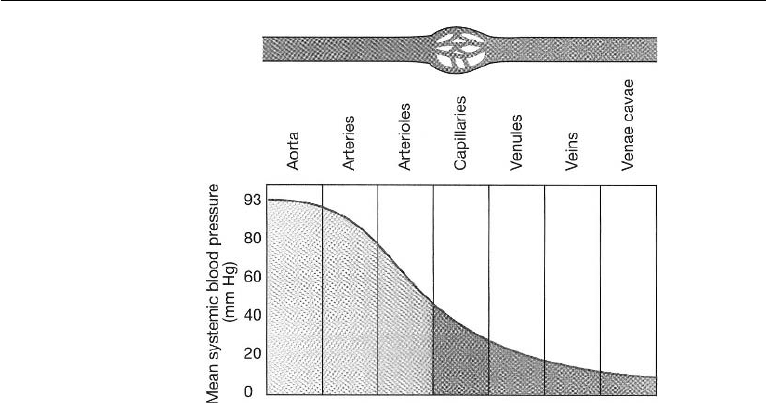
the right atrium. The mean blood pressure of the systemic circulation ranges from a
high of 93 mmHg in the arteries to a low of few mmHg in the venae cavae. Fig. 17.2
2. The Circulatory System in the Human Body 767
Figure 17.1 Schematic of blood flow in systemic and pulmonary circulation. (Reproduced with permis-
sion from Silverthorn, D.U. (2001) Human Physiology: An Integrated Approach, 2nd ed., Prentice Hall,
Upper Saddle River, NJ.).
shows that pressure falls continuously as blood moves farther from the heart. The
highest pressure in the vessels of the circulatory system is in the aorta and in the
systemic arteries while the lowest pressure is in the venae cavae.
In pulmonary circulation, contraction of the right atrium ejects carbon dioxide
rich blood through the tricuspid valve into the right ventricle. Contraction of the right
ventricle pumps the blood through the pulmonic valve (also called semilunar valve)
into the pulmonary arteries. These arteries bifurcate and transport blood into the
complex network of pulmonary capillaries in the lungs. These capillaries lie between
and around the alveoli walls. During respiratory inhalation, the concentration of oxy-
gen in the air is greater in the air sacs of the alveolar region than in the capillary blood.
Oxygen diffuses across capillary walls into blood. Simultaneously, the concentration
of carbon dioxide in the blood is higher than in the air and carbon dioxide diffuses
from the blood into the alveoli. Carbon dioxide exits through the mouth and nostrils.
Oxygenated blood leaves the lungs through the pulmonary veins and enters the left
768 Introduction to Biofluid Mechanics
Figure 17.2 Pressure gradient in the blood vessels. (Reproduced with permission from Silverthorn,
D.U. (2001) Human Physiology: An Integrated Approach, 2nd ed., Prentice Hall, Upper Saddle
River, NJ.).
atrium. When the left atrium contracts, it pumps blood through the bicuspid (mitral)
valve into the left ventricle. Figs. 17.3 and 17.4 provide an overview of external and
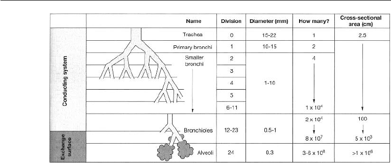
cellular respiration and the branching of the airways, respectively.
Blood is pumped through the systemic and pulmonary circulations at a rate of
about 5.2 liters per minute under normal conditions. The systemic and pulmonary
circulations described above constitute one cardiac cycle. The cardiac cycle denotes
any one or all of such events related to the flow of blood that occur from the beginning
of one heartbeat to the beginning of the next. Throughout the cardiac cycle, the blood
pressure increases and decreases. The frequency of the cardiac cycle is the heart rate.
The cardiac cycle is controlled by a portion of the autonomic nervous system (that
part of the nervous system which does not require the brain’s involvement in order to
function).
In coronary circulation, blood is supplied to and from the heart muscle itself.
The muscle tissue of the heart, or myocardium, is thick and it requires coronary blood
vessels to deliver blood deep into the myocardium. The vessels that supply blood with
a high concentration of oxygen to the myocardium are known as coronary arteries.
The main coronary artery arises from the root of the aorta and branches into the left
and right coronary arteries. Up to about seventy five percent of the coronary blood
supply goes to the left coronary artery, the remainder going to the right coronary artery.
Blood flows through the capillaries of the heart and returns through the cardiac veins
which remove the deoxygenated blood from the heart muscle. The coronary arteries
that run on the surface of the heart are relatively narrow vessels and are commonly
affected by atherosclerosis and can become blocked, causing angina or a heart attack.
The coronary arteries are classified as “end circulation,” since they represent the only
source of blood supply to the myocardium.

2. The Circulatory System in the Human Body 769
Figure 17.3 Overview of external and cellular respiration. (Reproduced with permission from Silver-
thorn, D.U. (2001) Human Physiology: An Integrated Approach, 2nd ed., Prentice Hall, Upper Saddle
River, NJ.).
The Heart as a Pump
The heart has four pumping chambers–two atria (upper) and two ventricles (lower).
The left and right parts of the heart are separated by a muscle called the septum which
keeps the blood volumes in each part separate. The upper chambers interact with
the lower chambers via the heart valves. The heart has four valves which ensure that
blood flows only in the desired direction. The atrio-ventricular valves (AV) consist
of the tricuspid (three flaps) valve between the right atrium and the right ventricle,
and the bicuspid (two flaps, also called the mitral) valve between the left atrium
and the left ventricle. The pulmonary valve is between the right ventricle and the
pulmonary artery, and the aortic valve is between the left ventricle and the aorta. Both
the pulmonary and aortic valves have three symmetrical half moon shaped valve flaps
(cusps), and are called the semilunar valves. The function of the four chambers in
770 Introduction to Biofluid Mechanics
Figure 17.4 Branching of the airways. Areas have units of cm
2
. (Reproduced with permission from
Silverthorn, D.U. (2001) Human Physiology: An Integrated Approach, 2nd ed., Prentice Hall, Upper
Saddle River, NJ.).
the heart is to pump blood through pulmonary and systemic circulations. The atria
receive blood from the veins–right atrium receives carbon dioxide rich blood from
the SVC and IVC, and the left atrium receives oxygen rich blood from the pulmonary
veins. The heart is controlled by a single electrical impulse and both sides of the
heart act synchronously. Electrical activity stimulates the heart muscle (myocardium)
of the chambers of the heart to make them contract. This is immediately followed
by mechanical contraction of the heart. Both atria contract at the same time. The
contraction of the atria moves the blood from the upper chambers through the valves
into the ventricles. The atrial muscles are electrically separated from the ventricular
muscles except for one pathway through which an electrical impulse is conducted
from the atria to the ventricles. The impulse reaching the ventricles is delayed by
about 110 ms while the conduction occurs through the pathway. This delay allows the
ventricles to be filled before they contract. The left ventricle is a high pressure pump
and its contraction supplies systemic circulation while the right ventricle is a low
pressure pump supplying pulmonary circulation (Lungs offer much less resistance to
flow than systemic organs).
From the above discussions, we see that the pumping action of the heart can be
regarded as a two phase process–a contraction phase (systole) and a filling (relaxation)
phase (diastole). Systole describes that portion of the heartbeat during which contrac-
tion of the heart muscle and hence ejection of blood takes place. A single “beat” of
the heart involves three operations: atrial systole, ventricular systole and complete
cardiac diastole. Atrial systole is the contraction of the heart muscle of the left and
right atria, and occurs over a period of 0.1s. As the atria contract, the blood pressure
in each atrium increases, which forces the mitral and tricuspid valves to open forcing
blood into the ventricles. The AV valves remain open during atrial systole. Following
atrial systole, ventricular systole which is the contraction of the muscles of the left
and right ventricles occurs over a period of 0.3s. The ventricular systole generates
enough pressure to force the AV valves to close, and the aortic and pulmonic valves
open. (The aortic and pulmonic valves are always closed except for the short period
of ventricular systole when the pressure in the ventricle rises above the pressure in
2. The Circulatory System in the Human Body 771
the aorta for the left ventricle and above the pressure in the pulmonary artery for the
right ventricle.) During systole, the typical pressures in the aorta and the pulmonary
artery rise to 120 mmHg and 24 mmHg, respectively, (note conversion, 1 mmHg =
133 Pa). In normal adults, blood flow through the aortic valve begins at the start of
ventricular systole, and rapidly accelerates to a peak value of approximately 1.35
m/s during the first one-third of systole. Thereafter, the blood flow begins to decel-
erate. Pulmonic valve peak velocities are lower and in normal adults, they are about
0.75 m/s. Contraction of the ventricles in systole ejects about two thirds of the blood
from these chambers. As the left ventricle empties, its pressure falls below the pressure
in the aorta, and the aortic valve closes. Similarly, as the pressure in the right ventricle
falls below the pressure in the pulmonary artery, the pulmonic valve closes. Thus, at
the end of the the ventricular systole, the aortic and pulmonic valves close, with the
aortic valve closing a little earlier than the pulmonic valve. Diastole describes that
portion of the heart beat during which the chamber refilling takes place. The cardiac
diastole is the period of time when the heart relaxes after contraction in preparation
for refilling with circulating blood. The ventricles refill or ventricular diastole occurs
during atrial systole. When the ventricle is filled and ventricular systole begins, then
the AV valves are closed and the atria begin refilling with blood or atrial diastole
occurs. About a period of 0.4s following ventricular systole, both the atria and the
ventricles begin refilling and both chambers are in diastole. During this period, both
AV valves are open and aortic and pulmonic valves are closed. The typical diastolic
pressure in the aorta is 80 mmHg and, in the pulmonary artery, it is 8 mmHg. Thus, the
typical systolic and diastolic pressure ratios are 120/80 mmHg for the aorta and 24/8
mmHg for the pulmonary artery. The systolic pressure minus the diastolic pressure is
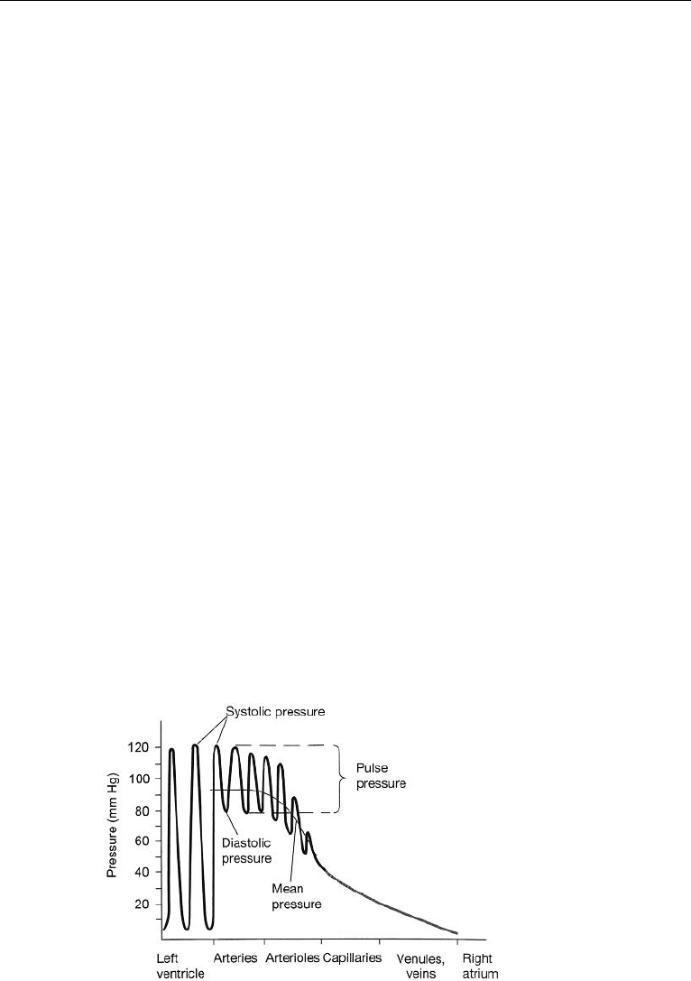
called the pressure pulse, and for the aorta (left ventricle) it is 40 mmHg. The pulse
pressure is a measure of the strength of the pressure wave. It increases with increased
stroke volume (say, due to activity or exercise). Pressure waves created by the ven-
tricular contraction diminish in amplitude with the distance and are not perceptible
in the capillaries. Fig. 17.5 shows the pressure throughout the systemic circulation.
Figure 17.5 Pressure throughout the systemic circulation. (Reproduced with permission from Silverthorn,
D.U. (2001) Human Physiology: An Integrated Approach, 2nd ed., Prentice Hall, Upper Saddle River, NJ.).