Budras Klaus-Dieter, Habel Robert E. Bovine Аnatomy
Подождите немного. Документ загружается.

e"1
e"3
e"2
r"1
r"2
(See p. 64)
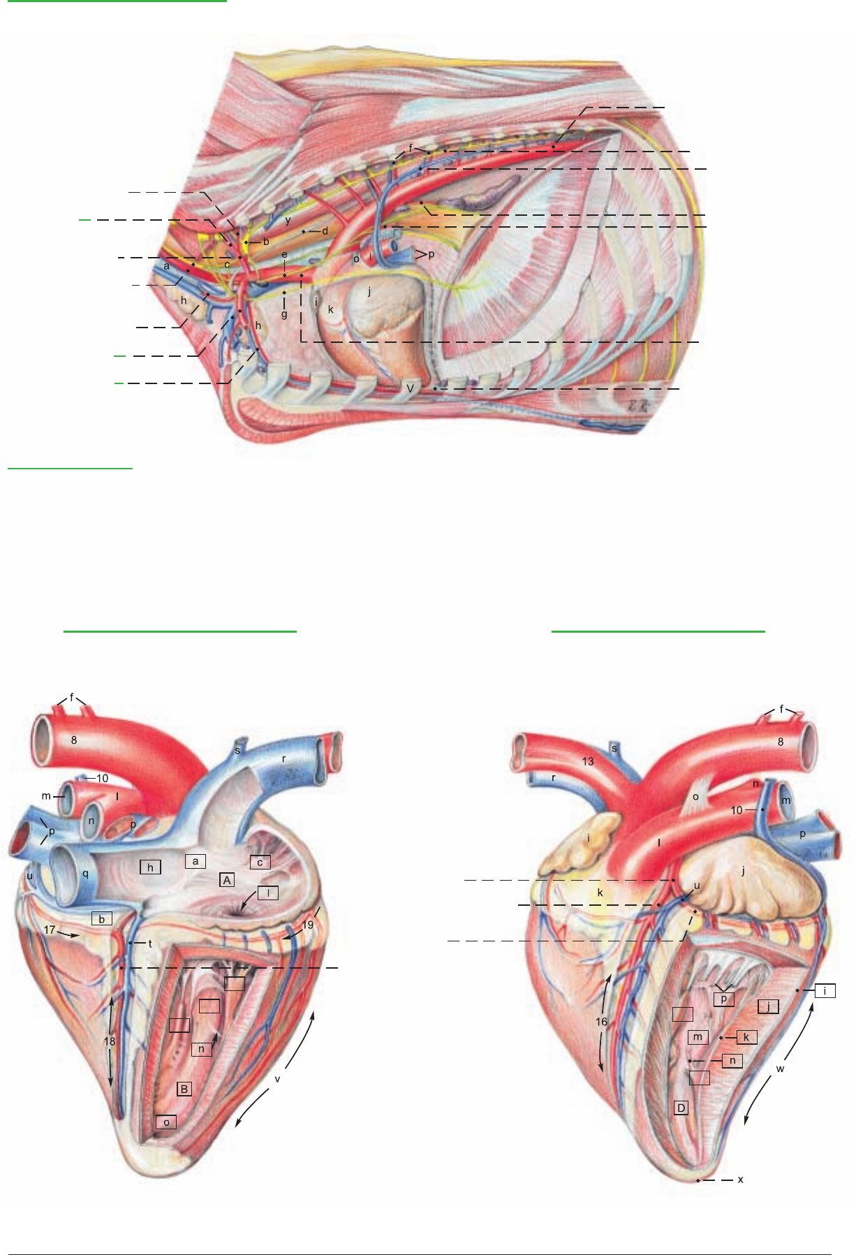
Left Thoracic cavity and Heart
1 Dorsal scapular a.
2 Vertebral and deep
cervical aa.
3 Costocervical trunk
4 Left common carotid a. and
vagosympathetic trunk
5 Supf. cervical a. and v.
6 Left subclavian a.
and v.
7 Internal thoracic a.
and v.
8 Thoracic aorta
9 Sympathetic trunk
10 Left azygos v.
Bronchoesophageal a.:
11 Esophageal br.
12 Bronchial br.
13 Brachiocephalic trunk
14 Sternopericardiac ligg.
(See pp. 61, 63, 67)
Legend: (Lnn. see p. 63)
a Trachea and int. jugular v.
b Cervicothoracic ganglion
c Middle cervical ganglion
d Thoracic duct
e Vagus n.
f Intercostal aa. and vv.
g Left phrenic n.
h Thymus
i Right auricle
j Left auricle
k Conus arteriosus
l Pulmonary trunk
m Left pulmonary a.
n Right pulmonary a.
o Lig. arteriosum
p Pulmonary vv.
q Caud. vena cava
r Cran. vena cava
s Costocervical v.
t Middle cardiac v.
u Great cardiac v.
v Right ventricular border
w Left ventricular border and
intermediate groove
x Apex of heart
y Longus colli m.
Right atrium and Right ventricle
(Atrial surface)
Left auricle and Left ventricle
(Auricular surface)
15 Left coronary a.
16 Paraconal interventricular
br. and groove
17 Circumflex br.
18 Subsinuosal interventricular br.
and groove
19 Right coronary a. and
coronary groove
65
Anatomie des Rindes englisch 09.09.2003 14:11 Uhr Seite 65

a) The SKIN (1) of the lateral abdominal wall (flank) is easily
moveable. Dorsally the surgically important triangular paralumbar
fossa (b) is outlined by the ends of the transverse processes of the
lumbar vertebrae, the last rib, and the prominent ridge formed by
the part of the internal oblique that extends from the tuber coxae
to the knee of the last rib. Ventrally, the subcutaneous cranial super-
ficial epigastric v. (“milk vein”—3) in the cow, is conspicuous,
meandering, and 2–3 cm thick. It comes from the int. thoracic v.
and emerges from the “milk well” (anulus venae subcutaneae abdo-
minis) at the second tendinous intersection of the rectus, ventral to
the 7th to 9th intercostal spaces. It joins the cranial mammary v.
(caudal superficial epigastric v., p. 91, 12) at the udder.
b) The SYSTEM OF THE EXTERNAL FASCIA OF THE
TRUNK includes the superficial fascia and deep fascia.
I. The superficial fascia of the trunk envelops the cutaneus trunci
and the cranially related cutaneus omobrachialis, which are essen-
tially the same as in the horse. The strong cranial preputial muscles,
present in the dog, but not in the horse, originate mainly from the
region of the xiphoid cartilage and secondarily from the ventral
border of the cutaneus trunci, and form a loop around the preputial
orifice. The caudal preputial muscles (see text figure p. 80) are
inconspicuous in the dog and absent in the horse and polled breeds
of cattle.* They originate from the deep fascia, mainly lateral to the
tunica vaginalis, but often also medial to it, and terminate at the
loop formed by the cranial preputial muscles.
The 8–12 cm long subiliac lymph node (5), absent in the dog, dif-
fers from the multiple nodes of the horse. It is a single large node
above the patella on the abdominal wall near the cranial border of
the tensor fasciae latae, easily palpable in the live ox. A small acces-
sory node may be present.
II. The deep fascia of the trunk covers the external oblique, and on
the ventrolateral abdomen is also known as the yellow abdominal
tunic (4) due to the inclusion of yellow elastic fibers. With its col-
lagenous laminae the deep fascia completely envelops the two
abdominal obliques; whereas it covers only the external surface of
the rectus and transversus. On both sides of the ventral median line
the yellow tunic gives off the elastic medial laminae of the udder, or
in the bull, radiates into the prepuce. The linea alba is the ventro-
median fixation and interwoven seam of the fasciae and aponeu-
roses of the abdominal muscles. It extends from the sternum
through the prepubic tendon to the pecten pubis and passes around
both sides of the umbilicus.
c) The NERVES OF THE ABDOMINAL WALL
I. The dorsal branches of spinal nerves T12–L3 divide into medi-
al and lateral mixed motor and sensory branches. The lateral brr.
(Tdl, Ldl) pass out between the longissimus and iliocostalis muscles
and divide into dorsomedial cutaneous brr. and dorsolateral cuta-
neous brr. The latter innervate the skin of the abdomen down to the
level of the patella. On p. 67 the small dorsomed. cut. brr. are mis-
labeled Ldl. The dorsolat. cut. brr. are cut off short. Those of T13
and L1 and L2 cross the paralumbar fossa to a line from the ven-
tral end of the last rib to the patella, but cannot be traced that far
by gross dissection. They must be blocked with the ventral brr. to
provide anesthesia for flank incisions.
II. The ventral branches of spinal nerves T12–L2 innervate the
skin, abdominal mm., and peritoneum. The ventral br. of L1 is the
iliohypogastric n. The ventral br. of L2, together with a communi-
cation from L3, forms the ilioinguinal n. The ventral brr. give off
lateral cutaneous branches (Tvl, Lvl) which emerge through the
external oblique on a line extending from the knees of the ribs to a
point ventral to the tuber coxae at the level of the hip joint. Passing
caudoventrally, they innervate the skin of the ventrolateral
abdomen. The ventral brr. of T12–L2 communicate with each oth-
er at the origins of the lateral cutaneous brr. and continue ventral-
ly on the external surface of the transversus. Near the milk vein
they give off ventral cutaneous branches (Tvc, Lvc)** extending to
the ventral midline and cranial portions of the prepuce or udder,
and terminate in the rectus and parietal peritoneum. The relations
of nerves T13–L2 to the transverse processes of the vertebrae are of
great clinical importance for anesthesia of the abdominal wall. The
lateral cutaneous femoral n. (11) comes from L3 and L4 through
the lumbar plexus. It accompanies the caudal branches of the deep
circumflex iliac a. and v., at first medial then craniolateral to the
tensor fasciae latae, down to the stifle. (For the innervation of the
udder see p. 90.)
d) The SKELETAL MUSCLE LAYER consists of four broad mus-
cles.
I. The external oblique abdominal m. (2). The lumbar part origi-
nates on the last rib and thoracolumbar fascia and runs to the tuber
coxae, and caudoventrally to the inguinal lig. and prepubic tendon
(see p. 80). The costal part begins with its digitations on the last 8–9
ribs, touching part of the ventral border of the latissimus dorsi. It
ends with the aponeurosis mainly on the linea alba, but also on the
prepubic tendon by means of its abdominal and pelvic tendons (see
pp. 79, 80). The transition of the muscle to its aponeurosis follows
the curve of the costal arch and continues to the tuber coxae. The
aponeurosis is a component of the external lamina of the sheath of
the rectus.
II. The internal oblique abdominal m. (10) originates mainly from
the tuber coxae and the iliac fascia (see p. 81). It also takes origin
from the thoracolumbar fascia and the lumbar transverse process-
es. The dorsal part ends on the last rib, and the portion running
from the tuber coxae to the knee of the last rib forms the cau-
doventral border of the paralumbar fossa. The main termination is
by its aponeurosis on the linea alba; the caudal border of the
aponeurosis joins the abdominal and pelvic tendons of the ext.
oblique and the tendon of the rectus in the prepubic tendon. The
aponeurosis, unlike that of the dog, is involved only in the external
lamina of the sheath of the rectus. (For its contribution to the deep
inguinal ring see p. 80.)
III. The transversus abdominis (7) originates with a tendinous lum-
bar part from the lumbar transverse processes, and a fleshy costal
part interdigitating with the diaphragm on the last 7–8 costal car-
tilages. It terminates on the linea alba, its aponeurosis forming the
internal lamina of the sheath of the rectus. Its caudal extent is at the
transverse plane of the tuber coxae.
IV. The rectus abdominis (6) takes origin from the 4th–9th costal
cartilages and has five tendinous intersections. The terminal ten-
dons of the recti become abruptly narrower near the inguinal
region and turn their inner surfaces toward each other, forming in
the cow a narrow median trough. Near the prepubic tendon the rec-
tus tendons twist into sagittal planes and fuse by decussation cau-
dal to the intertendinous fossa (see p. 78 c). They form a common
median tendon incorporated in the prepubic tendon and continu-
ous with the symphyseal tendon.
e) The INTERNAL FASCIA OF THE TRUNK (see p. 80) lines the
transversus and rectus on the lateral and ventral abdominal wall as
the fascia transversalis. Dorsally it covers the psoas and iliacus as
iliac fascia. It joins the pelvic fascia on the pelvic wall.
f) The PERITONEUM (see also p. 80). The peritoneum extends
into the pelvic cavity as the rectogenital, vesicogenital, and pubo-
vesical pouches (excavations) and in the bull is evaginated into the
scrotum as the vaginal tunic.
66
CHAPTER 7: ABDOMINAL WALL AND ABDOMINAL CAVITY
1. THE ABDOMINAL WALL
* Long, and Hignett, 1970
** Schaller, 1956
For demonstration of the five layers of the abdominal wall (a, b, d, e, f), the remaining skin is cut along the dorsomedian line and along
the transverse plane of the tuber coxae, and reflected ventrally to the base of the udder or prepuce. Remnants of the cutaneus trunci,
abdominal muscles, and internal fascia of the trunk are cut just ventral to the iliocostalis lumborum and reflected ventrally, one after
the other, to the subcutaneus abdominal vein and the lateral border of the rectus abdominis.
Anatomie des Rindes englisch 09.09.2003 14:11 Uhr Seite 66

Lvl
Tvl
a Supf. cervical ln.
b Paralumbar fossa
c Rectus thoracis
d Int. intercostal mm.
Pectoral and abdominal regions
(lateral)
1 Skin
2 External oblique abd. m.
3 Cran. supf. epigastric v.
(Milk v.)
4 Yellow abdominal tunic
5 Subiliac ln.
(See p. 61)
6 Rectus abdominis and
tendinous intersections
7 Transversus
8 Iliohypogastric n.
9 Ilioinguinal n.
10 Int. oblique abd. m.
11 Lat. cut. femoral n.
Legend:
67
Anatomie des Rindes englisch 09.09.2003 14:12 Uhr Seite 67

68
2. TOPOGRAPHY AND PROJECTION OF THE ABDOMINAL ORGANS ON THE BODY WALL
Study of the abdominal organs is carried out by the students on both sides of the body at the same time. On the left side the stomach
and spleen are studied and the adhesions of the organs with the abdominal wall and with other organs and structures are noted. The
interior relations of the compartments of the stomach are exposed by fenestration of the dorsal sac of the rumen from the lumbar trans-
verse processes to the left longitudinal groove of the rumen, and removal of the contents. Prepared demonstrations of the stomach are
also studied. On the right side, before the study of the liver and intestines, the special relations of the greater omentum are examined
and the omental foramen is explored. Then the superficial wall (21) of the greater omentum is cut ventral and parallel to the descend-
ing duodenum, opening the caudal recess of the omental bursa (22). The duodenum with the mesoduodenum and pancreas are care-
fully reflected dorsally to the ventral surface of the right kidney. After study of the liver and its vessels, nerves, and ducts, the common
bile duct, hepatic a., portal v., portal lnn., and nerves are severed at the porta of the liver, and the hepatic ligaments and caudal vena
cava, cranial and caudal to the liver, are cut and the liver is removed. After complete transection of the superficial wall down to the
pylorus, the deep wall of the greater omentum is cut ventral to the distal loop of the ascending colon and the transverse colon, and the
supraomental recess (23) is opened for study of the remaining intestines. The blood vessels, nerves, and lymph nodes are identified with
attention to species-specific peculiarities. For final exenteration the duodenum between the cranial and descending parts, and the rec-
tum caudal to the caudal mesenteric a. are double-ligated and cut. Also the cranial and caudal mesenteric aa. ventral to the aorta, and
the splenic and gastroduodenal vv. at the portal v. are cut. While separating the mesentery and mesocolon from the dorsal abdominal
wall, the intestinal mass is removed from the abdominal cavity and the parts of the intestines are identified on the isolated intestinal
tract.
A knowledge of the topographic relations of the abdominal organs
to the body wall is essential for their examination from the exteri-
or as well as for laparotomy and rectal examination. The abdomi-
nal wall is divided into cranial, middle, and caudal regions, and
these are subdivided on each side as follows: I. The large cranial
abdominal region consists of a) left, and b) right caudoventral parts
of the costal regions and the hypochondriac regions (covered by the
costal cartilages), and c) the xiphoid region between the costal
arches. II. The middle abdominal region consists of the a) left and
b) right lateral abdominal regions (flanks) with the paralumbar fos-
sae, and the c) umbilical region. III. The caudal abdominal region
consists of right and left inguinal regions and the pubic region. In
the costal and hypochondriac regions the intrathoracic abdominal
organs are not in contact with the thoracic wall, but are separated
from it by the lungs and diaphragm. The rumen extends from the
diaphragm to the pelvic inlet. It takes up most the left half of the
abdominal cavity. Its extension to the right and toward the pelvic
inlet depends on the age of the animal, the kind of feed, and, if preg-
nant, the stage of gestation. These factors also affect the position
and relations of all other abdominal organs.
I. a) In the left costal and hypochondriac regions the atrium (3)
and recess (6) of the rumen are projected on the thoracic wall, as
well as the spleen (4), adherent to the dorsolateral surface of the
atrium from the vertebral ends of the 12th and 13th ribs, over the
middle of the 10th rib, to the level of the knees of the 7th and 8th
ribs. The reticulum (2) is in contact with the left abdominal wall in
the ventral third of the 6th and 7th intercostal spaces. Near the
median plane it may extend caudally as far as the transverse plane
of the 9th intercostal space, and ventrally to the level of the xiphoid
cartilage. Of the liver (1), only the left border is projected in the
narrow space between the diaphragm and reticulum in the ventral
3rd of the 6th intercostal space. The fundus of the abomasum (5)
lies on the left side between the reticulum and the atrium of the
rumen.
I. b) In the right costal and hypochondriac regions the liver (25),
covered by the diaphragm, and mostly also by the lung, is project-
ed on the thoracic wall, its border forming a caudally convex curve.
It lies almost entirely on the right side, including the left lobe (1),
right lobe (25), and caudate process (24). It extends from the ven-
tral end of the 6th intercostal space to the dorsal end of the 13th
rib. The percussion field of the liver, however, is limited to a zone
about a hand’s breadth wide along the border of the lung in the last
four intercostal spaces.
Ventral to the caudate process is the cranial part of the descending
duodenum (13) with the right lobe of the pancreas (15) in the meso-
duodenum. Ventral to the descending duodenum, covered by the
greater omentum, are cranial loops of the jejunum (19) and cranial
to them, the gall bladder (27) in the ventral part of the 10th inter-
costal space. Directly cranial to the gall bladder is the cranial part
of the duodenum (26), continuous ventrocaudally with the pylorus,
which varies in position from the ventral end of the 9th to that of
the 12th intercostal space.
Cranial to the pyloric part of the abomasum (28) is the omasum
(30), covered by the lesser omentum, between the transverse planes
of the 7th and 11th ribs, but because of its spherical shape, it press-
es the lesser omentum against the thoracic wall in the 7th–9th inter-
costal spaces only.
II. a) In the left lateral abdominal region only rumen compart-
ments adjoin the abdominal wall. The dorsolateral abdominal wall
in the region of the paralumbar fossa is in contact with the dorsal
sac (7) and the caudodorsal blind sac (8). The ventrolateral abdom-
inal wall is indirectly in contact, through the superficial wall (21) of
the greater omentum, with the ventral sac (9) and the caudoventral
blind sac (10) of the rumen.
II. b) In the right lateral abdominal region, projected from dorsal
to ventral on the dorsolateral abdominal wall, are the right kidney
(14) from the last rib to the 3rd lumbar vertebra, the right lobe of
the pancreas (15) with the descending duodenum (13), which pass-
es into the caudal flexure (12) at the level of the tuber coxae, and
immediately caudal to that, the sigmoid part of the descending
colon (11). Ventral to the duodenum, in the supraomental recess
(23) of the greater omentum, are the proximal loop of the ascend-
ing colon (16) and the cecum (17). The latter extends from the mid-
dle of the lumbar region to the pelvic inlet. The apex of the cecum
projects caudally from the supraomental recess. (Relations between
the descending duodenum and the parts of the large intestine may
vary but are not necessarily abnormal.) The ventrolateral abdomi-
nal wall covers the pyloric part of the abomasum (28) ventrally
along the right costal arch back to the knee of the 12th rib, and
middle and caudal parts of the jejunum (19) from the last rib to the
plane of the last lumbar vertebra. The jejunum overreaches the
greater omentum caudally and passes into the straight ileum (18)
just ventral to the cecum.
III. Ventrally in the xiphoid region, are the reticulum (2) cranially,
more on the left than on the right; the fundus of the abomasum (5)
caudal to the reticulum; the omasum (30) ventral to the right costal
arch, covered by the lesser omentum, between the transverse planes
of the 7th and 11th intercostal spaces; the fundus of the abomasum
caudal to the reticulum; and the atrium of the rumen on the left.
Ventrally in the middle abdominal region the body of the aboma-
sum (29) lies on the median line with more of it on the left than on
the right. At the angle of the abomasum the pyloric part (28) curves
to the right around the omasum, with the greater curvature cross-
ing the median line at the transverse plane of the last rib. The
jejunum (19) is caudal to the abomasum on the right as far caudal-
ly as the last lumbar vertebra, partly within the supraomental
recess. On the left of the median plane, caudally and also slightly to
the right, the ventral sac (9) and caudoventral blind sac (10), cov-
ered by the greater omentum, lie on the abdominal floor.
The costal part of the diaphragm is detached from the ribs on both sides. A dorsoventral incision is made through the diaphragm on
the right of the caudal vena cava and on the left of the adhesion with the spleen, and the severed parts of the diaphragm are removed.
In the process, the falciform ligament and the round ligament of the liver, still intact in the young animal, can be seen on the right.
Anatomie des Rindes englisch 09.09.2003 14:12 Uhr Seite 68
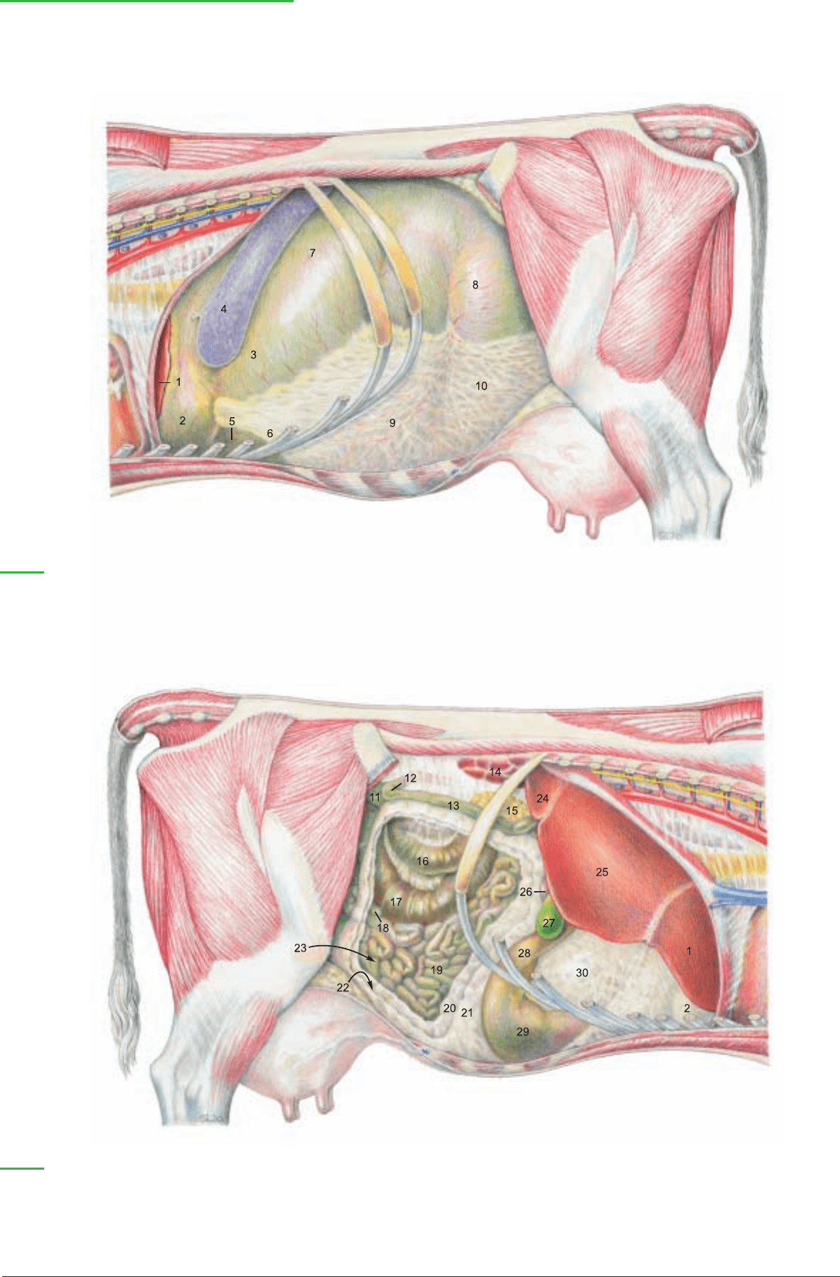
27 Gall bladder
28 Pyloric part of abomasum
29 Body of abomasum
30 Omasum covered by lesser omentum
Abdominal cavity and Digestive system
(Left side)
Legend:
1 Left lobe of liver
2 Reticulum
3 Atrium of rumen
4 Spleen
5 Fundus of abomasum
6 Recess of ventral sac of rumen
covered by omentum
7 Dorsal sac of rumen
8 Caudodorsal blind sac of rumen
9 Ventral sac of rumen
covered by omentum
10 Caudoventral blind sac of rumen
covered by omentum
11 Sigmoid part of descending colon
12 Caudal flexure of duodenum
13 Descending duodenum
14 Right kidney
15 Right lobe of pancreas
(Right side)
Legend:
(See pp. 17, 63, 65, 67)
16 Prox. loop of ascending colon
17 Cecum
18 Ileum
19 Jejunum
Greater omentum:
20 Deep wall
21 Supf. wall
22 Caudal recess
23 Supraomental recess
24 Caudate process of liver
25 Right lobe of liver
26 Cranial part of duodenum
69
Anatomie des Rindes englisch 09.09.2003 14:12 Uhr Seite 69

The ruminant stomach is one compartmentalized complex stomach
which consists of three nonglandular compartments lined with
stratified squamous epithelium (rumen, reticulum, and omasum)
and one compartment with glandular mucosa (abomasum). The
individual compartments all develop from one spindle-shaped gas-
tric primordium like that of the simple stomach. The total capacity
of the stomach varies with body size from 100 to 200 l.
At about 18 months the compartments have reached the following
approximate percentages of total stomach capacity: rumen 80 per-
cent, reticulum 5 percent, omasum 7 percent, and abomasum 8 per-
cent.* These postmortem measurements on isolated stomachs are
not reliable indications of capacity in the live animal.
a) The capacity of the RUMEN (A) is 102–148 l. Most of the inte-
rior bears papillae (21). Its parietal surface lies against the left and
ventral abdominal wall and its visceral surface is in contact with the
intestines, liver, omasum, and abomasum. The wide ruminoreticu-
lar orifice (22) and close functional relationship has given rise to the
term ruminoreticulum. The dorsal curvature (1) is adherent to the
internal lumbar muscles, right and left crura of the diaphragm,
spleen, pancreas, and left adrenal gland. The left kidney with its fat
capsule, almost completely surrounded by peritoneum, and pendu-
lous, is pushed over to the right of the median plane by the rumen.
The ventral curvature (2) lies on the ventral abdominal wall.
The surfaces of the rumen are divided by right (16) and left (3) lon-
gitudinal grooves connected by cranial (5) and caudal (6) grooves
into a dorsal sac (7) and a ventral sac (9). The dorsal sac contains
a large gas bubble during life and its dorsal wall is free of papillae.
The right longitudinal groove gives off dorsally a right accessory
groove (17) that rejoins the main groove and with it surrounds an
elongated bulge, the insula ruminis (18). The left longitudinal
groove gives off a dorsal branch, the left accessory groove (4). Dor-
sal and ventral rumen sacs communicate through the wide intraru-
minal orifice (19).
At the caudal end of the rumen on both sides the two rumen sacs
are divided by dorsal (11) and ventral (12) coronary grooves from
the caudodorsal (13) and caudoventral (14) blind sacs, both of
which extend about the same distance toward the pelvis.
At its cranial end there are no coronary grooves; however the atri-
um of the rumen (8) can be recognized craniodorsal to the cranial
groove, and the large recess of the rumen (10) is the cranial part of
the ventral sac.
The external grooves of the rumen correspond to the internal mus-
cular pillars (20) of the same names, covered by nonpapillated
mucosa. The ruminoreticular groove (15) forms the internal rumi-
noreticular fold (23).
b) The RETICULUM (B) has its cranial diaphragmatic surface in
contact with the diaphragm and left lobe of the liver. Its caudal vis-
ceral surface is in contact with the rumen, omasum, and aboma-
sum. Its greater curvature lies against the left abdominal wall, while
its lesser curvature contains the reticular groove. The fundus of the
reticulum is in the xiphoid region.
The mucosa forms a network of crests (29) in three orders of
height. The crests contain muscle, are covered with papillae, and
enclose four- to six-sided cells (29), which become smaller and
more irregular toward the reticular groove.
c) The GASTRIC GROOVE is the shortest route between the
esophagus and the pylorus. It consists of three segments: the retic-
ular groove, the omasal groove, and the abomasal groove.
It begins at the cardia (24), which opens caudally, as determined by
transruminal palpation in the live ox. Boluses expelled from the
esophagus go directly over the ruminoreticular fold (23) into the
atrium (8). From the cardia the 15–20 cm long reticular groove (25)
runs ventrally along the lesser curvature (right wall) of the reticu-
lum. Its muscular right (26) and left (27) lips, are named for their
relation to the cardia, over which they are continuous. As the lips
descend, the right lip becomes caudal and the left lip cranial, and
they run parallel and straight to the reticulo-omasal orifice (28),
where the right lip overlaps the left. The floor of the reticular
groove has longitudinal folds that increase in height toward the
omasum and at the orifice bear long sharp claw-like papillae which
continue into the omasum.
d) The OMASUM (C) is almost spherical with slightly flattened
sides and lies on the right on the floor of the intrathoracic part of
the abdominal cavity. The parietal surface is cranioventrolateral
(see p. 69); the visceral surface is caudodorsomedial; and the cur-
vature (30) is between them facing dorsally, caudally, and to the
right. All of the omasum except the ventral part of the parietal sur-
face is covered on the right by the lesser omentum (p. 69, 30). Cran-
ioventrally the base of the omasum (31), containing the omasal
groove, contacts the reticulum, rumen, and abomasum. Cranially
and dorsolaterally the omasum adjoins the liver, and medially, the
rumen. From the externally visible neck of the omasum the internal
omasal groove (35) leads to the omasoabomasal orifice (36). This
is bounded by two folds of mucosa, the vela abomasica (45), which
are covered on the omasal side by stratified squamous epithelium
and on the abomasal side by glandular mucosa. The thick muscu-
lar omasal pillar runs across the floor of the groove.
About 100 omasal laminae (32) in four orders of size project from
the curvature and the sides of the omasum toward the omasal
groove. The groove and the free borders of the largest laminae form
the omasal canal. Between the laminae are the interlaminar recess-
es (33). The laminae are covered by conical papillae (34).
e) The ABOMASUM is thin-walled and capable of great distension
and displacement. It has a capacity of up to 28 l. The drawing of the
right surface of the stomach (p. 71) shows the organ after removal
from the abdominal cavity and inflation, which distorts the relation
of abomasum to omasum. Its parietal surface and part of the greater
curvature (37) lie on the ventral abdominal wall. The caudal part of
the greater curvature is separated from the intestines by the greater
omentum. The visceral surface is in contact with the rumen. The
lesser curvature (38) bends around the omasum. The fundus of the
abomasum (39) is a cranial recess in the left xiphoid region. It is con-
tinuous with the body of the abomasum (40) and both have internal
permanent oblique, but not spiral, abomasal folds (44) of reddish-
gray mucosa containing proper gastric glands. The folds begin at the
omasoabomasal orifice and from the sides of the abomasal groove
(46) and reach their greatest size in the body. The more lateral folds
diverge toward the greater curvature, whereas the folds near the
abomasal groove run more nearly parallel to it. The folds diminish
toward the pyloric part which begins at the angle of the abomasum
and consists of the pyloric antrum, pyloric canal, and pylorus. It is
lined by wrinkled yellowish mucosa containing pyloric glands.
The pyloric sphincter (43) and the torus pyloricus (42) that bulges
from the lesser curvature into the pylorus can close off the flow
from the abomasum to the duodenum.
The abomasal groove runs along the lesser curvature, bordered by
low mucosal folds, from the omasoabomasal orifice to the pylorus.
70
3. STOMACH WITH RUMEN, RETICULUM, OMASUM, AND ABOMASUM
* Getty, 1975
Anatomie des Rindes englisch 09.09.2003 14:12 Uhr Seite 70
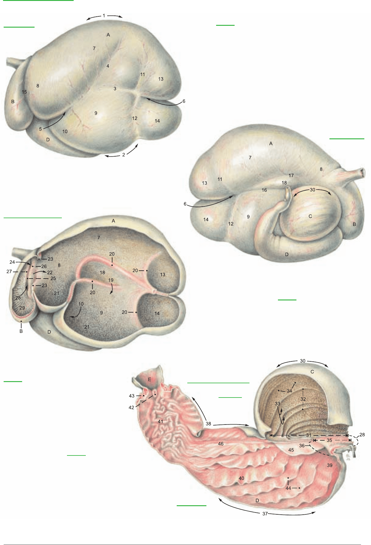
Abomasum
Stomach [Ventriculus]
Left surface
Legend:
A Rumen
1 Dorsal curvature
2 Ventral curvature
3 Left longitudinal groove
4 Left accessory groove
5 Cranial groove
6 Caudal groove
7 Dorsal sac
8 Atrium
9 Ventral sac
10 Recess of ventr. sac of rumen
11 Dorsal coronary groove
12 Ventral coronary groove
13 Caudodorsal blind sac
14 Caudoventral blind sac
15 Ruminoreticular groove
16 Right longitudinal groove
17 Right accessory groove
18 Insula
19 Intraruminal orifice
20 Pillars
21 Papillae
Right surface
Left surface of section
Legend:
Legend:
B Reticulum
22 Ruminoreticular orifice
23 Ruminoreticular fold
24 Cardia
25 Reticular groove
26 Right lip
27 Left lip
28 Reticulo-omasal orifice
29 Reticular crests and cells
C Omasum
30 Curvature
31 Base
32 Omasal laminae
33 Interlaminar recesses
34 Papillae
35 Omasal groove
36 Omasoabomasal orifice
Omasum
Right surface of section
Legend:
D Abomasum
37 Greater curvature
38 Lesser curvature
39 Fundus
40 Body
41 Pyloric part
42 Torus pyloricus
43 Pyloric sphincter
44 Abomasal folds
45 Velum
46 Abomasal groove
E Duodenum
(See pp. 69, 73)
71
Anatomie des Rindes englisch 09.09.2003 14:18 Uhr Seite 71

a) The CELIAC A. (1) originates from the aorta at the level of the
first lumbar vertebra. It has a relatively long course, and after giv-
ing off phrenic arteries and adrenal branches, divides on the right
dorsal surface of the rumen into hepatic, splenic, and left gastric aa.
The arteries of the rumen and reticulum correspond to small
branches of the splenic and left gastric aa. on the simple stomach.
The splenic a. (3) enters the dorsal part of the spleen. Near its ori-
gin it gives off the large right ruminal a. (4) to the right accessory
groove as the main artery of the rumen. This gives off right dorsal
and ventral coronary aa., goes through the caudal groove, and
comes out on the left side of the rumen, where it gives off left dor-
sal and ventral coronary aa. and anastomoses with the left ruminal
a. (5), which passes through the cranial groove of the rumen from
right to left. Near its origin it gives off the reticular a. (6), which
passes over the rumen, then ventrally in the ruminoreticular groove
on the left side, and through the groove from left to right. The right
and left ruminal aa. may originate either from the splenic or left
gastric aa.
The left gastric a. (8) supplies the omasum and goes to the lesser
curvature of the abomasum, where it anastomoses with the right
gastric a. (11) from the hepatic a. (2). On the greater curvature of
the abomasum, the left (9) and right (12) gastroepiploic aa. anas-
tomose. They come from the left gastric a. and the gastroduodenal
a. (a br. of the hepatic), respectively. The accessory reticular a. (10)
arises from the left gastric or from the first part of the left gas-
troepiploic. It runs dorsally on the diaphragmatic surface of the
lesser curvature of the reticulum. The veins, branches of the portal
v., have a predominantly corresponding course.
b) The innervation by AUTONOMIC NERVES is accomplished in
general as in the dog and horse.
The dorsal and ventral trunks of the vagus nn. are of special clini-
cal interest in regulating the functions of each compartment of the
stomach. The rumen is innervated mainly by the dorsal vagal trunk
(a), but the atrium of the rumen and the other three compartments
are innervated by both vagal trunks. Individual brr. of these nerves
may vary in location or extent. The dorsal vagal trunk supplies the
right side of the atrium (h), the brr. to the celiac plexus (c), the dor-
sal ruminal brr. (d), and the right ruminal br. (b), which runs back
in the right accessory groove, giving brr. to the dorsal and ventral
sacs, and passing around in the caudal ruminal groove to the left
side. A branch of the dorsal trunk is also given off to the cranial
ruminal groove and left longitudinal groove (e) and to the greater
curvature of the abomasum (g). Branches of the dorsal trunk (f)
pass over the omasum and the visceral side of the lesser curvature
of the abomasum, innervating the right lip of the reticular groove,
the caudal (visceral) surface of the reticulum, both sides of the oma-
sum, and the visceral surface of the abomasum to the pylorus.
The ventral vagal trunk (j) gives branches to the left side of the atri-
um (l), the diaphragmatic surface of the reticulum (k), and branch-
es that run in the lesser omentum to the liver, cranial part of the
duodenum, and pylorus (p). Branches of the ventral trunk (m)
innervate the left lip of the reticular groove (see p. 70, c), and con-
tinue across the parietal side of the neck of the omasum and run in
the lesser omentum along the parietal surface of the base of the
omasum and the lesser curvature of the abomasum to the pylorus,
innervating the parietal surface of the omasum and abomasum.
c) The LYMPH NODES of the stomach and spleen include the fol-
lowing: Celiac Inn. (p. 76, A) 2–5 are found with the cran. mesen-
teric lnn. (p. 77) at the origin of the aa. of the same names. Splenic
(or atrial) Inn. (E) 1–7 are grouped dorsocranial to the spleen
between the atrium of the rumen and the left crus of the diaphragm.
Among the numerous gastric lymph nodes, the reticuloabomasal
(A), ruminoabomasal (B), left ruminal (C), right ruminal (D), cra-
nial ruminal (not illustrated), reticular (F), omasal (not illustrated),
dorsal abomasal (G), and ventral abomasal (H) lie in the grooves
and in the omental attachments of the stomach compartments.
Their efferent lymphatic vessels go to the splenic nodes or nodes
preceding them, gastric trunks, visceral trunks, or the cisterna chyli
(p. 74).
d) OMENTA. The embryonic dorsal mesogastrium and ventral
mesogastrium undergo important changes in form and position
with the development of the four compartments of the stomach.
After the rotation of the spindle-shaped stomach primordium
through about 90° to the left, with the axis of the stomach directed
at first from craniodorsal to caudoventral, three protuberances
appear on the greater curvature. In craniocaudal order they are the
primordia of the rumen, reticulum, and greater curvature of the
abomasum. The craniodorsal end of the rumen tube is divided by
the future caudal groove into the future dorsal and ventral caudal
blind sacs.
The only protuberance on the lesser curvature is the primordium of
the omasum. In the course of further development the reticulum
moves cranially; the two blind sacs of the rumen turn dorsally and
then caudally, so that cranial and caudal blind sacs become dorsal
and ventral. The caudal groove is extended on both sides of the
rumen as the right and left longitudinal grooves, and a flexure in
the rumen tube becomes the cranial groove. The abomasum
approaches the rumen and reticulum, and its greater curvature
becomes ventral as it continues the rotation clockwise as viewed
from the head. The omasum comes up on the right side.
In spite of these complicated translocations, the attachments of the
dorsal and ventral mesogastria to the greater and lesser curvatures
of the stomach primordium are maintained. The line of attachment
of the dorsal mesogastrium on the stomach in the adult runs from
the dorsal surface of the esophagus at the hiatus to the right longi-
tudinal groove, through the caudal groove and the left longitudinal
groove of the rumen, across a part of the left surface of the atrium
and reticulum, and along the greater curvature of the abomasum to
the cranial part of the duodenum.
The greater omentum (see the lower left figure) with its deep wall
(15) and superficial wall (14), together with the omental bursa, is
the main derivative of the dorsal mesogastrium. It extends caudal-
ly, ventrally, and to the right. Caudally near the pelvis, as in the dog,
the deep wall is reflected as the supf. wall, forming a fold enclosing
the caudal recess of the omental bursa (16). Ventrally, because the
attachment of the dorsal mesogastrium to the rumen followed the
right longitudinal, caudal, and left longitudinal grooves, the ventral
sac is enclosed by the greater omentum and forms a part of the wall
of the omental bursa. On the right, the greater omentum is adher-
ent to the medial surface of the mesoduodenum from the cranial
flexure, along the descending part, to the caudal flexure of the duo-
denum (p. 69, 12). In the sling formed by the deep and supf. walls
of the greater omentum between the mesoduodenum and the right
longitudinal groove of the rumen, is the supraomental recess (13),
open caudally and containing the bulk of the intestines. The deep
wall of the greater omentum passes from the mesoduodenum, ven-
tral to the intestines, to its attachment in the right longitudinal
groove of the rumen, whereas the supf. wall passes ventral to the
intestines and the ventral sac of the rumen to the left longitudinal
groove. Both walls of the omentum meet in the caudal groove. Cra-
nial parts of the dorsal mesogastrium disappear or are shortened in
the adult by expansion of the atrium and adhesion with its sur-
roundings. The spleen on the left and the left lobe of the pancreas
are held between the rumen and the diaphragm by adhesions. The
line of origin of the dorsal mesogastrium is displaced to the right
and runs obliquely craniocaudally from the level of the esophageal
hiatus through the origin of the celiac a. to the level of the distal
loop of the ascending colon.
The ventral mesogastrium is divided by the developing liver into the
lesser omentum on the visceral surface of the liver and the falciform
lig. (see p. 75, 13) on the diaphragmatic surface. The lesser omen-
tum extends, as the hepatogastric lig., from the porta of the liver
ventrally to the esophageal hiatus, the lesser curvature of the re-
ticulum, the base of the omasum, and the lesser curvature of the
abomasum, covering the right surface of the omasum (p. 69, 30).
The lesser omentum ends as a free border, the hepatoduodenal lig.,
from the porta of the liver to the cranial flexure of the duodenum.
It contains the portal vein and forms the ventral border of the
omental (epiploic) foramen, which leads to the vestibule of the
omental bursa. The vestibule opens into the caudal recess.
72
4. BLOOD SUPPLY AND INNERVATION OF THE STOMACH; LYMPH NODES AND OMENTA
Anatomie des Rindes englisch 09.09.2003 14:18 Uhr Seite 72
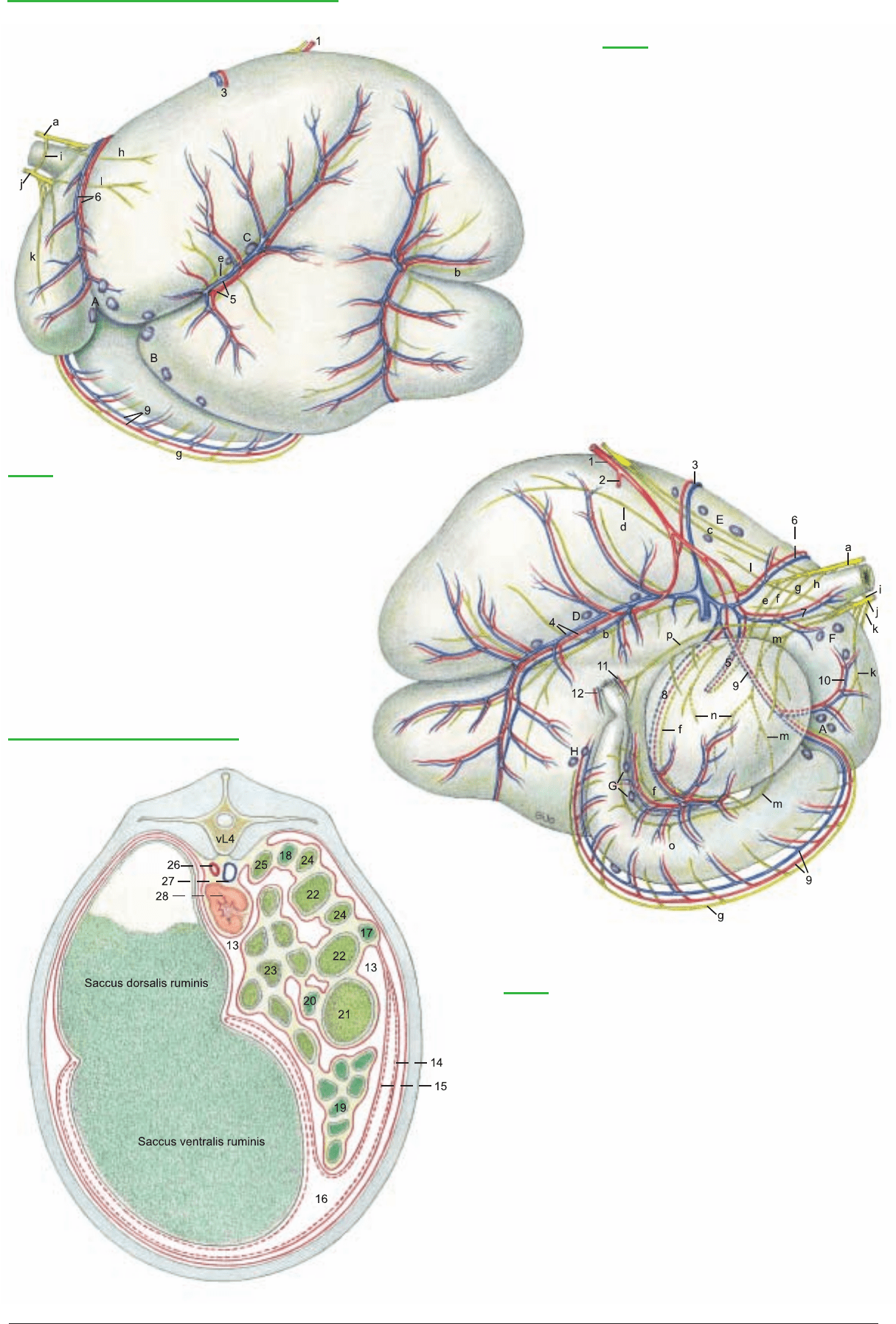
13 Supraomental recess
Greater omentum:
14 Superficial wall
15 Deep wall
Omental bursa:
16 Caudal recess
Duodenum:
17 Descending part
18 Ascending part
19 Jejunum
20 Ileum
21 Cecum
Ascending colon:
22 Proximal loop
23 Spiral loop
24 Distal loop
25 Descending colon
26 Aorta
27 Caudal vena cava
28 Left kidney
Gastric Vessels, Nerves, and Lymph nodes
(Left surface)
Legend:
A Reticuloabomasal lnn.
B Ruminoabomasal lnn.
C Left ruminal lnn.
D Right ruminal lnn.
E Splenic (or atrial) lnn.
F Reticular lnn.
G Dorsal abomasal lnn.
H Ventral abomasal lnn.
a Dorsal vagal trunk
b Right ruminal br.
c Brr. to celiac plexus
d Dorsal ruminal brr.
e Left ruminal br.
f Brr. of the dorsal vagal trunk
g Br. to greater curvature of abomasum
h Atrial brr.
i Communicating br.
j Ventral vagal trunk
k Cran. reticular brr.
l Atrial brr.
m Brr. of the ventral vagal trunk
n Omasal brr.
o Parietal abomasal brr.
p Long pyloric br.
(Right surface)
Legend:
1 Celiac a.
2 Hepatic a.
3 Splenic a. and v.
4 Right ruminal a. and v.
5 Left ruminal a.
6 Reticular a. and v.
7 Caud. esophageal brr.
8 Left gastric a. and v.
9 Left gastroepiploic a. and v.
10 Accessory reticular a. and v.
11 Right gastric a. and v.
12 Right gastroepiploic a. and v.
Greater omentum and Viscera
(Caudal surface of section)
Legend:
(See pp. 65, 67)
73
Anatomie des Rindes englisch 09.09.2003 14:18 Uhr Seite 73
a) The SPLEEN is relatively small, red-brown in the bull and blue-
gray in the cow. It is up to 50 cm long and its average weight varies
with sex, age, and body size from 390 to 2000 g. It is an elongated
oval, tongue-shaped organ of about equal width throughout. Its
position is almost vertical (see p. 69, 4). The dorsal end (2) is near
the vertebral column and the ventral end (6) is a hand’s breadth
dorsal to the 7th–8th costochondral junction. The cranial (4) and
caudal (5) borders are rounded in the bull, acute in the cow. The
spleen does not extend caudal to the line of pleural reflection. The
diaphragmatic surface is applied to the diaphragm; the visceral sur-
face, dorsomedially to the atrium of the rumen and cranioventrally
to the reticulum. Both surfaces of the dorsal part are more or less
extensively fused with the surroundings, so that a phrenicosplenic
lig. (1) and a gastrosplenic lig. are only vestigial. The rather small
hilus (3) is in the dorsal third of the cranial border in the area of
adhesion to the rumen.
b) The LIVER reaches its adult size by the third year and after that
its weight ranges from 4–10 kg depending on breed, age, and nutri-
tional condition. The weight is relatively greater in the calf. Its col-
or varies from yellowish in the calf to reddish-brown in the adult.
Because of the enlargement of the rumen it is almost entirely dis-
placed to the right (see p. 69), except for a small portion ventral to
the esophagus.
The right lobe is caudodorsal and the left lobe is cranioventral. The
thick dorsal border (28) is almost in the median plane. Here the
caudal vena cava (h) runs in a groove inclined ventrally to the fora-
men venae cavae. Between the caudate lobe and the left lobe is the
esophageal impression (w), distinct only in fixed livers. The acute
ventral border (27) is caudoventral on the right. The fixed specimen
shows a large omasal impression (q) and ventral to it, a reticular
impression (r). In contrast to the dog and horse the liver is not dis-
tinctly lobated. Except for the fissure for the round ligament (p),
interlobar notches are absent. The left lobe (26) is not divided. The
gallbladder fossa separates the right lobe (17), undivided as in the
horse, from the quadrate lobe (22). The caudate lobe lies between
the vena cava and the left (intrahepatic) branch of the portal vein.
As in the dog it has a papillary process (24), which overlaps the left
branch of the portal vein. The short caudate process (15) overlaps
the right lobe and is partially fused with it. Together they form the
renal impression for the cranial end of the right kidney. On the vis-
ceral surface is the porta hepatis where the portal v., hepatic a., and
autonomic nn. enter the liver, and the bile-carrying hepatic duct
and lymph vessels leave the liver. Of the hepatic ligaments, the right
triangular lig. (7) goes to the dorsal abdominal wall, and dorsome-
dial to it, the hepato-renal lig. (8) connects the caudate process to
the right kidney. The left triangular lig. (14) is found on the
diaphragm near the esophageal hiatus. The coronary lig. (21)
attaches the liver to the diaphragm and connects the triangular ligg.
and the falciform lig. Its line of attachment to the liver passes from
the left triangular lig. around ventral to the caud. v. cava and along
the right side of the caud. v. cava. On the right lobe it divides into
two laminae that surround the area nuda (16). The falciform lig.
(13) with the round lig. in its free border is attached to the
diaphragmatic surface of the liver on a line from the coronary lig.
at the foramen venae cavae to the fissure for the round lig. It is
attached to the diaphragm on a horizontal line from the foramen
venae cavae to the costochondral junction. Unlike that of the horse,
it does not go to the umbilicus. The diaphragmatic attachment is a
secondary adhesion resulting from the displacement of the liver to
the right, and in many adults the falciform and round ligg. have dis-
appeared. The gallbladder (25) is pear-shaped with a total length of
10–15 cm. It extends beyond the ventral (right) border of the liver.
The right and left hepatic ducts join to form the common hepatic
duct (18), which receives the cystic duct (20) and becomes the
short, wide common bile duct (19, ductus choledochus), which
opens into the duodenum about 60 cm from the pylorus on the
oblique greater duodenal papilla. Hepatocystic ducts open directly
into the gall bladder.
c) The main duct of the bovine PANCREAS is the accessory pan-
creatic duct (m), which opens in the descending duodenum
30–40 cm from the greater duodenal papilla. The pancreatic duct is
represented in the ox by small ducts that open into the common
hepatic duct in its course across the pancreas. The left lobe (10)
extends to the spleen and is attached by connective tissue to the
rumen and the left crus of the diaphragm. The body of the pancreas
(12) lies between the liver and the omasum ventral to the portal
vein, which passes dorsally through the pancreatic notch (11) to the
liver. The right lobe (9) is enclosed in the mesoduodenum descen-
dens and extends to the plane of the right kidney.
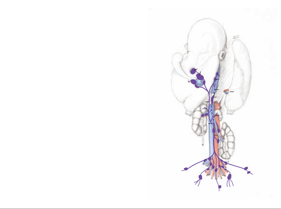
d) The LYMPH NODES of the spleen, liver, and pancreas.
The 1–7 splenic lnn. (p. 73) lie dorsocranial to the spleen between
the atrium of the rumen and the left crus of the diaphragm, and are
regularly examined in meat inspection.
The 6–15 hepatic (portal) lnn. (23) are grouped around the porta
of the liver and are regularly examined in meat inspection. The
accessory hepatic lnn. (29) are found on the dorsal border of the liv-
er near the caudal vena cava. The outflow of lymph occurs, togeth-
er with that of the dorsal and ventral abomasal lnn., through the
hepatic trunk. The pancreaticoduodenal lnn. (see p. 76, I) lie
between the pancreas and descending duodenum and between the
pancreas and transverse colon.
The lymph drainage is through the intestinal trunk (A) which joins
the hepatic trunk (B), and after receiving the gastric trunk (C) with
lymph from the stomach and spleen, becomes the visceral trunk (D)
and enters the cisterna chyli (E). The valveless cisterna chyli receives
the lumbar trunk (F), which drains the lymph from the pelvic limbs,
genital organs, and the pelvis.
The thoracic duct (G), emerging cranially from the cisterna chyli,
passes in the ox through a slit in the muscle of the right crus of the
diaphragm into the thorax. It does not pass through the aortic hia-
tus as in the horse and dog. For lymph nodes of the pelvic cavity,
see also pp. 82–83.
74
5. SPLEEN, LIVER, PANCREAS, AND LYMPH NODES
* see also Baum, 1912
E
B
23
29
C
F
A
D
G
25
3
Lymph nodes and Lymphatic vessels*
(ventral)
(See p. 82)
Anatomie des Rindes englisch 09.09.2003 14:18 Uhr Seite 74
