Budras Klaus-Dieter, Habel Robert E. Bovine Аnatomy
Подождите немного. Документ загружается.

d'
e'
e"
e"
h'
33'
h'
m'
q"
q'
r'
45'
50"
50'
P'
N'
r' Spiral gangl. of cochlea
s Sympathetic trunk
s' Cranial cervical gangl.
t Vagosympathetic trunk
u Spinal root of accessory n.
v Ansa cervicalis
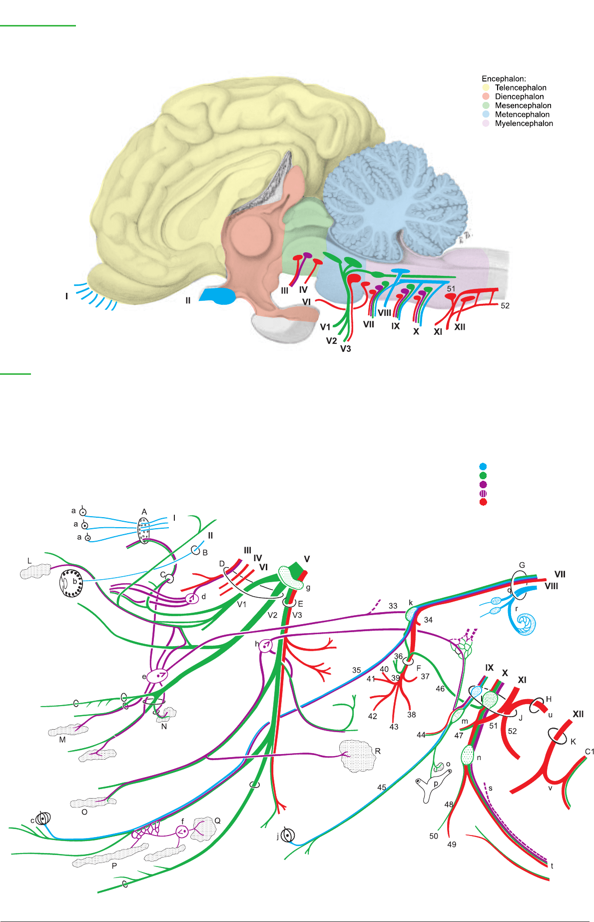
Cranial nerves
Legend:
A Cribriform plate
B Optic canal
C Ethmoid foramen
D For. orbitorotundum
E Oval foramen
F Stylomastoid for.
G Int. acoustic meatus
H Foramen magnum
J Jugular foramen
K Hypoglossal canal
L Lacrimal gl.
M Nasal gll.
N Palatine gll. (soft palate)
N' Palatine gll. (hard palate)
O Buccal gll.
P Monostomatic sublingual gl.
P' Polystomatic sublingual gl.
Q Mandibular gl.
R Parotid gl.
a Olfactory region
b Retina
c Fungiform papillae
d Ciliary ganglion
d' Short ciliary nn.
e Pterygopalatine gangl.
e' Orbital brr.
e" N. of pterygoid canal
(major and deep petrosal nn.)
f Mandibular ganglion
g Trigeminal ganglion
h Otic ganglion
h' Minor petrosal n.
j Vallate papillae
k Geniculate ganglion
l Proximal ganglia
m Distal gangl. (petrosal)
m' Tympanic n.
n Distal gangl. (nodose)
o Carotid glomus
p Carotid sinus
q Vestibular n.
q' Sup. vestibular gangl.
q'' Inf. vestibular gangl.
r Cochlear n.
Special sensory neuron
Sensory neuron
Parasympathetic neuron
Sympathetic neuron
Motor neuron
55
Anatomie des Rindes englisch 09.09.2003 13:50 Uhr Seite 55

The following statements concern only a few specific characteristics
of the ox. For the rest, the generally applicable textbook descrip-
tions and the detailed illustrations in the neurological literature
may be consulted.
a) The SPINAL CORD (MEDULLA SPINALIS) is surrounded by
the meninges in the vertebral canal. In animals it has a greater bio-
logical importance than in man, and in the ox its mass is almost as
great as that of the brain. The spinal cord presents a cervical
enlargement and a lumbar enlargement. The central canal is pre-
dominantly transversely oval, as in the horse. The cord ends as the
conus medullaris (16), containing the sacral and caudal segments.
This extends in the two-month-old calf through vert. S3, and at ten
months, through vert. S2*, but in the adult the conus extends only
into vert. S1. The difference is caused by the so-called “ascent of the
cord,” really by the continued growth of the vertebral column after
the growth of the cord has slowed. This results in a longer course
of the spinal nerves within the vertebral canal before they reach
their intervertebral foramina, forming the cauda equina (18), which
is composed of the conus medullaris, the terminal filament (17) of
connective tissue, and the sacral and caudal nerves. The clinical
importance is in the danger of injury to the cord by lumbosacral
puncture. The space between the spine of vert. L6 and the sacral
crest overlies the intervertebral disc and the cranial part of the body
of vert. S1. In the mature ox, although the sacral segments of the
cord are all in vert. L6, the caudal segments, the last lumbar nerve,
the sacral nerves, and some caudal nerves are vulnerable. Epidural
anesthesia is performed in the ox by injection between the first and
second caudal vertebrae, and lumbosacral puncture is restricted to
diagnostic withdrawal of cerebrospinal fluid.
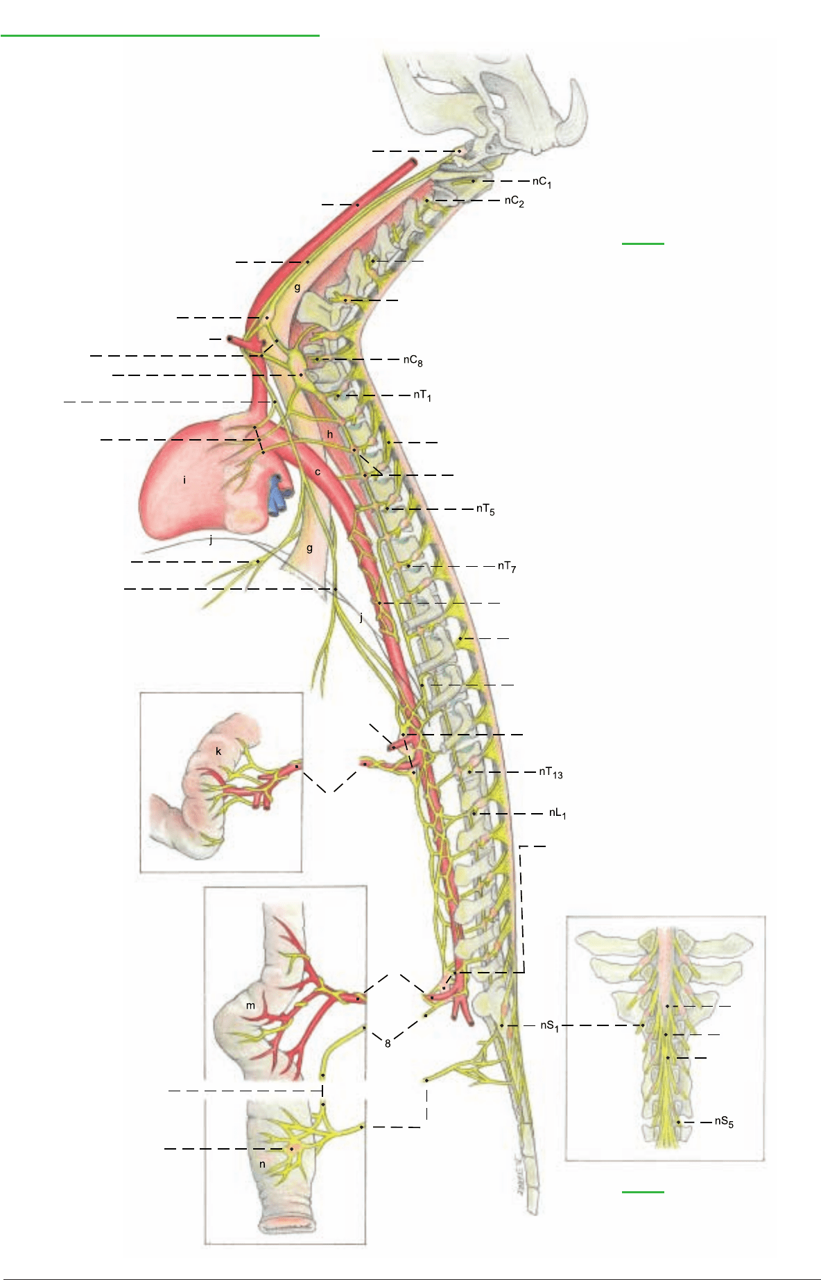
b) THE AUTONOMIC NERVOUS SYSTEM includes the sympa-
thetic part, the paraympathetic part, and the intramural intestinal
plexuses.
The efferent nerve fibers:
I. The sympathetic part consists mainly of efferents with pre- and
postsynaptic neurons, and also contains afferents with only one
neuron. It is also called the thoracolumbar nervous system because
the nerve cell bodies of the efferents are in the lateral horns of the
corresponding segments of the spinal cord. However, the sympa-
thetic trunk (12) extends farther caudally, to the first caudal verte-
bra, where the paired ganglia unite in the ganglion impar. The tho-
racolumbar body parts and organs are supplied by relatively short
(nearly transverse) communicating brr. to the ganglia of the sym-
pathetic trunk.
1. The thoracic organs are supplied by postsynaptic unmyelinated
neurons that come from the cervicothoracic ggl. (5) or from the
ansa subclavia (4) or from the middle cervical ggl. (3) and go, e.g.
as cardiac nn. or pulmonary nn., to the corresponding organs. They
form with branches of the vagus n. (e.g. cardiac brr. or pulmonary
brr.) autonomic plexuses for the thoracic organs (e.g. cardiac
plexus, 7).
2. The abdominal organs are mainly supplied through the major
splanchnic n. (13), which leaves the sympathetic trunk at the level
of vert.T 10, and passes over the lumbocostal arch of the dia-
phragm to the celiac ggl. and cran. mesenteric ggl. (14). In addition,
the minor splanchnic nn. and the lumbar splanchnic nn. from the
lumbar sympathetic trunk go to the solar plexus or to the caud.
mesenteric ggl. (15). The myelinated presynaptic first neurons come
mainly without synapse through the ggll. of the sympathetic trunk,
and most of the neurons synapse first in the following prevertebral
ggll.: celiac ggl. (14), cran. (14), and caud. (15) mesenteric ggll. The
unmyelinated postsynaptic second neurons reach the areas they
supply through periarterial plexuses of the visceral aa., e.g. those of
the intestinal wall.
The communicating brr. to the somatic thoracic and lumbar nn.
(white communicating brr.) synapse in the ggll. of the sympathetic
trunk (12), and the second neurons (gray communicating brr.) con-
duct sympathetic impulses to those nn. The body parts and organs
(Nos. 3–6) cranial or caudal to the thoracolumbar body segments
are supplied by relatively long (longitudinal) nerves.
3. The head is supplied by efferent sympathetic neurons from the
cervicothoracic ggl. that pass through the ansa subclavia and the
middle cervical ggl. and the vagosympathetic trunk (2) to the cran.
cervical ggl. (1). This ggl. at the level of the base of the skull is the
last synaptic transfer station. From here only postsynaptic
unmyelinated neurons, as perivascular plexuses, reach, with blood
vessels of the same name, their areas of innervation in the head (e.g.
int. carotid plexus, maxillary plexus).
4. The neck is supplied by the vertebral n. (11). It leaves the cervi-
cothoracic ggl. and passes through the foramina transversaria of
the cervical vertebrae as far as the third. It gives gray rami commu-
nicantes to the 2nd to 6th cervical nn.
5. The pelvic cavity receives sympathetic neurons over two differ-
ent pathways. The dorsal path goes through the lumbar and sacral
sympathetic trunk and into the sacral splanchnic nn. which run
together with the pelvic n. (10) to the pelvic plexus (9). The ventral
path goes from the lumbar sympathetic trunk through the lumbar
splanchnic nn. (15) to the caud. mesenteric ggl. (15) and over the
hypogastric n. (18) to the mixed autonomic pelvic plexus. Here at
the pelvic inlet, on the lateral wall of the rectum, is the transfer to
the postsynaptic neurons which supply the pelvic organs and the
descending colon.
6. The limbs are supplied by postganglionic unmyelinated neu-
rons. From the cervicothoracic ggl. at the cran. end of the thoracic
sympathetic trunk, they reach the thoracic limb, and from the caud.
end of the lumbar sympathetic trunk they reach the pelvic limb.
They first pass through the brachial plexus or lumbosacral plexus
in the somatic nn., and more distally enter the adventitia of blood
vessels.
II. The parasympathetic part to which cranial nn. III, VII, IX, and
X and the pelvic n. (10) belong, supplies with its efferents the
glands and smooth muscle cells in e.g. the gut, and also in the eye
and in the salivary and lacrimal gll. The efferents are connected
through two neurons in series to carry the impulse from the CNS to
the target organ. In the vagus, the presynaptic axon is very long,
extending from the CNS to the synapse with the second neuron in
the target organ. Vagal fibers extend as far as the transverse colon.
For the origin and distribution of the vagus, see pp. 48, 54, and 72.
The afferent nerve fibers:
The sympathetic and parasympathetic nn. contain afferents of sen-
sory neurons that measure the contraction or distention of hollow
organs and transmit pain. The vagus at the diaphragm contains
more than 80 % afferent fibers. The cell bodies of the sympathetic
afferents lie in the spinal ganglia, and those of the vagus are in the
proximal (jugular) and distal (nodose) ganglia near the base of the
skull (see p. 48).
56
4. SPINAL CORD AND AUTONOMIC NERVOUS SYSTEM
* Weber, 1942
Demonstration specimens are provided for the study of the spinal cord. The arches of the vertebrae and portions of the meninges have
been removed to show the dorsal surface of the cord. Transverse sections are studied to see the distribution of gray and white matter,
the course of the central canal, and the positions of the fiber tracts.
Anatomie des Rindes englisch 09.09.2003 13:50 Uhr Seite 56
b
d
e
f
16 Conus medullaris
17 Filum terminale
18 Cauda equina
Spinal cord and Autonomic nervous system
(lateral)
1 Cran. cervical ggl.
2 Vagosympathetic trunk
3 Middle cervical ggl.
4 Ansa subclavia
5 Cervicothoracic ggl.
[Stellate ggl.]
6 Vagus n.
7 Cardiac plexus
8 Hypogastric n.
9 Pelvic plexus
10 Pelvic n.
11 Vertebral n.
12 Ggll. of sympathetic trunk
13 Major splanchnic n.
14 Celiac ggl. and
cran. mesenteric ggl.
15 Caud. mesenteric ggl. and
lumbar splanchnic nn.
Spinal ggl.
Dorsal root
Ventral vagal trunk
Dorsal vagal trunk
Aortic plexus
Ventral root
16
17
18
Legend:
a Left common carotid a.
b Left subclavian a.
c Aorta
d Celiac a.
e Cran. mesenteric a.
f Caud. mesenteric a.
g Esophagus
h Longus colli
i Heart
j Diaphragm
k Small intestine
m Large intestine
n Rectum
a
Legend:
(dorsal)
57
Anatomie des Rindes englisch 09.09.2003 13:50 Uhr Seite 57
a) The VERTEBRAL COLUMN is composed of seven cervical
vertebrae, thirteen (12–14) thoracic vertebrae, six (7) lumbar ver-
tebrae, five sacral vertebrae, and eighteen to twenty (16–21) caudal
vertebrae.
The vertebrae are joined by fibrocartilaginous intervertebral discs,
and surround the vertebral canal (7). The basic parts: body (1), arch
(8), and processes are developed differently according to function.
I. The cervical vertebrae (C1–C7) are generally shorter than in the
horse. The spinous process (12) is longer than in the horse, and
inclined cranially. Only on the seventh is it almost vertical, and on
the third and fourth cervical vertebrae the free end is split. On the
first, the spinous process is represented by a tubercle (29'). The
massive transverse process (13) of the third to the fifth bears a cra-
nial ventral tubercle (13') and a caudal dorsal tubercle (13") as in
the dog and horse. On the sixth cervical vertebra the ventral tuber-
cle is replaced by a sagittal quadrilateral plate, the ventral lamina
(13'). The cranial (16) and caudal (17) articular processes are very
small compared to those of the horse. The atlas (C1) lacks a trans-
verse foramen (15); the dorsal arch bears a large dorsal tubercle
(29'); and the ventral arch, a large ventral tubercle (30'), which is
sometimes bifid. The axis (C2) is shorter than in the horse; its dens
(32) is semicylindrical; the spinous process (12) is a high and
straight crest, but not split caudally as it is in the horse. The lateral
vertebral foramen (31'), absent in the dog, is very large.
II. The thoracic vertebrae (T1–T13) have relatively long bodies
compared to the dog and horse. The spinous process (12) of the
first to the fifth thoracic vertebra is broad with sharp cranial and
caudal borders, and provided on the free end with a cartilaginous
cap until about the third year. These ossify by the eighth year. The
withers (interscapular region) is not as high as in the horse. The sev-
enth to eleventh thoracic spines are strongly inclined caudally. The
spine is vertical on the last thoracic (anticlinal) vertebra. In most
thoracic vertebrae the caudal vertebral notch (11) is closed by a
bridge of bone to form a lateral vertebral foramen (11'). The
mamillary processes (20) are not very prominent; on the last two
thoracic vertebrae they merge with the cranial articular processes
(16).
III.The lumbar vertebrae (L1–L6) have a long body and a flat arch
with an almost square, cranially and caudally extended, spinous
process (12). The horizontal transverse processes (13) are curved
cranially and separated by wide spaces. The cranial lumbar verte-
brae often have lateral vertebral foramina as in the thoracic verte-
brae. The mamillary processes are always fused with the cranial
articular processes.
IV.The sacral vertebrae (S1–S5) are completely fused to form the
sacrum after 3–4 years. Depending on breed, the sacrum is more or
less arched dorsally. Ventrally it has a distinct groove for the medi-
an sacral artery. The spinous processes are fused to form a median
sacral crest (35) (as in the dog, but unlike the horse) with an occa-
sional interruption between the fourth and fifth vertebrae. The
sacral promontory (38) is the cranial ventral prominence of the first
sacral vertebra. It is palpable per rectum. The auricular surfaces of
the alae face caudodorsally. The fused articular processes form a
ridge, the intermediate sacral crest (37), which bridges over the nar-
row dorsal sacral foramina (39), and lies medial to the last sacral
foramen. This is very large and not divided into dorsal (39) and
ventral (40) foramina because the last two transverse processes are
not completely fused.
V. The caudal [coccygeal] vertebrae (Cd1+) and their processes are
significantly larger and better developed than in the horse. The
progressively narrowing vertebral canal (7) extends to the fifth cau-
dal vertebra. The paired hemal processes (21) (present as in the dog,
unlike the horse) may be closed to form hemal arches (22) from the
second to the fifth caudal vertebra.
b) Of the thirteen RIBS, eight are sternal ribs (41) and five are
asternal (42). They increase in length to the tenth rib and, especial-
ly in the middle of the thorax, they are flat toward the sternal end
with sharp caudal borders, and wider than in the dog and horse,
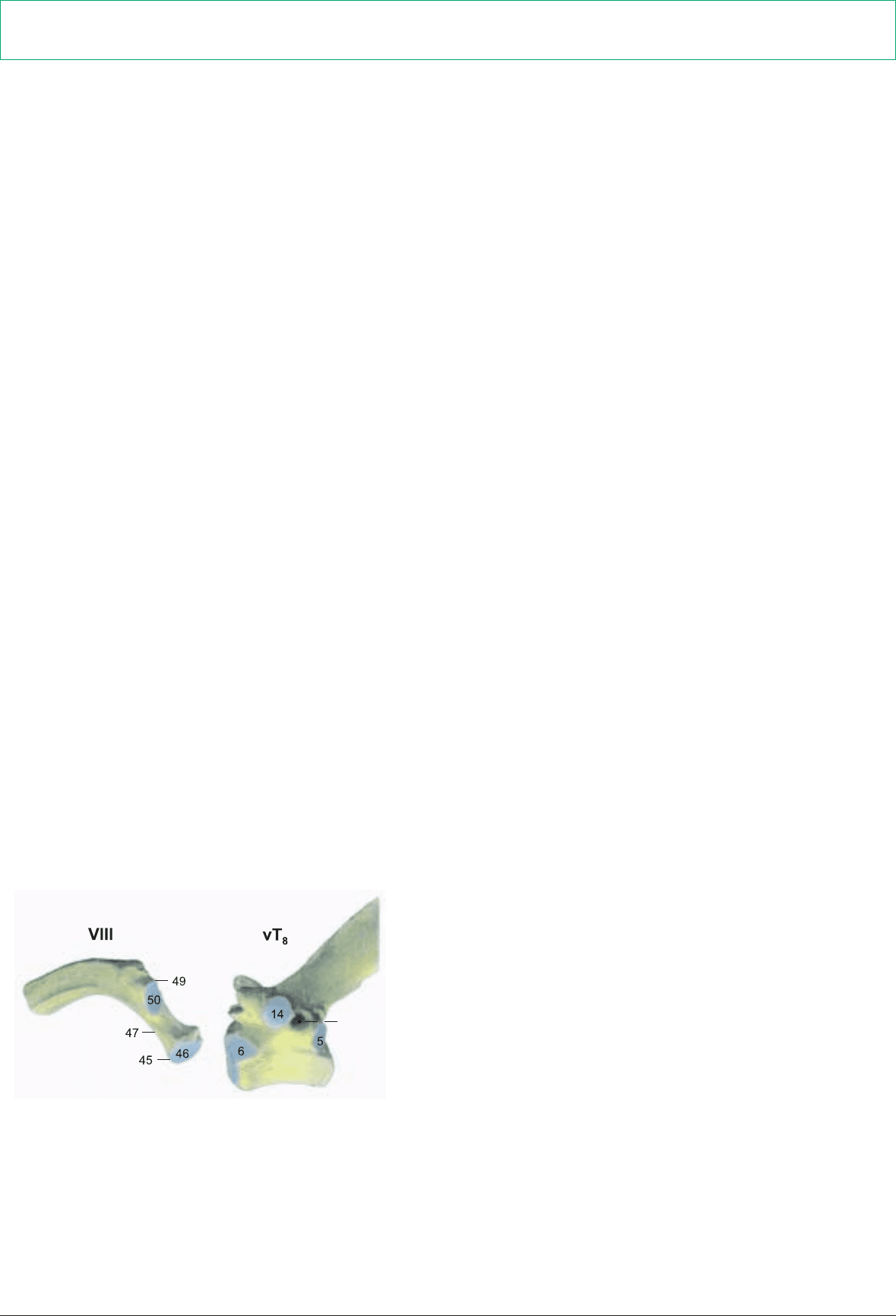
whereby the intercostal spaces become narrower. The head (45)
and the tubercle (49) are well developed and separated by a long
neck (47). The knee [genu costae, 53] is at the costochondral junc-
tion.
c) The body of the STERNUM, formed by five sternebrae (56), is
slightly arched dorsally and flattened dorsoventrally. The triangu-
lar manubrium sterni (54) is raised craniodorsally and has no
manubrial cartilage. It is attached to the body of the sternum by a
true joint. The xiphoid process (57) is smaller than in the horse. A
sternal crest is absent, as in the dog.
d) The elastic NUCHAL LIGAMENT is generally better devel-
oped than in the dog and horse. It consists of a paired funiculus (A)
and a lamina (B), which is paired in the cranial part and unpaired
in the caudal part.
The funiculus is divided into right and left halves attached to the
external occipital protuberance. They extend, without attachment
to the cervical vertebrae, to the withers, and become gradually
wider to form the sagittally positioned, flat, wide parts lateral to the
first to fifth thoracic spinous processes, but not capping them. The
wide parts gradually become narrower and unite to form the
supraspinous ligament (C), which extends to the sacrum. It is elas-
tic cranially, but becomes collagenous in the midlumbar region. The
lamina arises with its cranial paired part from spinous processes
C2–C4 and fuses with the funiculus. The caudal unpaired part, also
elastic, which in the horse is thin and contains few elastic fibers,
arises from vertebrae C5–C7 and terminates on the first thoracic
spinous process under the wide parts of the funiculus. A
supraspinous bursa may be present between the first few thoracic
spines and the wide parts of the funiculus.
58
CHAPTER 5: VERTEBRAL COLUMN, THORACIC SKELETON, AND NECK
1. VERTEBRAL COLUMN, LIGAMENTUM NUCHAE, RIBS, AND STERNUM
Review the basic parts of the bones on individual bones and mounted skeletons, and study the special features in the ox mentioned
below.
11'
(cranial)
(caudal)
Costovertebral articulations
Anatomie des Rindes englisch 09.09.2003 13:50 Uhr Seite 58

48
51
9
27'
29'
30'
13'
13"
13'
31'
13"
13'
Vertebral column, Thoracic skeleton, and Nuchal ligament
Vertebral column and Bones of the thorax
Cervical vertebrae (C1–C7)
Thoracic vertebrae (T1–T13, 14)
Lumbar vertebrae (L1–L6)
Sacral vertebrae (S1–S5)
Caudal [Coccygeal] vertebrae (Cd1–Cd16, 21)
Body of vertebra (1)
Ventral crest (2)
Cranial end (3)
Caudal end (4)
Caud. costal fovea (5)
Cran. costal fovea (6)
Vertebral canal (7)
Vertebral arch (8)
Intervertebral foramen (9):
Cran. vertebral notch (10)
Caud. vertebral notch (11)
Lat. vertebral foramen (11')
Spinous process (12)
Transverse process (13)
Ventral tubercle (C3–C5) (13') [Ventral lamina C6]
Dorsal tubercle (C3–C5) (13")
Costal fovea (T1–T13) (14)
Transverse foramen (C2–C6) (15)
Cran. articular process (16)
Caud. articular process (17)
Costal process (18)
[Transverse proc.] (L1–L6)
[Ventr. tubercle] (C3–C5)]
Mamillary process (T+Cd) (20)
Hemal process (Cd2–Cd15) (21)
Hemal arch (Cd4 + Cd5) (22)
Interarcuate space:
Lumbosacral (23)
Sacrocaudal (24)
Atlas [C1]
Lateral mass
Transverse proc. [Wing of atlas, Ala] (26)
Alar foramen (27')
Lat. vertebral foramen (28)
Dorsal arch (29)
Dorsal tubercle (29')
Ventral arch (30)
Ventral tubercle (30')
Axis [C2]
Lat. vertebral foramen (31')
Dens (32)
Sacrum [S1–S5]
Wing [Ala] of sacrum (33)
Median sacral crest (35)
Lat. sacral crest (36)
Intermediate sacral crest (37)
Promontory (38)
Dorsal sacral foramen (39)
Ventral sacral foramen (40)
Ribs [Costae]
Sternal ribs (41)
Asternal ribs (42)
Costal bone [Os costale] (44)
Head of rib (45)
Artic. surface of head (46)
Neck of rib (47)
Body of rib (48)
Costal tubercle (49)
Artic. surf. of tubercle (50)
Angle of rib (51)
Costal cartilage (52)
Knee of rib [Genu costae] (53)
Sternum
Manubrium (54)
Body of sternum (55)
Sternebrae (56)
Xiphoid process (57)
Legend:
Nuchal ligament:
A Funiculus nuchae
B Lamina nuchae
C Supraspinous lig.
C1
(caudodorsal)
C1
C2
C7
(lateral)
C6
(lateral)
T13
(dorsolateral)
L5 + L6
S1–S5
Cd1
Cd5
(caudal)
Cd1
(ventral)
L5 + L6
S1–S5
59
Anatomie des Rindes englisch 09.09.2003 13:50 Uhr Seite 59

a) Of the CUTANEOUS MUSCLES, the cutaneus colli is thin and
often impossible to demonstrate. It originates from the ventro-medi-
an cervical fascia. The cutaneus trunci resembles that of the horse;
whereas the cranially attached cutaneus omobrachialis, absent in
the dog, is thinner than in the horse, and occasionally unconnected
to the cutaneus trunci. For the preputial muscles, see p.66.
b) The SUPERFICIAL SHOULDER GIRDLE MUSCLES
(TRUNK—THORACIC LIMB MUSCLES):
The trapezius with its cervical part (11) and thoracic part (11') is sig-
nificantly better developed than in the horse. This fan-shaped mus-
cle originates from the funicular nuchal lig. and supraspinous lig.
between the atlas and the 12th (10th) thoracic vertebra and ends on
the spine of the scapula. The cervical part is connected ventrally to
the omotransversarius (8), which, as in the dog, extends between the
acromion and the transverse process of the atlas (axis), where it is
fused with the tendon of the splenius. The brachiocephalicus con-
sists of the cleidobrachialis (clavicular part of deltoideus, p. 4) and
the cleidocephalicus. The two parts of the latter in the ox are the clei-
do-occipitalis and the cleidomastoideus. The cleido-occipitalis (7),
and the cleidomastoideus, originate from the clavicular intersec-
tion—an indistinct line of connective tissue across the brachio-
cephalicus cranial to the shoulder joint. The cleido-occipitalis is
joined to the cleidomastoideus as far as the middle of the neck, sep-
arates from it, adjoins the ventrocranial border of the trapezius, and
ends on the funicular nuchal lig. and occipital bone. The cleidomas-
toideus (6) lies ventral to the cleidooccipitalis, is partially covered by
it, and ends as a thin muscle with a slender tendon on the mastoid
process and the tendon of the longus capitis. The sternocephalicus
consists of the sternomastoideus and sternomandibularis.
The sternomastoideus mm. (4) originate from the the manubrium
sterni only, are fused in the caudal third of the neck, and terminate
in common with the cleidomastoideus. The sternomandibularis (5)
originates laterally from the manubrium and from the first rib; and,
crossing the sternomastoideus, runs ventral to the jugular groove
and ends with a thin tendon on the rostral border of the masseter
and aponeurotically on the mandible and the depressor labii inferi-
oris. The sternomastoideus and cleidomastoideus are homologous
to the human sternocleidomastoideus.
The latissimus dorsi (12) arises from the thoracolumbar fascia and
from the 11th and 12th ribs. The fibers run cranioventrally to a
common termination with the teres major and an aponeurotic con-
nection with the coracobrachialis and deep pectoral as well as the
long head of the triceps.
Of the superficial pectoral muscles, the flat transverse pectoral (25')
originates from the sternum and ends on the medial deep fascia of the
forearm. The descending pectoral (25) is a thick muscle originating
from the manubrium and ending with the brachiocephalicus on the
crest of the humerus. It is not as visible under the skin as in the horse.
c) JUGULAR GROOVE AND LATERAL PECTORAL
GROOVE: The jugular groove is bounded dorsally by the cleido-
mastoideus, ventrally by the sternomandibularis, and, in the cranial
half of the neck, medially by the sternomastoideus. The ext. jugu-
lar vein (3) lies in the groove. At the junction of the head and neck
it bifurcates, giving rise to the maxillary (2) and linguofacial (1)
veins. At the thoracic inlet it gives off a dorsal branch, the superfi-
cial cervical vein (21); and gives off the cephalic vein (10) to the lat-
eral pectoral groove between the brachiocephalicus and the
descending pectoral muscle.
a) DEEP SHOULDER GIRDLE MUSCLES: The rhomboideus
consists of the rhomboideus cervicis (28) and thoracis (28') but no
rhomboideus capitis, unlike the dog. These are covered by the
trapezius, originate from the funicular nuchal lig. and supraspinous
lig. between C2 and T7 (T8), and terminate on the medial surface of
the scapular cartilage. The deep pectoral (26 and p. 5, t) is a strong
unified muscle which ends primarily on the major and minor tuber-
cles. A branch of the tendon fuses with the latissimus dorsi and ends
on the tendon of origin of the coracobrachialis. The subclavius (26'),
absent in the dog, is not well developed. It extends from the first
costal cartilage to the deep surface of the clavicular intersection. The
serratus ventralis extends from the 2nd (3rd) cervical vertebra to the
9th rib, and is clearly divided into serratus ventralis cervicis (27) and
thoracis. The serratus ventralis thoracis (27') arises by distinct mus-
cle slips and is interspersed with strong tendinous layers. It is
attached not only to the facies serrata of the scapula, but penetrates
with a thick broad tendon between the parts of the subscapularis to
end in the subscapular fossa.
b) LONG HYOID MUSCLES: The sternohyoideus (14), ster-
nothyroideus (15), and omohyoideus do not belong to the shoulder
girdle muscles, but are long muscles of the hyoid bone and thyroid
cartilage. The first two resemble those of the horse, but do not have
a tendinous intersection; they are, however, connected by a tendi-
nous band in the middle of the neck. The omohyoideus (13) is thin
and does not come from the shoulder, but from the deep cervical
fascia, and thereby indirectly from the transverse processes of the
3rd and 4th cervical vertebrae. In the angle between the ster-
nomastoideus and sternomandibularis, and crossed laterally by the
external jugular vein, it passes medially under the mandibular
gland to end with the sternohyoideus on the basihyoid.
c) VISCERA AND CONDUCTING STRUCTURES OF THE
NECK: In the middle of the space for the viscera and conducting
structures is the trachea (19). In life the tracheal cartilages are arched
to give it a vertical oval section, but after death it has a tear-drop
shape. Dorsolateral to the trachea is the common carotid artery (16),
with the vagosympathetic trunk (17), The latter is accompanied by
the small int. jugular v. This may be absent. The esophagus (18) is
dorsal in the first third of the neck; in the other two thirds it is on the
left side of the trachea and at the thoracic inlet it is dorsolateral. The
left recurrent n. (18) accompanies the esophagus ventrally; the right
recurrent n. accompanies the trachea dorsolaterally.
d) LYMPHATIC SYSTEM AND THYMUS: The superficial cervi-
cal lymph node (9) lies in the groove cranial to the supraspinatus,
covered by the omotransversarius and cleido-occipitalis. It receives
lymph from the neck, thoracic limb, and thoracic wall back to the
12th rib. Its efferent lymphatics go to the tracheal trunk; on the left,
also to the thoracic duct. The cranial deep cervical lnn. (22) lie near
the thyroid gland; the middle deep cervical lnn. (23), in the middle
third of the neck on the right of the trachea and on the left of the
esophagus. The caudal deep cervical lnn. (24) are placed around the
trachea near the first rib. They receive lymph from the cervical vis-
cera, ventral cervical muscles and preceding lymph nodes of the
head, neck, and thoracic limb. (See the table of lymph nodes.) Some
of their efferents have the same termination as those of the superfi-
cial cervical ln.; others end in the cran. vena cava. The thymus (20)
is fully developed only in the fetus. It consists of an unpaired left
thoracic part (may be maintained to six years of age), a V-shaped
paired cervical part with the unpaired apex directed toward the
thoracic cavity, and a paired cranial part (already retrogressed at
birth).
60
2. NECK AND CUTANEOUS MUSCLES
A dorsomedian skin incision is made from the skull to the level of the last rib, and laterally along the last rib to its costochondral junc-
tion. A skin incision from the cranial end of the first incision is directed ventrally behind the base of the ear and across the angle of the
mandible to the ventromedian line. The skin is reflected ventrally, sparing the cutaneous muscles, ext. jugular v., and cutaneous nerves,
and continuing to the ventromedian line of the neck, on the lateral surface of the limb to the level of the sternum, and to a line extend-
ing from the axilla to the last costochondral junction. This flap of skin is removed. Note the dewlap [Palear], a breed-variable ventro-
median fold of skin on the neck and presternal region.
3. DEEP SHOULDER GIRDLE MUSCLES, VISCERA AND CONDUCTING STRUCTURES OF THE NECK
The superficial shoulder girdle muscles and the sternomastoideus and sternomandibularis are transected near their attachments on the
limb and sternum and removed, leaving short stumps. The accessory n. (c) and the roots of the phrenic nerve (C5 to C7, q) must be
spared in the dissection.
Anatomie des Rindes englisch 09.09.2003 13:50 Uhr Seite 60
r'
n'
o'
n"
o'
p'
26'
r'
o"
n'''
(See pp. 5, 65, 67)
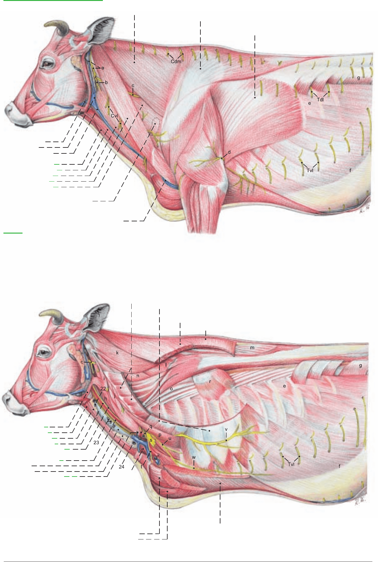
Regions of the neck and chest
1 Linguofacial v.
2 Maxillary v.
3 External jugular v.
Sternocleidomastoideus:
4 Sternomastoideus
5 Sternomandibularis
6 Cleidomastoideus
7 Cleidooccipitalis
8 Omotransversarius
9 Supf. cervical ln.
10 Cephalic v.
Trapezius:
11 Cervical part
11' Thoracic part
12 Latissimus dorsi
Legend:
a Great auricular n. and
caud. auricular v.
b Transverse n. of the neck
c Accessory n.
d Intercostobrachial n.
e External intercostal mm.
f External oblique abd. m.
g Internal oblique abd. m.
h Longus capitis
i Intertransversarius longus
j Ventral cervical
intertransversarii
k Splenius
l Semispinalis capitis
m Spinalis et semispinalis
thoracis et cervicis
Longissimus:
n Longissimus capitis et atlantis
n' Longissimus cervicis
n' Longissimus thoracis
n''' Longissimus lumborum
Iliocostalis:
o Iliocostalis cervicis
o' Iliocostalis thoracis
o" Iliocostalis lumborum
Scalenus:
p Scalenus dorsalis
p' Scalenus ventralis
q C6 root of phrenic n.
r Serratus dors. cranialis
r' Serratus dors. caudalis
s Brachial plexus
t Cran. pectoral nn.
u Caud. pectoral nn.
v Long thoracic n.
w Lat. thoracic n.
Serratus ventralis:
27 Serratus vent. cervicis
27' Serratus vent. thoracis
Rhomboideus:
28 Rhomboideus cervicis
28' Rhomboideus thoracis
13 Omohyoideus
14 Sternohyoideus
15 Sternothyroideus
16 Common carotid a.
17 Vagosympathetic trunk
18 Esophagus and left
recurrent laryngeal n.
19 Trachea
20 Thymus
21 Supf. cervical a. and v.
22 Cran. deep cervical lnn.
23 Middle deep cervical lnn.
24 Caud. deep cervical lnn.
Supf. pectoral mm.:
25 Descending pectoral m.
25' Transverse pectoral m.
26 Deep pectoral m.
26' Subclavius
61
Cdm = Med. dors. cut. brr. of cervical nn. Tdl = Dorsolat. cut. brr. of thoracic nn. Tvl = Ventrolat. cut. brr. of thoracic nn.
Anatomie des Rindes englisch 09.09.2003 14:11 Uhr Seite 61

a) The RESPIRATORY MUSCLES (see appendix on myology)
belong partly to the muscles of the back and partly to those of the
thorax. They function as expiratory muscles in the contraction of
the thorax or as inspiratory muscles in the expansion of it. The
obligate respiratory muscles are aided by the auxiliary respiratory
muscles. The diaphragm is the primary respiratory muscle and the
partition between the thoracic and abdominal cavities.
The line of diaphragmatic attachment rises steeply, running across
the ribs from the knee of the 8th, across the 11th rib below its mid-
dle to the vertebral end of the 13th rib. In ruminants the two costal
parts (3) of the diaphragm are clearly separated from the 13–15 cm
wide sternal part (not illustrated) by clefts between muscle fibers.
The lumbar part (2) resembles that of the horse in its relation to the
aortic hiatus and esophageal hiatus, but sends muscle fiber bundles
from the right and left crura, sometimes with fibrocartilaginous
inlays, to the foramen venae cavae (5). This lies on the right in a rel-
atively large tendinous center (4), which on inspiration is at the lev-
el of the 7th rib.
b) The THORACIC CAVITY is protected by the bony thoracic
cage [thorax] and extends from the especially narrow cranial tho-
racic aperture [thoracic inlet] to the diaphragm. It contains the two
pleural cavities of unequal size. The pleural sacs project into the
thoracic inlet as the cupulae pleurae (15). The left one does not
extend beyond the first rib. The right one projects 4–5 cm cranial
to the first rib. The parietal pleura includes the costal pleura (6),
diaphragmatic pleura (8), and the mediastinal pleura (16), where
right and left pleural sacs adjoin and where they cover the peri-
cardium as pericardial pleura (18). The visceral pleura covers the
lungs as the pulmonary pleura, which is connected to the mediasti-
nal pleura by the short pulmonary ligament. This is present only in
the caudal area. The mediastinal recess (9) is a diverticulum of the
right pleural cavity containing the accessory lobe of the right lung.
The costodiaphragmatic recess (7) is the potential space between
the basal border of the lung and the diaphragmatic line of pleural
reflection. The latter runs slightly craniodorsal to the line of
diaphragmatic attachment, dipping ventrally at every intercostal
space.
c) The MEDIASTINUM is thicker than in the horse. The heart
occupies the middle mediastinum and divides the rest of the media-
stinum into cranial (16), caudal, dorsal, and ventral parts. The
mediastinum is composed of the two mediastinal pleural layers and
the fibrous substantia propria between them. It encloses the usual
organs and structures: the esophagus, trachea, blood and lymph
vessels, lymph nodes, nerves, and the pericardium. The cranial
mediastinum is pushed against the left thoracic wall in the first and
second intercostal spaces, ventral to the great vessels, by the cranial
lobe of the right lung. The caudal mediastinum, containing the left
phrenic nerve, is attached to the left side of the the diaphragm.
Together with a fold on the right, the plica venae cavae (h), they
enclose the mediastinal recess (9), containing the accessory lobe of
the right lung. Perforations of the mediastinum, allowing commu-
nication between right and left pleural cavities, as described in the
dog and horse, do not occur in the ox.
d) The LUNGS are accessible for percussion and auscultation in a
cranial and a caudal lung field. The total area is relatively small.
The cranial lung field is of lesser significance for clinical examina-
tion. It lies cranial to the thoracic limb in the first three intercostal
spaces. The caudal lung field is bounded cranially by the tricipital
line and dorsally by the muscles of the back. The basal border as
determined by percussion or auscultation is 3–4 cm above the actu-
al border of the lung, which is too thin for clinical examination. It
is almost straight in contrast to the curvature in the dog and horse.
It intersects the cranial border at the knee of the 6th rib. In the 7th
intercostal space it intersects the dorsal plane through the shoulder
joint. In the 11th space it meets the dorsal border.
The right lung is considerably larger than the left lung. The interlo-
bar and intralobar fissures are distinctly marked so that both the
right and left cranial lobes are divided into cranial (19) and caudal
(20) parts, unlike the dog and horse. In addition to the caudal lobe
(30) of both lungs, the right lung has an accessory lobe (29), as in
all domestic mammals, and a middle lobe (23), absent in the horse.
In addition, the right cranial lobe has a special tracheal bronchus
(22) that comes from the trachea cranial to the bifurcation (26).
Also, the bovine lung has a distinctly visible lobular structure out-
lined by an increase in the amount of connective tissue.
e) The LYMPHATIC SYSTEM is not only clinically important (as
in the dog and especially in the horse), but also of great practical
interest in meat inspection; therefore a knowledge of it is indispen-
sable. (See the appendix on the lymphatic system.) Lymph nodes
routinely examined in meat inspection are: the left (24), middle
(27), and cranial (21) tracheobronchial lnn., the latter lying cranial
to the origin of the tracheal bronchus; and the small, inconstant
right tracheobronchial lnn. (25), called the supervisor’s node. Rou-
tinely palpated for enlargement are the pulmonary lnn. (28) con-
cealed in the lung near the main bronchi. Also routinely examined
are the cranial (14), middle (12), and caudal (13) mediastinal lnn.
The latter consist of a group of small nodes between the esophagus
and aorta and one 15–25 cm long ln. that extends dorsal to the
esophagus to the diaphragm and drains a large area on both sides
of the latter. Finally, included in the routine examination are the
thoracic aortic lnn. (11) dorsal to the aorta and medial to the sym-
pathetic trunk.
In special cases the following are examined: the intercostal lnn. (10)
lateral to the sympathetic trunk, and the cranial sternal ln. (17) dor-
sal to the manubrium sterni and ventral to the internal thoracic ves-
sels.
The caudal sternal lnn. and the phrenic ln. on the thoracic side of
the foramen venae cavae are unimportant for meat inspection.
Most of the lymphatic drainage passes through the mediastinal lnn.
and the terminal part of the tracheal duct, as well as the thoracic
duct (1), which does not go through the aortic hiatus, but through
the right crus of the diaphragm. At T5 it crosses to the left side of
the esophagus and trachea. It may be enlarged to form an ampulla
before it opens into the bijugular trunk.
62
CHAPTER 6: THORACIC CAVITY
1. RESPIRATORY MUSCLES AND THORACIC CAVITY WITH LUNGS
The deep shoulder girdle muscles and the vessels and nerves of the limb, with attention to their roots, are cut as close as possible to
the thoracic wall, and the limb is removed. The diaphragmatic line of pleural reflection, where the costal pleura is reflected as the
diaphragmatic pleura, is clinically important as the caudoventral boundary of the pleural cavity. In the dorsal end of the 11th inter-
costal space, a small opening is made through the intercostal muscles into the pleural cavity; then the caudoventral limits of the cos-
todiaphragmatic recess (7) are probed and marked on the ribs as the intercostal muscles are removed. The line extends from the knee
of the 7th or 8th rib, through the middle of the 11th, to the angle of the 13th rib at the lateral border of the muscles of the back. The
basal border of the lung is also marked on the ribs. After study of the lung field, the ribs, with the exception of the 3rd, 6th, and 13th,
are cut above the line of pleural reflection and removed, sparing the diaphragm and noting the slips of origin of the ext. oblique abdom-
inal muscle.
Anatomie des Rindes englisch 09.09.2003 14:11 Uhr Seite 62
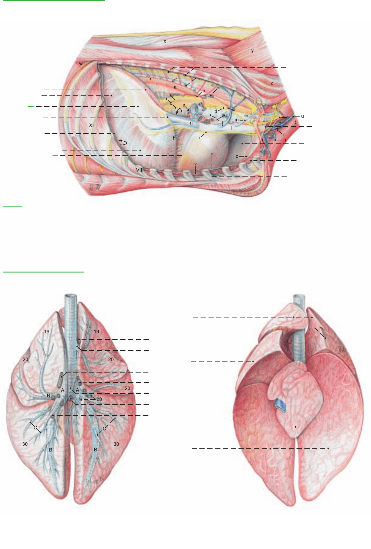
Right thoracic cavity and Lungs
1 Thoracic duct
Diaphragm:
2 Lumbar part
3 Costal part
4 Tendinous
center
5 Foramen
for vena cava
Pleural cavity:
6 Costal pleura
7 Costodiaphragmatic
recess
8 Diaphragmatic pleura
9 Mediastinal recess
10 Intercostal lnn.
11 Lnn. of thoracic aorta
12 Middle mediastinal lnn.
13 Caud. mediastinal lnn.
14 Cran. mediastinal lnn.
15 Pleural cupula
16 Cran. mediastinum
17 Cran. sternal ln.
18 Pericardial pleura
(See pp. 61, 65, 67)
Legend:
A Main bronchus
B Lobar bronchus
C Segmental bronchus
a Thoracic aorta
b Bronchoesophagial a.
c Dors. and vent. vagal trunks
d Right vagus n.
e Pulmonary vv.
f Pulmonary a.
g Caud. vena cava
h Plica venae cavae
i Phrenic n.
j Right azygos v.
k Trachea and tracheal bronchus
l Cran. vena cava
m Costocervical v.
n Right recurrent laryngeal n.
o Right subclavian a. and v.
p Internal thoracic a. and v.
q Cephalic v.
r Supf. cervical a. and v.
s Vagosympathetic trunk
t Common carotid a. and
internal jugular v.
u External jugular v.
v Transverse thoracic m.
w Retractor costae
x Spinalis et semispinalis
cervicis et capitis
y Semispinalis capitis
z Longissimus cervicis
Lungs and Bronchial lnn.
(Left lung)
(Right lung) (Right lung)
(Left lung)
Cranial lobes:
19 Cranial part
20 Caudal part
21 Cran. tracheobronchial ln.
22 Tracheal bronchus
23 Middle lobe
24 Left tracheobronchial ln.
25 Right tracheobronchial ln.
26 Bifurcation of the trachea
27 Middle tracheobronchial ln.
28 Pulmonary lnn.
29 Accessory lobe
30 Caudal lobes
(dorsal)
(ventral)
63
Anatomie des Rindes englisch 09.09.2003 14:11 Uhr Seite 63
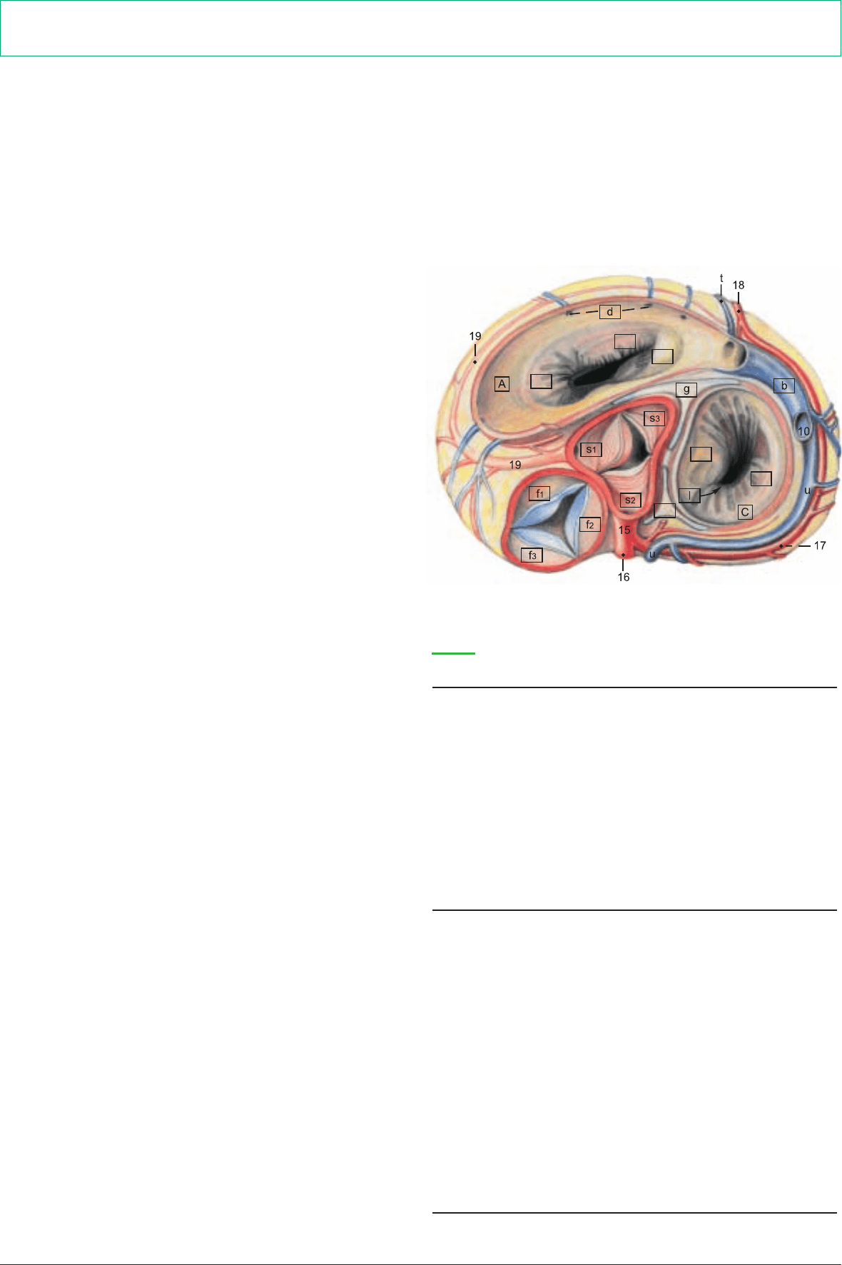
a) The HEART (COR) is relatively small in comparison to that of
the horse. Its weight varies between 0.4 and 0.5 percent of the body
weight. Its absolute weight in cows averages 2.4 kg and in bulls
2.6 kg.
The heart is located between the planes of the 3rd and 5th inter-
costal spaces in the ventral half of the thoracic cavity. The inclina-
tion of the cardiac axis is relatively steep, with the base of the heart
directed craniodorsally. The apex (x) of the heart is directed cau-
doventrally, but does not reach the sternum. The greater part of the
heart lies on the left of the median plane and brings the pericardi-
um into contact with the left thoracic wall in the 3rd and 4th inter-
costal spaces. Its left ventricular border (w) presses the pericardium
into contact with the left side of the diaphragm close to the median
plane, and this is clinically significant because of the proximity of
the reticulum, with its penetrating hardware. The heart field, clini-
cally important for auscultation and percussion, is an outline of the
heart projected on the left thoracic wall from the 3rd to the 5th
intercostal space. On the surface of the heart in addition to the
paraconal (16) and subsinuosal (18) interventricular grooves, there
is an intermediate groove on the left ventricular border that does
not reach the apex. Also species-specific are the distinctly dentate
margins of the auricles, which overhang the base of the heart, but
are smaller than those of the horse. The friable white structural fat
(suet) that can make up as much as 24 percent of the weight of the
heart lies in four interconnected lobes on the right and left atria
between the great vessels and in the coronary grooves.
The pericardium is attached by two divergent sternopericardiac lig-
aments (14) to the sternum at the level of the notches for the 6th
costal cartilages.
Of the coronary arteries, the left coronary a. (15) is substantially
larger (left coronary supply type as in the dog, but unlike the horse
and pig.) It gives off the paraconal interventricular branch (16) in
the groove of the same name, as well as the circumflex branch (17)
which runs around the caudal surface of the heart in the coronary
groove, and ends as the subsinuosal interventricular branch (18) in
the groove of the same name. The small right coronary a. (19) takes
a circumflex course in the coronary groove between the right atri-
um and ventricle.
The heart bones are remarkable features of the heart skeleton—the
fibrous rings around and between the valves. The large, 3–6 cm,
three-pronged right heart bone (g) and the small, 2 cm, left heart
bone (g') are in the aortic ring.
b) The remaining BLOOD VESSELS show greater differences
from the dog than from the horse.
The first branch of the aortic arch, as in the horse, is the brachio-
cephalic trunk (13), the common trunk of the vessels to cranial
parts of the thorax, to the thoracic limbs, and to the head and neck.
It gives off first the left subclavian a., then the right subclavian a.,
and continues as the bicarotid trunk for the left (4) and right (see p.
63) common carotid aa. The left (6) and right (see p. 63) subclavian
aa. give off cranially the costocervical trunk (3) for vessels to the
vertebrae, spinal cord, and brain (vertebral a. 2); to the neck (deep
cervical a., 2 and dorsal scapular a., 1); and to the ribs (supreme
intercostal a., which can also originate from the subclavian a. or the
aorta). Dorsocranially the subclavian gives off the superficial cervi-
cal a. (5), and caudally, the internal thoracic a. (7), which is the last
branch before the subclavian turns around the first rib and becomes
the axillary a. The thoracic aorta (8) gives off dorsal intercostal aa.
and on the right, dorsal to the base of the heart, the broncho-
esophageal a., whose bronchial (12) and esophageal (11) branches
may originate as separate arteries from the aorta or an intercostal
a. The tracheal bronchus is supplied by its own branch, either from
the aorta or from the bronchial branch.
The veins show a distribution similar to that of the arteries. A right
azygos v., (see p. 63), present in the dog and horse, is only rarely
developed as far as the last thoracic vertebra in the ox, and may be
absent caudal to the 5th dorsal intercostal v. The left azygos v. (10)
is always present. It drains into the coronary sinus of the right atri-
um. It does not occur in the dog and horse.
c) The NERVES in the thoracic cavity are the same as in the dog
and horse. The greater splanchnic n. takes origin from the sympa-
thetic trunk (9) at the 6th to 10th ganglia, unlike the dog and horse,
and separates from the trunk just before they pass over the
diaphragm in the lumbocostal arch.
64
2. HEART, BLOOD VESSELS, AND NERVES OF THE THORACIC CAVITY
The surface of the heart is studied in situ; the internal relations are studied on isolated hearts. The visible blood vessels and nerves are
identified.
e'1
e'2
e'3
r'2
r'1
g'
* The letters in this legend are framed in the heart illustrations (pp. 64, 65).
Section through the Base of the Heart
Legend:*
A Right atrium
a Sinus of venae cavae
b Coronary sinus
c Pectinate mm.
d Veins of right heart
C Left atrium
Pulmonary vv.
(See p. 65 p)
g Right heart bone
g' Left heart bone
h Fossa ovalis
i Epicardium
j Myocardium
k Endocardium
B Right ventricle
e Right atrioventricular valve
[Tricuspid valve]
e'1 Parietal cusp
e'2 Septal cusp
e'3 Angular cusp
e"1 Small papillary mm.
e"2 Great papillary m.
e"3 Subarterial papillary m.
f Pulmonary valve
f1 Right semilunar valvule
f2 Left semilunar valvule
f3 Intermediate semilunar valvule
D Left ventricle
r Left atrioventricular valve
[Mitral valve]
r'1 Parietal cusp
r'2 Septal cusp
r"1 Subauricular papillary m.
r"2 Subatrial papillary m.
s Aortic valve
s1 Right semilunar valvule
s2 Left semilunar valvule
s3 Septal semilunar valvule
l Atrioventricular orifice
m Interventricular septum
n Septomarginal trabeculae
o Trabeculae carneae
p Tendinous cords
Anatomie des Rindes englisch 09.09.2003 14:11 Uhr Seite 64
