Budras Klaus-Dieter, Habel Robert E. Bovine Аnatomy
Подождите немного. Документ загружается.

B'
XIV
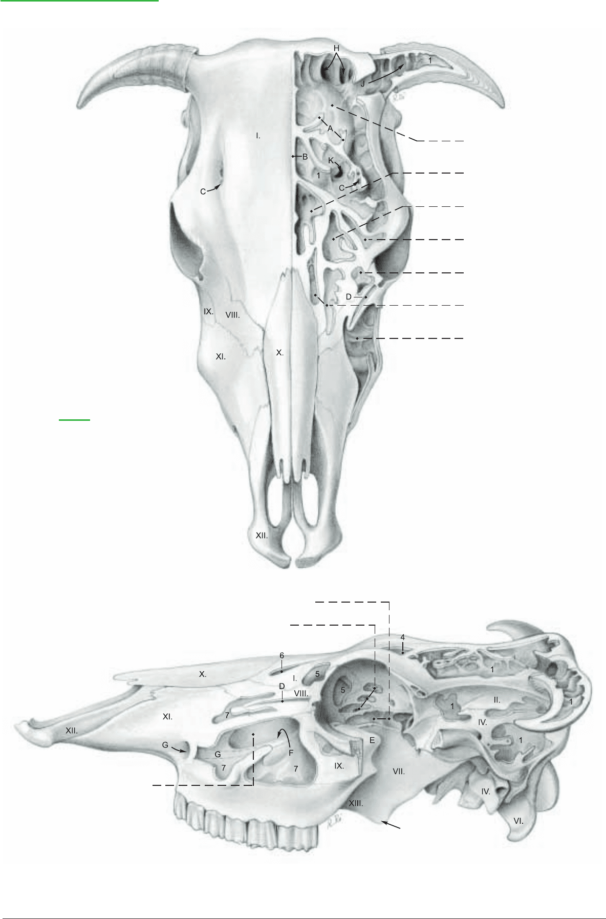
Paranasal Sinuses and Horns
(dorsal)
1 Caudal frontal sinus
2 Med. rostral frontal sinus
3 Intermediate rostral frontal sinus
4 Lat. rostral frontal sinus
5 Lacrimal sinus
6 Dorsal conchal sinus
7 Maxillary sinus
Legend:
A Intrasinual lamellae
B Median septum between frontal sinuses
B' Oblique transverse septum
C Supraorbital canal
D Lacrimal canal
E Lacrimal bulla
F Maxillopalatine opening
G Infraorbital canal
H Nuchal diverticulum
J Cornual diverticulum
K Postorbital diverticulum
(See p. 45)
(lateral)
8 Sphenoid sinus
9 Ethmoid cells
10 Palatine sinus
35
The Roman numerals refer to the bones of the skull on pp. 31 and 33.
Anatomie des Rindes englisch 09.09.2003 13:14 Uhr Seite 35
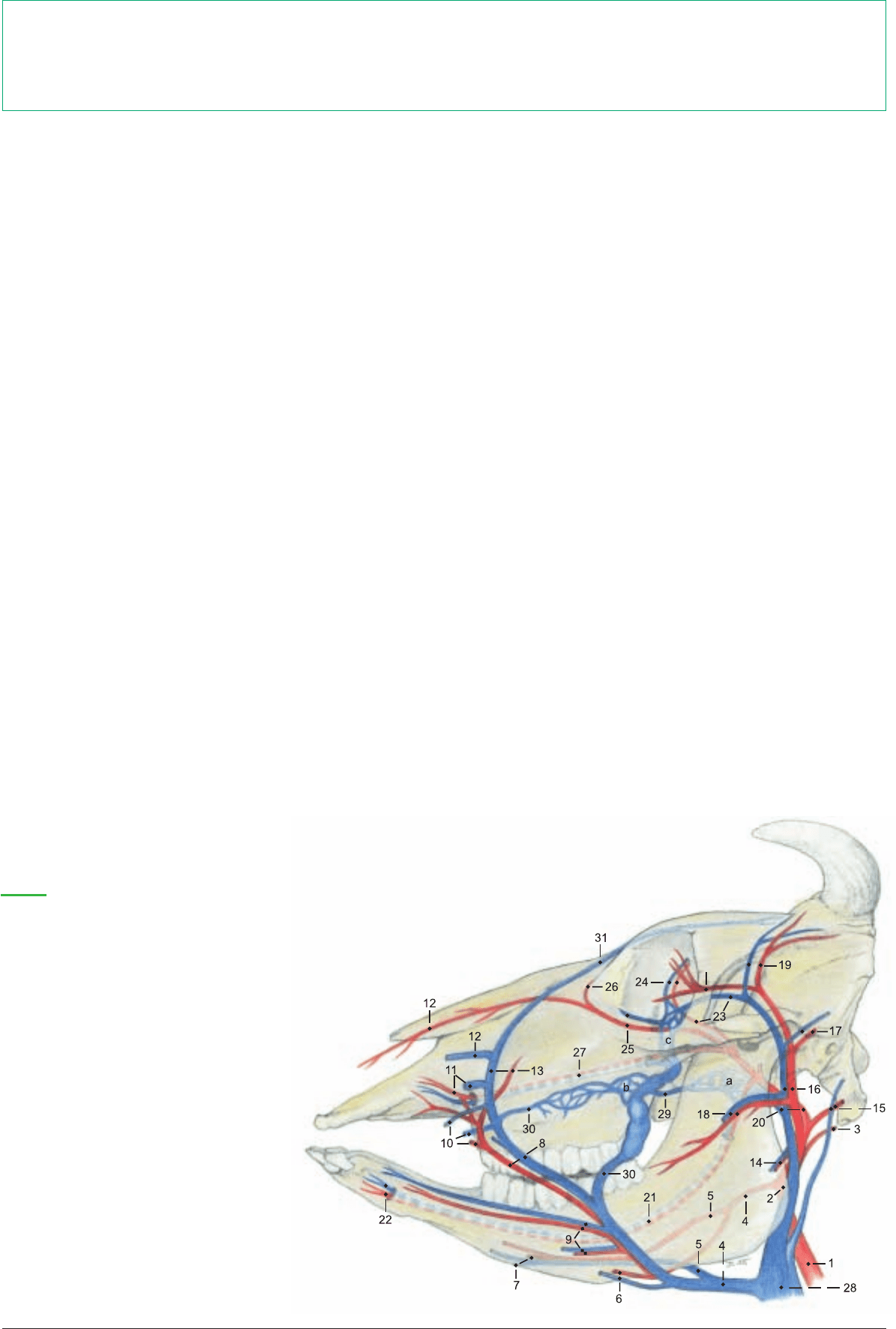
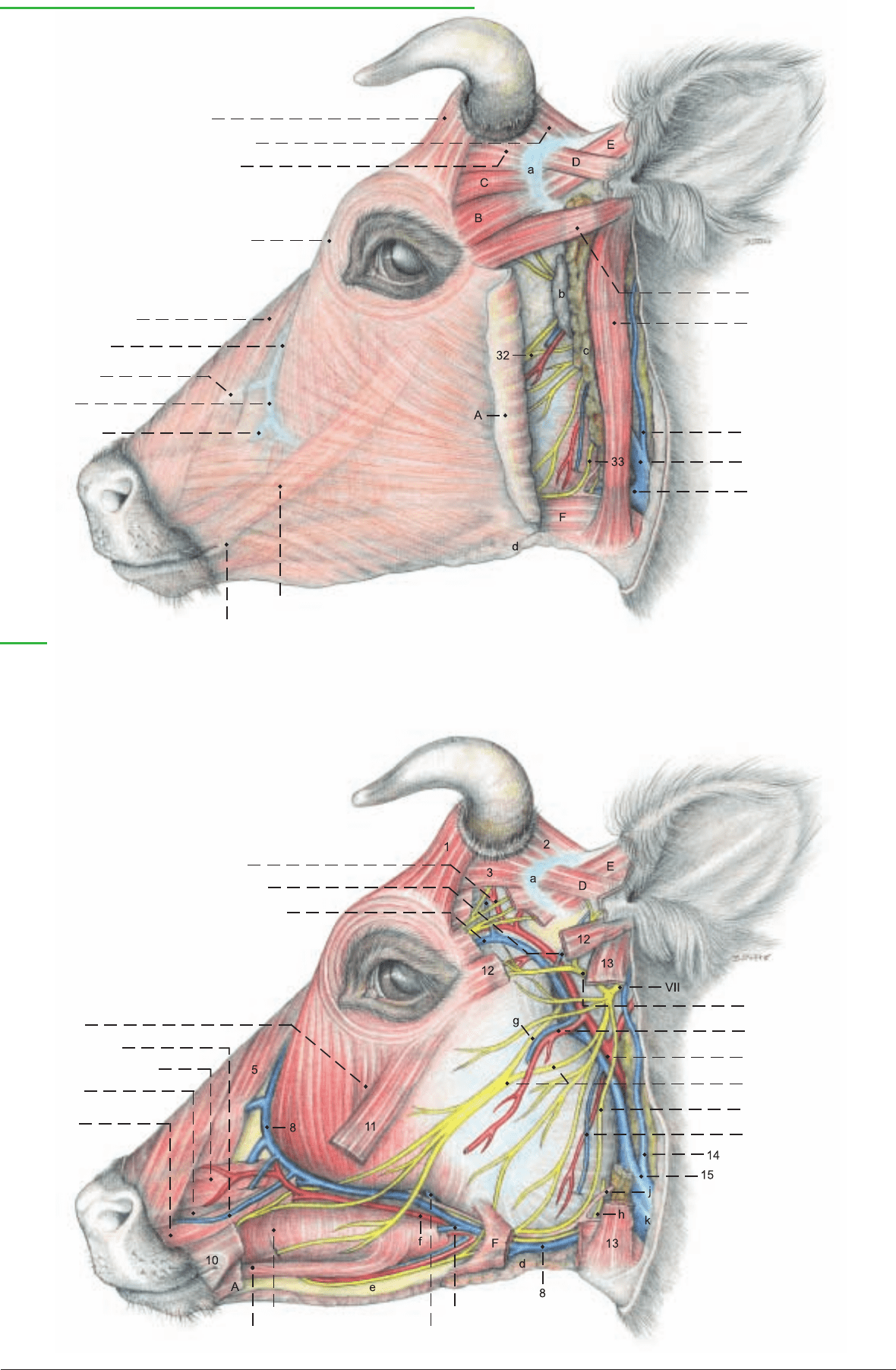
a) The SUPERFICIAL VEINS (refer to p. 37) are drained by the
external jugular v. (k) whose main branches, the linguofacial and
maxillary vv., cross the lateral surface of the mandibular gland. The
linguo-facial v. (16), after giving off the lingual v., is continued as
the facial v. (8). The lingual v. gives off the sublingual v. as in the
dog. The sternomandibularis (F) must be reflected to see the facial
v. where it crosses the ventral border of the mandible in the vascu-
lar groove with the facial a. (f), ventr. buccal br. (33) of the facial
n., parotid br. (h) of the buccal n. (V3),* and parotid duct (j). On
the lateral surface of the mandible the supf. and deep vv. of the low-
er lip (28) are given off. From the caudal side of the facial v. at this
level, the deep facial v. (27) passes deep to the masseter to the deep
facial plexus (text fig. b) and to the orbit. The facial vessels contin-
ue dorsally, supplying deep and superficial vessels of the upper lip
(21). The vein supplies the lat. nasal v. (9) and dorsal nasal v. (7),
and is continued by the v. of the angle of the eye (6). The latter pass-
es dorsomedial to the orbit and becomes the frontal v., which
courses in the supraorbital groove (p. 31, 1') to the supraorbital
foramen.
Caudal to the angle of the mandible, medial to the parotid gl., the
maxillary v. (15) gives off the caud. auricular v. (14) and the ventral
masseteric v. (34). (The occipital v. comes from the int. jugular v.)
Before the maxillary v. turns deep to the ramus of the mandible it
gives rise to the large supf. temporal v. (31), which gives off the
short transverse facial v. (30), the rostral auricular v. (18), and the
cornual v. (17), and turns rostrally into the orbit to become the dor-
sal ext. ophthalmic v. (19).
b) The FACIAL N. (VII) as it leaves the stylomastoid foramen,
gives off the caud. auricular n. and internal auricular br., which
does not give off the cutaneous brr. that go to the base and inner
surface of the auricle in the horse and dog; these are supplied exclu-
sively by the auricular branch of the vagus n. Dorsally, the facial n.
gives off the auriculopalpebral n. (29), which divides into the ros-
tral auricular brr. and the zygomatic br. The latter runs forward on
the surface of the zygomatic arch to the eyelids and ends in palpe-
bral brr. In the parotid gland the facial n. divides into dorsal and
ventral buccal brr. The dorsal buccal br. (32) emerges at the ventral
end of the parotid ln. under the parotid gland. It is joined by a large
branch of the sensory auriculotemporal n. (V3, g) and courses
toward the upper lip, supplying facial muscles and cutaneous sen-
sation. The ventral buccal br. (33) is more slender than the dorsal
br. It follows the caudal and ventral borders of the masseter (unlike
that of the horse) to the vascular groove, whence it runs along the
ventral border of the buccinator and depressor labii inferioris to the
lower lip. The cervical br. (Ramus colli) is absent in the ox.
c) The FACIAL MUSCLES include lip and cheek muscles, the mus-
cles of the eyelids and nose, and ear muscles. The levator nasolabi-
alis (5) is a broad thin muscle originating from the frontal bone and
the frontalis. Between its superficial and deep layers pass the leva-
tor labii superioris (22) and caninus (23). These two muscles and
the depressor labii superioris (24) originate close together from the
facial tuber. The levator labii superioris covers the ventral part of
the infraorbital foramen, which is nevertheless palpable. The
depressor labii inferioris (25) originates deep to the masseter from
the caudal part of the alveolar border of the mandible. The zygo-
maticus (11) originates from the masseteric fascia ventral to the
orbit and runs obliquely across the masseter and buccinator to the
orbicularis oris (10) at the corner of the mouth. The buccinator (26)
forms the muscular layer of the cheek. The molar part is covered by
the masseter and the depressor labii inferioris. The buccal part is a
thin layer of mostly vertical fibers.
The muscles of the eyelids are the orbicularis oculi (4), frontalis (1),
levator palpebrae superioris (see p. 41, 13), and malaris (20). The
frontalis (not present in the horse) takes over the function of the
absent retractor anguli oculi lat., and augments the action of the
levator palpebrae superioris. Of the ear muscles, the parotidoauric-
ularis (13) extends on the surface of the parotid gland from the ven-
tral part of the parotid fascia to the intertragic notch. The zygo-
maticoauricularis (12) begins on the zygomatic arch and runs back
to end at the intertragic notch. The cervicoscutularis (2) originates
from the lig. nuchae and the skull behind the intercornual protu-
berance. The short interscutularis (3) comes from the cornual
process and the temporal line, and has no connection with the con-
tralateral muscle.
36
4. SUPERFICIAL VEINS OF THE HEAD, FACIAL N. (VII), AND FACIAL MUSCLES
* V = Trigeminal n., V1 = Ophthalmic n. V2 = Maxillary n. V3 = Mandibular n.
To demonstrate the superficial veins and nerves, the head is split in the median plane and the skin is removed, except for a narrow strip
of skin around the horn, eye, nose, and mouth, noting the cutaneus faciei (A) and the frontalis, which is spread superficially over the
frontal region. The parotidoauricularis and zygomaticoauricularis are transected and reflected to expose the parotid gland. The dor-
sal part of the gland above the maxillary v. is removed piecemeal, sparing the vessels and nerves in the gland, and the large parotid
lymph node ventral to the temporo-mandibular joint.
19'
24 Supraorbital a. and v.
25 Malar a. and v.
26 A. of angle of eye
27 Infraorbital a. and v.
28 Ext. jugular v.
29 Buccal v.
30 Deep facial v.
31 Frontal v.
Arteries and Veins of the Head
Legend: (Numbers differ from those in text.)
a Pterygoid plexus
b Deep facial plexus
c Ophthalmic plexus
1 Common carotid a.
2 External carotid a.
3 Occipital a.
4 Linguofacial tr. and v.
5 Lingual a. and v.
6 Submental a. and v.
7 Sublingual a. and v.
8 Facial a. and v.
9 Supf. and deep
inf. labial a. and v.
10 Superior labial a., supf.
and deep sup. labial vv.
11 Rostral lat. nasal a.
and lat. nasal v.
12 Dors. nasal a. and v.
13 Arterial br. and
v. of angle of eye
14 Ventr. masseteric br. and v.
15 Caud. auricular a. and v.
16 Supf. temporal a. and v.
17 Rostr. auricular a. and v.
18 Transverse facial a. and v.
19 Cornual a. and v.
19' Inf. and sup. palpebral aa.
20 Maxillary a. and v.
21 Inferior alveolar a. and v.
22 Mental a. and v.
23 Ext. ophth. a.,
dors. ext. ophth. v.
Anatomie des Rindes englisch 09.09.2003 13:14 Uhr Seite 36
(See p. 39)
Arteries and Veins of the head, Facial n. and Facial muscles
1 Frontalis
2 Cervicoscutularis
3 Interscutularis
4 Orbicularis oculi
5 Levator nasolabialis
6 V. of angle of eye
7 Dorsal nasal v.
8 Facial v.
9 Lateral nasal v.
10 Orbicularis oris
11 Zygomaticus
12 Zygomaticoauricularis
13 Parotidoauricularis
14 Caudal auricular v.
15 Maxillary v.
16 Linguofacial v.
Legend:
A Cutaneous faciei
B Zygomaticoscutularis
C Frontoscutularis
D Scutoloauricularis supf. dors.
E Scutoloauricularis supf. accessorius
F Sternomandibularis
a Scutiform cartilage
b Parotid ln.
c Parotid gl.
d Mandibular gl.
e Mandible
f Facial a.
g Communicating br. between
auriculotemporal n. (V3) and
dorsal buccal br. (VII)
h Parotid br. of buccal n. (V3)
j Parotid duct
k External jugular v.
17 Cornual a. and v.
18 Rostral auricular v.
19 Dors. ext. ophthalmic v.
20 Malaris
21 Superior labial v.
22 Levator labii superioris
23 Caninus
24 Depressor labii
superioris
25 Depressor labii inferioris
26 Buccinator
27 Deep facial v.
28 V. of lower lip
29 Auriculopalpebral n.
30 Transverse facial a. and v.
31 Supf. temporal a. and v.
32 Dorsal buccal br. of VII
33 Ventral buccal br.of VII
34 Ventral masseteric v.
37
Anatomie des Rindes englisch 09.09.2003 13:14 Uhr Seite 37

a) The TRIGEMINAL N. (V) of the ox exhibits no marked differ-
ences in its branches from that of the dog and horse.
I. The mandibular n. (V3) is sensory to the teeth, oral mucosa, and
skin of the lower jaw, as well as the tongue, parotid gl., and part of
the ear. Unlike the other divisions of the trigeminal n. (V1 and V2) it
also has somatic motor components. These are in the following
branches: the masticatory n. (20) divides into the deep temporal nn.
(18) and masseteric n. (19) which innervate the corresponding mus-
cles. Branches to the pterygoids, tensor tympani, and tensor veli
palatini have corresponding names. The inferior alveolar n. gives
off, before entering the mandibular foramen, the mylohyoid n. (29)
for the muscle of that name and for the rostral belly of the digastri-
cus, and sends cutaneous branches to the rostral part of the inter-
mandibular region. The following branches of the mandibular n.
have no somatic motor components: The many-branched buccal n.
(4) conducts sensory fibers and receives parasympathetic fibers from
the glossopharyngeal n. (IX) via the large oval otic ganglion to the
oral mucosa and the buccal salivary glands. Its parotid br. (16),
which occurs only in ruminants, turns around the rostral border of
the masseter and runs back to the parotid gland close to the duct.
The auriculotemporal n. (26) turns caudally to the ear, skin of the
temporal region, and parotid gland, supplying sensory branches and
parasympathetic innervation (from IX via the otic ganglion). The
nerve then turns rostrally and joins the dorsal buccal br. of VII as the
communicating br. with the facial n. (1) thereby supplying sensation
to the skin of the cheek. The lingual n. (30) is sensory to the sublin-
gual mucosa and tongue. From the chorda tympani (VII —27) it
receives taste fibers for the rostral 2/3 of the tongue, and parasym-
pathetic fibers for the sublingual and mandibular glands. Its sublin-
gual n. (33) runs as in the dog but not as in the horse, on the lat. sur-
face of the sublingual gll. to the floor of the mouth. The sensory infe-
rior alveolar n. (28) passes through the mandibular foramen to the
mandibular canal. It supplies the lower teeth and after emerging
from the mental foramen as the mental n. (5) it supplies the skin and
mucosa of the lower lip and chin.
II. The maxillary n. (V2—21) is sensory and contains parasympa-
thetic components from VII via the pterygopalatine ganglion. It
gives off the zygomatic n. and the pterygopalatine n. with the major
palatine, minor palatine, and caudal nasal nn. Its rostral continua-
tion is the infraorbital n. (6) which gives off sensory brr. in the
infraorbital canal for the upper teeth, and after emerging from the
foramen divides into numerous branches for the dorsum nasi, nos-
tril, planum nasolabiale, upper lip, and the nasal vestibule. (For the
ophthalmic n., V1, see p. 40.)
b) The MASTICATORY MM. INCLUDING THE SUPERFICIAL
INTERMANDIBULAR MM. are innervated by the mandibular n.
(V3). The caudal belly of the digastricus is innervated by the facial
n. (VII). Of the external masticatory mm., as in the horse, the mas-
seter (13) is larger than the temporalis (17), and, covered by a glis-
tening aponeurosis, presents a superficial layer with almost hori-
zontal muscle fibers, and a deep layer with caudoventral fiber direc-
tion. The internal masticatory mm.: the medial pterygoid (22) and
the lateral pterygoid (22), are clearly separate. The superficial inter-
mandibular mm. are the mylohyoideus (25) digastricus (31). There
is no occipitomandibularis in the ox. The digastricus, which does
not perforate the stylohyoideus, terminates rostral to the vascular
groove on the medial surface of the ventral border of the mandible.
Right and left digastrici are connected ventral to the lingual process
of the basihyoid by transverse muscle fibers.
c) The LARGE SALIVARY GLANDS are the parotid, mandibular,
monostomatic sublingual, and polystomatic sublingual gll.
I. The parotid gland (14, p. 37, c) is elongated and thick. It lies along
the caudal border of the masseter from the zygomatic arch to the
angle of the mandible. Numerous excretory ducts converge to the
parotid duct (15) at the ventral end of the gland. The duct runs with
the facial vessels from medial to lateral through the vascular groove
in the ventral border of the mandible, ascends in the groove along the
rostral border of the masseter, and enters the oral vestibule opposite
the fifth upper cheek tooth (M2). The deep surface of the gland is
related to the maxillary and linguofacial vv., the end of the ext.
carotid a., the mandibular gl., and the parotid ln. The facial n. with
the origin of its buccal branches is enveloped by the parotid gland.
II. The mandibular gland (9) is curved, lying medial to the angle of
the mandible and extending from the paracondylar process to the
basihyoid. Its enlarged bulbous end is palpable in the inter-
mandibular region, where it is in contact with the contralateral
gland. The deep surface is related to the lat. retropharyngeal ln.,
common carotid a., pharynx, and larynx. The mandibular duct
(32) leaves the middle of the concave border of the gland and cours-
es deep to the mylohyoideus and dorsal to the monostomatic sub-
lingual gl. to the sublingual caruncle on the floor of the oral cavity
rostral to the frenulum of the tongue.
III. The monostomatic sublingual gl. (24) is about 10 cm long. Its
major sublingual duct ends near the mandibular duct under the
sublingual caruncle.
IV. The polystomatic sublingual gl. (23) extends in a chain of lob-
ules from the palatoglossal arch to the incisive part of the mandible.
The microscopic sublingual ducts open under the tongue on each
side of a row of conical papillae extending caudally from the sub-
lingual caruncle .
The small salivary glands:
The buccal gll. are developed best in the ox.
The superficial layer of the dorsal buccal gll. (3) is on the surface of
the buccinator. The deep layer is covered by the muscle. They
extend from the angle of the mouth to the facial tuber and are cov-
ered caudally by the masseter. The middle buccal gll. (7) are found
in ruminants between two layers of the buccinator and dorsal to the
vein of the lower lip. The ventral buccal gll. (8) lie on the mandible
from the angle of the mouth to the rostral border of the masseter.
They are ventral to the vein of the lower lip and covered, except the
caudal part, by the buccinator. Small salivary gll. are present
throughout the oral mucosa. Total secretion of saliva in the ox is
about 50 liters in 24 hours.*
d) The LYMPHATIC SYSTEM. Ruminant lymph nodes differ
from those of the horse; they are usually single large nodes rather
than groups of small nodes. All of the following nodes are routine-
ly incised in meat inspection. The parotid ln. (12) lies between the
rostral border of the parotid gl. and the masseter, ventral to the
temporomandibular joint. It is palpable in the live animal. The
mandibular ln. (10) lies ventral to the mandible, halfway between
the rostral border of the masseter and the angle of the mandible, in
contact with the facial vein. It is covered laterally by the ster-
nomandibularis and the facial cutaneous m., but is palpable in the
live animal; it is lateral to the bulbous ventral end of the mandibu-
lar gl., which is in contact with the contralateral gl. and should not
be mistaken for the mandibular ln.
The medial retropharyngeal ln. is in the fat between the caudodor-
sal wall of the pharynx, through which it can be palpated, and the
longus capitis. Its lateral surface is related to the large (1.5 x 0.5
cm) cranial cervical ganglion and cranial nn. IX to XII.
The lateral retropharyngeal ln. (11) receives all of the lymph from
the other lymph nodes of the head and is drained by the tracheal
trunk. It lies in the fossa between the wing of the atlas and the
mandible, covered laterally by the mandibular gland.
38
5. TRIGEMINAL N. (V3 AND V2), MASTICATORY MM., SALIVARY GLL., AND LYMPHATIC SYSTEM
* Somers, 1957
For the dissection of the temporalis and masseter the covering facial muscles and superficial nerves and vessels are removed. The mas-
seter is removed in layers, showing its tough tendinous laminae, its almost horizontal and oblique fiber directions and its innervation
by the masseteric n. (V3) passing through the mandibular notch. Medial to the masseter is the large deep facial venous plexus (2). To
remove the zygomatic arch three sagittal cuts are made: I. at the temporomandib. joint, II. through the zygomatic bone rostral to its
frontal and temporal processes, and III. through the zygomatic process of the frontal bone. In the course of disarticulation of the tem-
poromandib. joint the temporalis is separated from its termination on the coronoid proc., whereby its innervation from the deep tem-
poral nn. is demonstrated. The mandible is sawed through rostral to the first cheek tooth. After severing all structures attached to the
medial surface of the mandible, the temporomandib. joint is disarticulated by strong lateral displacement of the mandible while the
joint capsule is cut. The fibrocartilaginous articular disc compensates for the incongruence of the articular surfaces.
Anatomie des Rindes englisch 09.09.2003 13:14 Uhr Seite 38
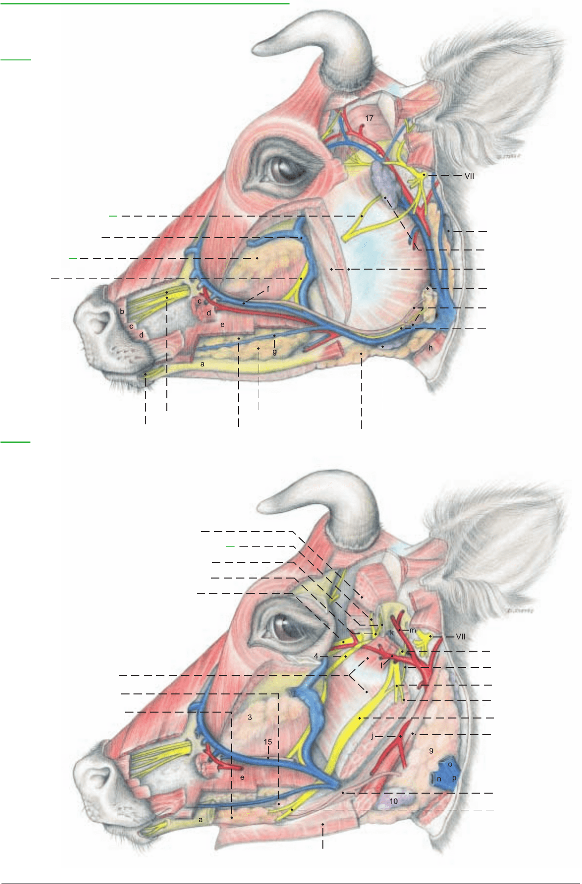
33 Sublingual n.
Mandibular n. (V3), Maxillary n. (V2), and Salivary glands
Legend:
a Mandible
b Levator labii superioris
c Caninus
d Depressor labii superioris
e Buccinator
f Facial vein
g Vein of lower lip
h Sternomandibularis
1 Br. communicating (V3) with
facial n.
2 Deep facial venous plexus
3 Dorsal buccal gll.
4 Buccal n.
5 Mental n.
6 Infraorbital n.
7 Middle buccal gll.
8 Ventral buccal gll.
9 Mandibular gl.
10 Mandibular ln.
11 Lat. retropharyngeal ln.
12 Parotid ln.
13 Masseter
14 Parotid gl.
15 Parotid duct
16 Parotid br. of buccal n.
(See pp. 37, 47, 49)
Legend:
j Linguofacial trunk
k Articular disc
l Maxillary a.
m Supf. temporal a.
n Linguofacial v.
o Maxillary v.
p Ext. jugular v.
17 Temporalis
18 Deep temporal nn.
19 Masseteric n.
20 Masticatory n.
21 Maxillary n.
22 Med. and lat. pterygoids
23 Polystomatic sublingual gl.
24 Monostomatic sublingual gl.
25 Mylohyoideus
26 Auriculotemporal n.
27 Chorda tympani
28 Inf. alveolar n.
29 Mylohyoid n.
30 Lingual n.
31 Digastricus
32 Mandibular duct
39
Anatomie des Rindes englisch 09.09.2003 13:15 Uhr Seite 39
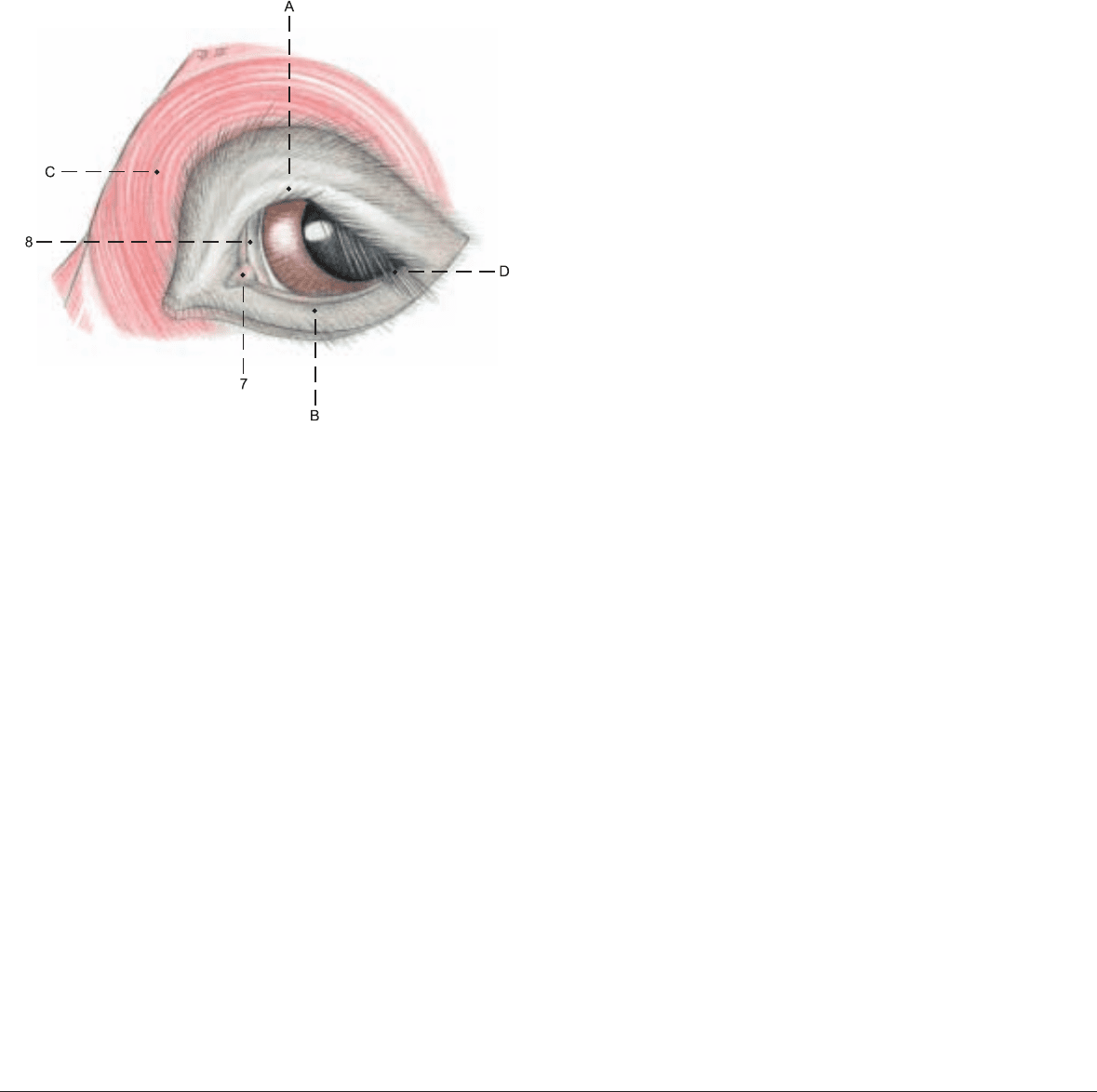
The ACCESSORY ORGANS include the eyelids and conjunctiva,
the lacrimal apparatus, and the cone of striated bulbar muscles
with their fasciae and nerves. They will be described in the order in
which they are exposed (see also the text figure and p. 43).
I. The upper and lower eyelids (palpebra superior, A, and inferior,
B) consist of an outer layer of haired skin, a middle fibromuscular
layer, and the palpebral conjunctiva. The fibrous part of the middle
layer is attached to the osseous orbital margin and increases in den-
sity toward the free border to form the tarsus, which contains the
tarsal glands.
The eyelashes (cilia, D) of the lower lid are fewer and shorter than
those of the upper lid, but they are present in the ox.
The striated muscles are: the strong orbicularis oculi (C), and in the
upper eyelid, the termination of the levator palpebrae superioris
(13) and fibers of the frontalis. The upper and lower tarsal mm. are
parts of the smooth muscle system of the orbit, which retracts the
eyelids and protrudes the eyeball under sympathetic stimulation.
The palpebral conjunctiva (10) is continuous at the fornix (11) with
the bulbar conjunctiva (12), which ends at the limbus of the cornea.
The third eyelid (8) consists of a fold of conjunctiva in the medial
angle, enclosing the T-shaped outer end of the cartilage of the third
eyelid.
The deep part of the cartilage is surrounded by the gland of the
third eyelid, larger than in the horse, extending about 5 cm straight
back into the fat medial to the eyeball and discharging tears
through orifices on the bulbar side of the third lid.
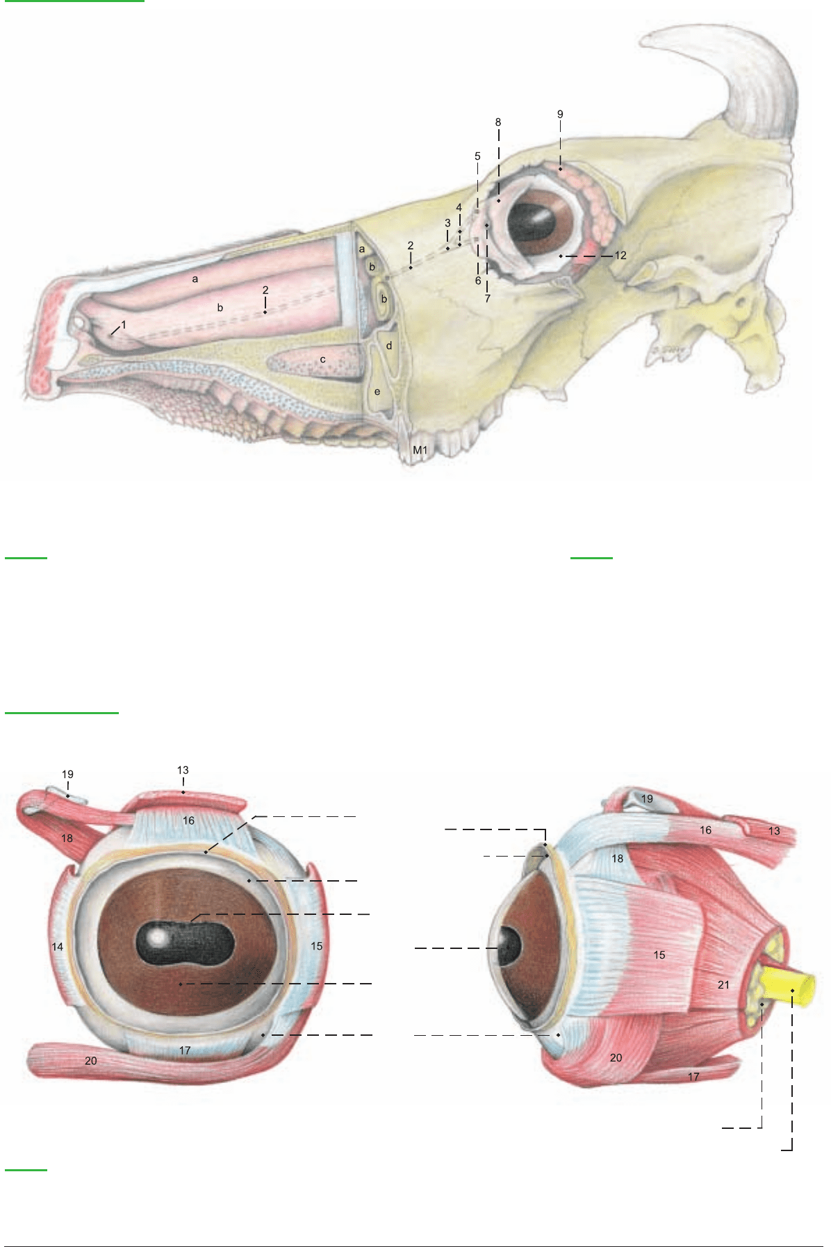
II. The lacrimal apparatus. The lacrimal gl. (9) lies in the dorsolat-
eral quadrant of the orbit, with the broad dorsal part under the root
of the zygomatic proc., and a long thin tail which extends around
the lateral margin of the orbit.
The lacrimal ducts pass from the ventral end of the gland to orifices
in the lateral fornix. The gland of the third eyelid is the largest
accessory lacrimal gland. The tears collect around the lacrimal
caruncle (7) in the lacrimal lake in the medial angle anterior to the
third eyelid. They are drained through the upper (5) and lower (6)
lacrimal puncta and lacrimal canaliculi (4) which join at the
lacrimal sac (3). This is drained by the nasolacrimal duct (2) to the
nasolacrimal orifice (1) concealed on the medioventral surface of
the alar fold.
III. The bulbar muscles are surrounded by the periorbita which, in
the osseous part of the orbit, is the periosteum, containing the
trochlea (19), but caudolaterally where the bony orbit is deficient
in domestic mammals, the periorbita alone forms the wall of the
orbit. It is a tough, fibrous, partially elastic membrane stretched
from the lateral margin of the orbit to the pterygoid crest. The
lacrimal gland and the levator palpebrae superioris are covered
only by the periorbita. The remaining structures are also enveloped
in the deep orbital fasciae: the fasciae of the muscles and the bulbar
fascia (vagina bulbi).
The ophthalmic n. (V 1) (see p. 53) divides while still in the for.
orbitorotundum into the following three nerves:
1. The usually double lacrimal n. runs along the lateral surface of
the lateral rectus and gives off branches to the lacrimal gl. and the
upper eyelid. The two strands of the lacrimal n. then unite and the
zygomaticotemporal br. so formed perforates the periorbita and
turns caudally under the zygomatic proc. of the frontal bone to the
temporal region, where it sends twigs to the skin and continues ven-
tral to the temporal line as the cornual branch to the skin on the
cornual process.
2. The frontal n. gives rise to the nerve to the frontal sinuses, which
perforates the wall of the orbit. The frontal n. then passes around
the dorsal margin of the orbit (unlike that of the horse) and
becomes the supraorbital n. to the frontal region.
3. The nasociliary n. gives off the long ciliary nn., which penetrate
the sclera and supply sensation to the vascular tunic (see p. 42) and
cornea; the ethmoidal n., with sensory and autonomic fibers to the
caudal nasal mucosa; and the infratrochlear n. The last turns
around the mediodorsal margin of the orbit to the skin of the medi-
al angle of the eye and the frontal region.
Almost all of the striated bulbar muscles: dorsal (16), medial (14),
and ventral (17) recti; ventral oblique (20), levator palpebrae sup.
(13), and retractor bulbi (21), except its lateral part, are innervated
by the oculomotor n. (III).
Only the dorsal oblique (18) is innervated by the trochlear n. (IV).
The lateral rectus (15) and the lateral part of the retractor bulbi
(21) are served by the abducent n. (VI).
The bulbar muscles originate around the optic canal, with the
exception of the ventral oblique, which comes from a fossa on the
medial wall of the orbit just above the lacrimal bulla. With the
exception of the levator palpebrae sup. all of the bulbar muscles ter-
minate on the sclera.
40
6. ACCESSORY ORGANS OF THE EYE
Anatomie des Rindes englisch 09.09.2003 13:15 Uhr Seite 40
21 Retractor bulbi
Lacrimal apparatus
(Dissection)
(medial)
(lateral)
Legend:
(See pp. 45, 47)
1 Nasolacrimal orifice
2 Nasolacrimal duct
3 Lacrimal sac
4 Lacrimal canaliculi
5 Superior lacrimal punctum
6 Inferior lacrimal punctum
7 Lacrimal caruncle
8 Third eyelid
9 Lacrimal gland
Legend:
a Dorsal nasal concha
b Ventral nasal concha
c Venous plexus
d Maxillary sinus
e Palatine sinus
Bulbar muscles (Left eye)
(anterior)
(lateral)
10 Palpebral
conjunctiva
11 Fornix of conjunctiva
12 Bulbar conjunctiva
Granula iridica
Pupil
Iris
Sclera
Retrobulbar fat
Optic nerve
Legend:
13 Levator palpebrae superioris
14 Medial rectus
15 Lateral rectus
16 Dorsal rectus
17 Ventral rectus
18 Dorsal oblique
19 Trochlea
20 Ventral oblique
41
Anatomie des Rindes englisch 09.09.2003 13:29 Uhr Seite 41

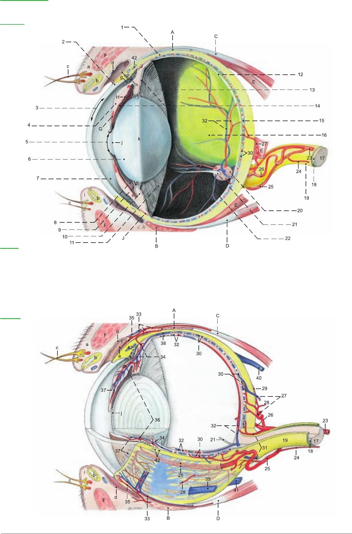
The eyeball of the ox is smaller than that of the horse, and is not
flattened so much anteroposteriorly. For orientation, the pupil and
the optic nerve are taken as reference points. The pupil is at the
anterior pole, and the optic n. is below and slightly lateral to the
posterior pole. Like other ungulates, the ox has a transversely ellip-
tical pupil (5). When it dilates, it becomes round. The black pro-
jections (granula iridica, 5) on the upper and lower margins of the
pupil are vascular appendages covered by pigmented epithelium
from the back of the iris. Those on the lower margin are small. On
eyeballs sectioned on the equator and meridionally, one can study
the external (fibrous) tunic, the middle (vascular) tunic, and the
internal tunic (retina).
I. The fibrous tunic comprises the sclera (1), enclosing the greater
part of the bulb in its dense white connective tissue, and the trans-
parent cornea (3). These parts join at the corneal limbus (2).
II. The vascular tunic consists of the choroid, ciliary body, and iris.
The choroid (15) is highly vascular and pigmented. In its posterior
part, just above the optic disc, is the blue-green, reflective tapetum
lucidum (16), a fibrous structure of roughly semicircular outline
with a horizontal base.
The ciliary body, containing the weak ciliary m. (J), is the anterior
continuation of the choroid. Its most prominent feature is the cil-
iary crown (corona ciliaris, 10), composed of vascular, radial ciliary
processes (10), from which the zonular fibers (9) extend to the
equator of the lens. Posterior to the ciliary processes is the ciliary
ring (orbiculus ciliaris, 11), a zone bearing minute ciliary folds (11).
It is narrower medially than elsewhere. The posterior epithelium is
the pars ciliaris retinae.
Between the ciliary body and the pupil is the iris (4) with the sphinc-
ter (G) and dilator (H) mm. of the pupil, The bovine iris is dark
because of the heavy pigmentation of the posterior epithelium (pars
iridica retinae).
III. The retina lines the entire vascular coat, so that each part of the
vascular coat has a double inner layer derived from the two-layered
ectodermal optic cup of the embryo. The greater part of the retina
is the optical part (12), extending from the optic disc (20) to the cil-
iary body at the ora serrata (13). It contains the visual elements in
its nervous layer and has an outer pigmented layer, which adheres
to the vascular tunic when the nervous layer is detached. The out-
er layer is free of pigment over the tapetum.
The blind part (pars ceca, 14) of the retina lines the iris and ciliary
body. In the iridial part the outer layer contributes the sphincter
and dilator mm., and the inner layer is pigmented; in the ciliary
part, the outer layer is pigmented.
At the optic disc (20) the nerve fibers of the retina exit through the
area cribrosa of the sclera, acquire a myelin sheath, but no neu-
rolemma, and form the optic n. (17), which is morphologically a
tract of the brain, covered by a thin internal sheath (18) corre-
sponding to the pia mater and arachnoidea, and a thick external
sheath (19) corresponding to the dura mater.
IV. The lens (6) is surrounded by the elastic lens capsule (j), which
is connected to the ciliary body by the zonular fibers. Under the
capsule, the anterior surface of the lens is covered by the lens
epithelium. Toward the equator (k) the epithelial cells elongate to
form the lens fibers—the substance of the lens. The fibers, held
together by an amorphous cement, meet on the anterior and poste-
rior surfaces of the lens in three sutures (radii lentis), which are
joined to form a Y (the lens star), best seen in the fresh state.
V. Inside the eyeball the anterior and posterior chambers lie before
the lens and the vitreous body lies behind it. The anterior chamber
(7) is between the cornea and iris. It communicates freely through
the pupil with the posterior chamber (8) which is between the iris
and the lens with its zonula. Viewed from the anterior chamber the
circular pectinate ligament (h) is seen in the iridocorneal angle (g),
attaching the iris by delicate radial trabeculae to the scleral ring at
the corneal limbus. Between these trabeculae are the spaces of the
iridocorneal angle (of Fontana), through which the aqueous humor
drains to the circular venous plexus of the sclera (42).
The vitreous chamber (22) lies between the lens and the retina, and
is filled by the vitreous body. Its stroma is a network that holds in
its meshes a cell-free jelly, the water content of which determines
the intraocular pressure.
VI.The blood supply of the eye comes from the int. and ext. oph-
thalmic aa. and the malar a. The small int. ophthalmic a. (24)
comes from the rostral epidural rete mirabile (see p. 50), accompa-
nies the optic n., and anastomoses with the ext. ophthalmic a. and
the post. ciliary aa. The ext. ophthalmic a. (23), from the maxillary
a., forms the ophthalmic rete mirabile deep in the orbit on the ven-
tral surface of the dorsal rectus. The supraorbital a. arises from the
rete, gives off in the orbit the ext. ethmoidal a. and ant. conjuncti-
val aa., and enters the supraorbital canal, supplying the frontal
sinus and emerging to supply the frontalis m. and skin. Also arising
from the rete are the muscular brr. (28) and the lacrimal a. The
muscular brr. supply the eye muscles and give off ant. ciliary aa.
(33) and posterior conjunctival aa. (35). The ext. ophthalmic a.
divides into two long post. ciliary aa. (25, 26), which give off short
post. ciliary aa. (27) near the eyeball, and continue to the equator
of the eyeball before they enter the sclera. In the ciliary region of the
iris they form the major arterial circle of the iris (36). Near the bul-
bar end of the optic n. the long post. ciliary aa. supply small
choroidoretinal aa. (31), which accompany the optic n. and supply
the four retinal arteries seen with the ophthalmoscope in the fun-
dus of the eye. Accompanied by the corresonding veins, they appear
near the center of the disc and spread out over the interior of the
retina in a pattern characteristic of the ox, with the largest vessels
directed dorsally. The venous blood of the eyeball is drained
through the vorticose vv. (38–41), ciliary vv. (27–33), and the
choroidoretinal vv. (31) to the intraorbital ophthalmic venous
plexus.
42
7. THE EYEBALL (BULBUS OCULI)
33 Ant. ciliary a. and v.
34 Aa. and vv. of ciliary body
35 Post. conjunctival a. and
conjunctival v.
36 Major arterial circle of the iris
37 Aa. and vv. of the iris
38 Lat. dorsal vorticose v.
39 Lat. ventral vorticose v.
40 Med. dorsal vorticose v.
41 Med. ventral vorticose v.
42 Venous plexus of the sclera
Muscles of the eye:
A Dorsal oblique
B Ventral oblique
C Dorsal rectus
D Ventral rectus
E Retractor bulbi
F Orbicularis oculi
G Sphincter pupillae
H Dilator pupillae
J Ciliaris
a Upper eyelid (palpebra superior)
b Tarsal gll.
c Eyelashes (cilia)
d Palpebral conjunctiva
e Bulbar conjunctiva
f Fornix of conjunctiva
g Iridocorneal angle
h Pectinate lig.
i Lower eyelid (palpebra inferior)
j Lens capsule
k Equator of lens
Legend: (See figures on p. 43)
23 Ext. ophthalmic a.
24 Int. ophthalmic a .
25 Lat. long post. ciliary a.
26 Med. long post. ciliary a.
27 Short post. ciliary a., and post.
ciliary v.
28 Muscular br.
29 Episcleral a.
30 Choroid aa. and vv.
31 Choroidoretinal a. and v.
32 Retinal arteries and veins
Anatomie des Rindes englisch 09.09.2003 13:29 Uhr Seite 42
43
(lateral)
Organ of vision
Right eye
(medial)
Legend:
Fibrous tunic:
1 Sclera
2 Limbus of cornea
3 Cornea
4Iris
5 Pupil with granula iridica
6 Lens
7 Anterior chamber
8 Posterior chamber
9 Zonular fibers
Ciliary body:
10 Ciliary crown and ciliary processes
11 Ciliary ring (orbiculus ciliaris) and ciliary folds
Retina:
12 Optical part of retina
13 Ora serrata
14 Blind part of retina (pars ceca)
15 Choroid
16 Tapetum lucidum
17 Optic n.
18 Internal sheath of optic n.
19 External sheath of optic n.
20 Optic disc
21 Hyaloid process
22 Vitreous chamber
(See pp. 40, 41)
Left eye
Anatomie des Rindes englisch 09.09.2003 13:29 Uhr Seite 43
a) NOSE.
I. The end of the nose and the upper lip are covered by hairless
skin—the planum nasolabiale (22), where the skin is marked by
minute grooves and raised areas with the openings of serous
nasolabial glands. Incision reveals a thick layer of glandular tissue.
The nostril (23) is rounded medioventrally and extends dorsolater-
ally as the alar groove (24) between the lateral border of the nostril
and the wing of the nose (ala nasi, 24). The wing is med. in the
horse, dorsomed. in ruminants, and lat. in man and dog. In the ox
it is held up by the rostral part of the dorsal lat. nasal cartilage (26).
The alar cartilage and nasal diverticulum of the horse are absent in
the ox. The ventrolateral border of the nostril is supported by the
lateral accessory nasal cartilage (27), attached to the dorsal lateral
nasal cartilage. In addition, a medial accessory nasal cartilage (25)
and a ventral lateral nasal cartilage (28) are present.
II. Each nasal cavity begins with the vestibule (12), a narrow zone
of hairless skin and stratified squamous epithelium. The rest of the
nasal cavity is lined by respiratory epithelium, except the olfactory
region in the caudal part. The dorsal concha (5) is between the dor-
sal (4) and middle (6) meatuses. The caudal part of the middle mea-
tus is divided into dorsal and ventral channels by the middle con-
cha (2). The ventral concha (7) is between the middle and ventral
(8) meatuses. The common meatus (3) is next to the nasal septum
and connects the other three meatuses. Because the vomer is not
attached to the caudal half of the hard palate, the right and left ven-
tral meatuses communicate caudal to the plane of the second cheek
tooth. The ventral concha is continued rostrally by the alar fold
(11) to the wing of the nose. The nasolacrimal orifice (10) is just
caudal to the mucocutaneous border, concealed on the medioven-
tral surface of the alar fold, but in the live ox the wing can be drawn
dorsolaterally to cannulate the nasolacrimal duct. The basal fold
(13) extends from the floor of the ventral meatus to the alar fold.
The ventral meatus is the only one through which a stomach tube
can be passed. The dorsal nasal concha is connected to the nostril
by the straight fold (9). Cavernous venous plexuses (29) are present
in the three nasal folds, in the conchae, and on the sides of the
vomer and ventral border of the nasal septum. In aged cattle the
rostral end of the nasal septum is ossified. A nasal concha is the
whole shell-like structure, including the inner and outer mucous
membranes, the submucosa containing cavernous venous plexuses,
and the middle lamina, or os conchae, of thin, partly cribriform,
bone. The caudal part of the nasal cavity, lined by olfactory epithe-
lium, contains the ethmoid conchae (1), which include the middle
concha (2). The bones of the ethmoid conchae are called turbinates.
The caudal part of the ventral concha encloses a single cavity—the
ventral conchal sinus (h). The rostral part forms dorsal and ventral
scrolls (7) which enclose several smaller cavities (h'). The dorsal
concha forms a single dorsal conchal sinus (f).
The incisive duct runs rostroventrally from the floor of the nasal
cavity through the palatine fissure to open into the mouth at the
incisive papilla just caudal to the dental pad (a).
The vomeronasal organ lies on the floor of the nasal cavity lateral
to the nasal septum. Its duct opens into the incisive duct within the
hard palate, and its caudal end is rostral to the first cheek tooth.
The lateral nasal gland is absent in the ox. (See the paranasal sinus-
es, p. 34.)
b) ORAL CAVITY.
The lips are not so mobile and selective as in the horse; they accept
nails and pieces of fence wire that cause traumatic reticulitis. Near
the angle of the mouth the cornified labial papillae (b) become long
and sharp and directed caudally like the buccal papillae (b) inside
the cheek. Together they serve to retain the cud during the wide lat-
eral jaw movements of rumination. The oral vestibule (14) is the
space between the teeth and the lips and cheeks. The oral cavity
proper (17) is enclosed by the teeth and dental pad (a) (see also p.
32), except at the diastema and at the palatoglossal arches, where
it opens into the pharynx. On the rostral two-thirds of the hard
palate (c, d, 16) are the transverse palatine ridges (16) whose raised
caudal borders bear a row of minute caudally directed spines. The
palatine venous plexus (c) is thickest between the premolars and
just rostral to them. Attached to the floor of the oral cavity (see text
figure) is the broad, double frenulum of the tongue (B). Rostrolat-
eral to the the frenulum is the large, flat sublingual caruncle (A),
which conceals the orifices of the ducts of the mandibular gl. and
the monostomatic sublingual gl. Caudal to the caruncle on each
side is a row of conical papillae. Med. and lat. to the papillae are
the minute orifices of the polystomatic sublingual gll. (p. 38).
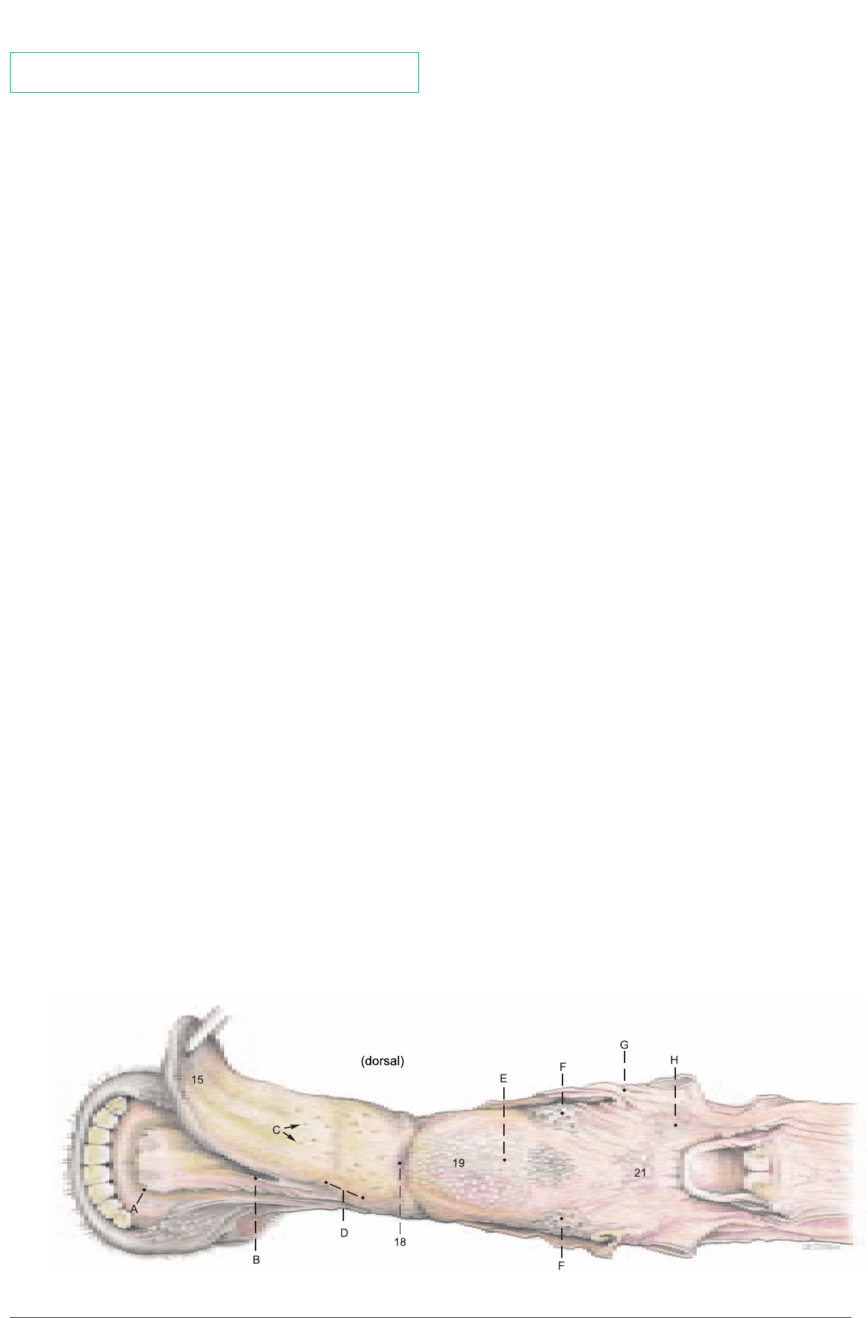
c) TONGUE.
The dorsal surface (dorsum linguae) is divided by the transverse lin-
gual fossa (18) into a flat apical part and a high, rounded torus lin-
guae (19). The tip (apex, 15) of the tongue is pointed. The apical
half of the tongue is covered on the dorsum and margin by fine,
sharp filiform papillae (D) directed backward and adapted to the
use of the tongue as an organ of prehension in grazing. Scattered
among the filiform papillae are round fungiform papillae (C),
which bear taste buds, as do the vallate papillae (F). The latter form
an irregular double row of about twelve on each side of the caudal
part of the torus, which is covered by large conical and lentiform
papillae (E). Foliate papillae are absent. The palatoglossal arches
(lat. to G) are attached to the sides of the root of the tongue (21).
On the root and on both sides of the median glossoepiglottic fold
are many small orifices of the crypts of the lingual tonsil (H) and its
glands.
44
8. NOSE AND NASAL CAVITIES, ORAL CAVITY AND TONGUE
Tongue
The nasal septum is removed to expose the nasal cavity.
Anatomie des Rindes englisch 09.09.2003 13:29 Uhr Seite 44
